Validation of the “Step-by-Step”
Approach in the Management
of Young Febrile Infants
Borja Gomez, MD, a, b Santiago Mintegi, MD, PhD, a, b Silvia Bressan, MD, PhD, c Liviana Da Dalt, MD, d Alain Gervaix, MD, e Laurence Lacroix, MD, e on behalf of the European Group for Validation of the Step-by-Step Approach
abstract
BACKGROUND: A sequential approach to young febrile infants on the basis of clinical and laboratory parameters, including procalcitonin, was recently described as an accurate tool in identifying patients at risk for invasive bacterial infection (IBI). Our aim was to prospectively validate the Step-by-Step approach and compare it with the Rochester criteria and the Lab-score.
METHODS: Prospective study including infants ≤90 days with fever without source presenting in 11 European pediatric emergency departments between September 2012 and August 2014. The accuracy of the Step-by-Step approach, the Rochester criteria, and the Lab-score in identifying patients at low risk of IBI (isolation of a bacterial pathogen in a blood or cerebrospinal fluid culture) was compared.
RESULTS: Eighty-seven of 2185 infants (4.0%) were diagnosed with an IBI. The prevalence of IBI was significantly higher in infants classified as high risk or intermediate risk according to the Step by Step than in low risk patients. Sensitivity and negative predictive value for ruling out an IBI were 92.0% and 99.3% for the Step by Step, 81.6% and 98.3% for the Rochester criteria, and 59.8% and 98.1% for the Lab-score. Seven infants with an IBI were misclassified by the Step by Step, 16 by Rochester criteria, and 35 by the Lab-score.
CONCLUSIONS: We validated the Step by Step as a valuable tool for the management of infants with fever without source in the emergency department and confirmed its superior
accuracy in identifying patients at low risk of IBI, compared with the Rochester criteria and the Lab-score.
aPediatric Emergency Department, Cruces University Hospital, Bilbao, Spain; bUniversity of the Basque Country, Bilbao, Spain; cPediatric Emergency Unit - Department of Woman's and Child Health, University of Padova, Italy; dCa’Foncello Hospital, Treviso, Italy; and ePediatric Emergency Division, Geneva University Hospitals and University of Geneva, Geneva, Switzerland
Dr Gomez conceptualized and designed the study, developed the design of the data collection forms, coordinated and supervised data collection, carried out the analyses, and drafted the initial manuscript; Dr Mintegi conceptualized and designed the study, collaborated in the revision of the data, and reviewed and revised the manuscript; Dr Bressan conceptualized and designed the study, was involved in data collection, and reviewed and revised the manuscript; Drs Da Dalt and Gervaix were involved in the design of the study, were involved in data collection, and reviewed and revised the manuscript; Dr Lacroix was involved in data collection and reviewed and revised the manuscript; and all authors approved the fi nal manuscript as submitted.
DOI: 10.1542/peds.2015-4381 Accepted for publication May 10, 2016
Address correspondence to Borja Gomez, MD, Pediatric Emergency Department, Cruces University Hospital, Plaza de Cruces s/n, Barakaldo, Spain. E-mail: [email protected]
To cite: Gomez B, Mintegi S, Bressan S, et al. Validation of the “Step-by-Step” Approach in the Management of Young Febrile Infants. Pediatrics. 2016;138(2):e20154381
WHAT’S KNOWN ON THIS SUBJECT: A sequential approach to young febrile infants on the basis of clinical and laboratory parameters, including procalcitonin, was recently described. When applied to retrospectively collected data, this tool revealed a good accuracy in identifying patients at low risk of invasive bacterial infection.
In the last 2 decades, several studies have been conducted to find the best set of criteria to identify those young febrile infants who are at a low risk of having a bacterial infection. These infants are candidates for outpatient management without receiving empirical antibiotic treatment. Since the classic Rochester, 1 Philadelphia, 2 and Boston 3 criteria were published, the management of infants younger than 90 days old with fever without source (FWS) has evolved. Regardless of the protocol used, current adherence to any of them in clinical practice is low. 4, 5 Changes in the epidemiology of bacterial pathogens in the last decades 6, 7 and introduction of biomarkers such as C-reactive protein (CRP) and, more recently, procalcitonin (PCT) could justify this low adherence rate and make several authors advocate for a more individualized approach. The latter includes new biomarkers and a reduction in lumbar puncture rates, antibiotic treatments, or in-hospital admission for many well-appearing infants outside the neonatal period. 8–10
The “Step by Step” is a new algorithm developed by a European group of pediatric emergency physicians. Its primary objective was to identify a low risk group of infants who could be safely managed as outpatients without lumbar puncture nor empirical antibiotic treatment. This approach ( Fig 1) evaluates sequentially the general appearance of the infant, the age, and result of the urinalysis and, lastly, the results of blood biomarkers, including PCT, CRP, and absolute neutrophil count (ANC). We retrospectively tested the Step-by-Step approach in 1123 infants 11 and found that it is able to accurately identify different groups of patients according to their risk of suffering from a noninvasive or invasive bacterial infection (IBI). In addition, this approach seemed to better identify low risk patients suitable for an outpatient
management compared with the Rochester criteria or the more recently developed Lab-score. 12, 13
The objective of this study was to prospectively validate these results in a larger multicenter population.
METHODS
Study Design
We conducted a multicenter prospective study including 11 European pediatric emergency departments (PEDs): 8 Spanish, 2 Italian, and 1 Swiss centers. Infants
≤90 days old attending with FWS between September 2012 and August 2014 were included. This study was approved by the Clinical Research Ethics Committee of the Basque Country and by the institutional review board at each study site. Written informed consent was requested from the parents or caregivers of the patient.
After data collection, the Step-by-Step approach was applied to the study sample to analyze its accuracy. The Rochester criteria and the Lab-score were also applied, and the diagnostic performances of the 3 sets of criteria were compared (Supplemental Table 6).
Clinical Management of the Patients
A urine dipstick, a urine culture collected by an aseptic technique (bladder catheterization or
suprapubic aspiration), white blood cell (WBC) count, CRP, PCT, and a blood culture were requested for each patient. The decision to perform any other test was made at the discretion of the physician in charge. The patients were admitted and/or received antibiotic treatment according to the management protocol of each center.
Exclusion Criteria
1. Clear source of fever identified after a careful medical history
and/or physical examination in the PED.
2. No fever on arrival at the PED and fever that had been only subjectively assessed by parents on touch, without the use of a thermometer.
3. Absence of 1 or more of the mandatory ancillary tests (blood culture, urine culture collected by an aseptic technique, urine dipstick, PCT, CRP, or WBC count).
4. Refusal of the parents or caregiver to participate
Data Collection
Deidentified data were collected through a standardized electronic form to be completed online and included age, sex, duration and degree of fever, general appearance of the patient on arrival at the PED, relevant medical history, results of laboratory tests, diagnosis, treatment, and site of care (managed as outpatient or admitted). The parents or caregivers of those infants managed as outpatients received a follow-up telephone call within 1 month after the initial visit at the PED to check the course of the episode. In case that after 3 telephone calls, it was not possible to contact with the caregivers, the electronic registries of the PED and the Public Health System were used to identify and review any posterior visit to the primary care center or to any other hospital.
Defi nitions
•FWS: Temperature measured at home or at the PED ≥38°C, in patients with a normal physical examination and no respiratory signs/symptoms or a diarrheal process.
previous hospitalization, and no chronic or underlying illness.
• Well-appearing: Defined by a normal Pediatric Assessment Triangle 14 in those PEDs in which this data are systematically recorded. For the other PEDs, infants were considered as not well-appearing if the findings of the physical examination documented in the medical record indicated any clinical suspicion of sepsis.
• IBI: Isolation of a bacterial pathogen in a blood or cerebrospinal fluid culture.
Staphylococcus epidermidis,
Propionibacterium acnes, Streptococcus viridans, or
Diphtheroides were considered contaminants.
• Non-IBI: Urinary tract infections (UTIs; urine culture with growth of ≥10 000 cfu/mL with leukocyturia associated) and bacterial gastroenteritis (isolation of bacteria in stool culture). When a registered patient received a diagnosis highly suggestive of having a bacterial etiology but with no positive bacterial culture, the case was discussed among the principal investigators to decide the most appropriate classification.
• Possible bacterial infection: Infants classified as possible UTI (positive urine culture without leukocyturia) and those finally diagnosed with a pneumonia or an acute otitis media with no positive bacterial culture.
• Sepsis: We used the sepsis criteria published by Goldstein et al 15 with only the following modification: Well-appearing patients with fever and leukocytosis were not diagnosed with a sepsis if they did not have any other sepsis criteria (tachycardia, bradycardia, tachypnea, or signs of organ dysfunction).
• Occult bacteremia: Presence of a pathogenic bacterium in the blood of a well-appearing infant with FWS.
FIGURE 1
Statistical Analysis
Normally distributed data were expressed as mean ± SD, nonnormally distributed data as median and interquartile range, and categorical variables were reported as percentages. We calculated the relative risk (RR) for presenting an IBI or a non-IBI in those infants presenting the risk factor evaluated on each step.
To compare the performance of this approach with the Rochester criteria and the Lab-score, we calculated the prevalence of IBI and non-IBI and the 95% confidence interval (95% CI) among those infants classified as low risk patients according to each protocol. We also calculated the sensitivity, specificity, positive and negative predictive values (PPVs and NPVs) and positive and negative
likelihood ratios (LRs) of the low risk criteria used on each protocol and the IBI missed according to each of the 3 approaches.
The statistical analysis was carried out using the IBM SPSS Statistics for Windows (version 21; IBM SPSS Statistics, IBM Corporation).
RESULTS
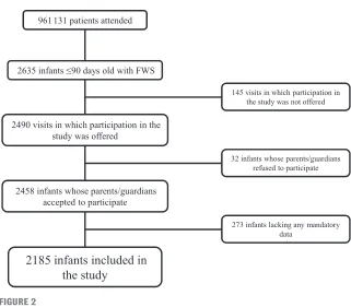
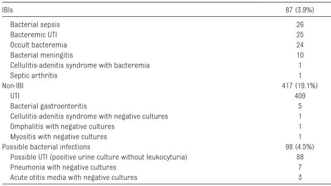
Overall, 966 413 patients attended, including 2635 infants ≤90 days old with FWS (0.27%). Of them, 2185 infants (82.9%) were finally included in the study ( Fig 2). Table 1 reports descriptive statistics for the main epidemiologic variables, complementary tests performed, and initial management. Of the 2185 included infants, 504 were diagnosed with a bacterial infection (23.1%), including 87 patients (3.9%) with an IBI and 417 (19.1%) with a non-IBI ( Table 2).
Applying the Step-by-Step approach, the prevalence of IBI and non-IBI in the different subgroups of patients is shown in Fig 3, as well as the corresponding RR for patients with each risk factor. The first part of the algorithm (evaluating general appearance, age, and presence of leukocyturia) identified 79.3% of the IBI (including 22 of 26 patients with sepsis and 9 of 10 with bacterial meningitis) and 98.5% of the non-IBI.
After taking into account PCT, CRP, and ANC values, we identified a subgroup of 991 low risk infants (45.3% of the studied population) with a prevalence of IBI of 0.7%. Supplemental Table 7 reveals the characteristics and initial management of the 7 infants who would have been classified as low risk patients and who were finally diagnosed with an IBI.
Nine other infants were diagnosed with a clinical sepsis without microbiological confirmation and 2 with a viral sepsis. All of them would FIGURE 2
Flow diagram to indicate the included and excluded patients.
TABLE 1 Epidemiologic and Clinical Characteristics, Complementary Tests, and Management of Patients
Age (median and interquartile range), d 47 (29–65)
≤21 d old, % 16.7
Sex (boy), % 59.5
Duration of fever (median and interquartile range), ha 5 (2–12) Highest temperature measured at home (median and interquartile range), °Cb 38.5 (38–38.8) Temperature upon arrival to the PED (median and interquartile range), °Cc 38.1 (37.8–38.5)
Previously healthy, % 85.9
Classifi ed as well appearing, % 87.7
PCT, CRP, WBC count, urine dipstick, urine culture collected by sterile method, blood culture, %
100
Lumbar puncture performed, % 27.4
Flu test, % 12.5
Antibiotic treatment, % 49.0
Admitted, % 58.5
Pediatric/neonatal ICU 1.6
a Evolution time was available in 2103 patients.
have been identified by the general appearance and age criteria.
Prevalence of bacterial infection among infants classified as low risk patients according to each set of criteria and number of IBIs that would have been misclassified are shown in Table 3. Prevalence of potentially missed IBI was higher when using the Lab-score or the Rochester criteria than the Step by Step (P < .05). Prevalence of non-IBI was also higher but differences only reach statistical significance when compared with the Lab-score. Prevalence of possible bacterial infection was similar in all the risk groups. Number needed to test with the Step by Step instead of with the Rochester criteria or the Lab-score to avoid missing an IBI was 102 infants and 81 infants, respectively. Table 4 reveals the diagnostic accuracy measures for each of the 3 approaches for identifying IBIs. The Step by Step had the lowest negative LR (0.17).
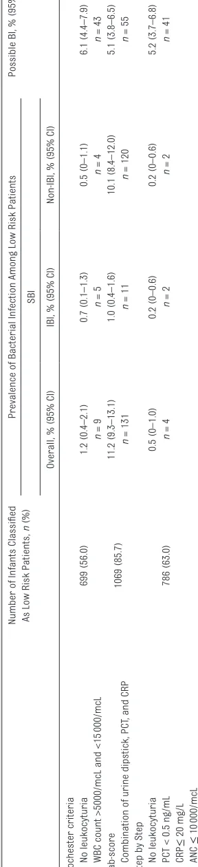
To compare specifically the performance of the complementary tests recommended by each set of criteria, we performed an “ad hoc” secondary analysis on those infants who met none of the clinical risk factors included in any of the 3 approaches (ie, well-appearing and previously healthy infants older than 28 days old). Although no lower age cutoff is defined by Rochester criteria, the Lab-score was validated for infants older than 7 days old, and the Step by Step considers 21 days old as a high risk cutoff. However, in clinical practice, and regardless of the protocol used, infants younger than 28 days old are usually more aggressively managed. Our results revealed that the Step by Step confirmed to be the most accurate tool of the 3 analyzed strategies to identify children at low risk of IBI ( Table 5).
DISCUSSION
Our results validate the Step-by-Step approach as an accurate tool to identify subgroups of young infants with FWS at different risk of IBI.
This approach includes both clinical and laboratory criteria, applying them in a sequential order, according to their clinical relevance, starting with the general appearance. Several studies have demonstrated that, as expected, febrile children who are not well appearing are at higher risk of bacterial infections, both in the general pediatric population 16 and in infants younger than 3 months. 17 In fact, clinical appearance is the factor that mostly increases this risk. 16, 17
The second item that the Step by Step evaluates is the age. Most classic guidelines consider 28 days old as the cutoff under which a complete sepsis workup and admission with empirical antibiotic treatment is recommended. 10, 18–20 However, more recent studies suggest alternative cutoff points. A descriptive study developed in 1 of our study participating centers evaluated retrospectively 1575 infants ≤90 days old with FWS analyzing the bacterial infection rate week by week.21 Infants 21 to 28 days old had a similar prevalence of bacterial infections compared with older patients and a lower rate than
infants ≤21 days old. Some authors even found a significant reduction in SBI rate after the second week of life, but without establishing different management strategies according to this cutoff. 22 Of note, in our study, 4 of the 7 patients finally diagnosed with an IBI and classified by the Step by Step as low risk patients were 22 to 28 days old (Supplemental Table 7). This finding, not observed in our previous retrospective study, 11 suggests to be cautious when assessing patients in the fourth week of age and recommends further studies to safely identify the best secondary age cutoff point.
Finally, leukocyturia identifies those infants with a high probability of having a UTI but also a subgroup of infants with an increased risk of having a bacteremia. 17 Indeed 1 of the most frequent IBI in young febrile infants is UTI with associated bacteremia. 23, 24
In our study, general appearance, age, and urine dipstick identified almost 80% of the IBI patients and, more interestingly, 85% of the sepsis and 90% of the bacterial meningitis. The bacterial meningitis and 3 of the 4 bacterial sepsis not detected by this first part of the algorithm would have been identified by an elevated PCT value. Several studies have compared the performance of PCT and CRP in the management of
TABLE 2 Bacterial Infections Diagnosed
IBIs 87 (3.9%)
Bacterial sepsis 26
Bacteremic UTI 25
Occult bacteremia 24
Bacterial meningitis 10
Cellulitis-adenitis syndrome with bacteremia 1
Septic arthritis 1
Non-IBI 417 (19.1%)
UTI 409
Bacterial gastroenteritis 5
Cellulitis-adenitis syndrome with negative cultures 1
Omphalitis with negative cultures 1
Myositis with negative cultures 1
Possible bacterial infections 98 (4.5%)
Possible UTI (positive urine culture without leukocyturia) 88
Pneumonia with negative cultures 7
young febrile infants. 25–27 PCT is a better biomarker to rule in an IBI, and, due its more rapid kinetic, 28 it is
a more suitable biomarker in young infants who, for the great majority, present to the PED with a very early
onset fever.17, 26, 27, 29 However, in 6 of 7 patients potentially missed by the Step by Step, fever duration FIGURE 3
was less than 2 hours, which is far too short even for PCT to rise. This very short fever duration makes the evaluation of these patients even more challenging and highlights the important role of a short-term PED observation in the management of these patients.
The intermediate risk group includes patients with an elevated CRP or ANC. We excluded the WBC count, because neither leukocytosis nor leukopenia have proved to be good predictors of bacterial infection in young infants. 30 –33
In comparing the Step-by-Step approach with previously developed sets of low risk criteria, we only focused on the Lab-score and Rochester criteria. This is because the Boston and Philadelphia criteria recommended performing systematically a lumbar puncture in all febrile infants. This would have implicated a change in the management protocols in use in the participating centers. In addition, most of the more recent guidelines do not recommend performing this test systematically, favoring an individualized approach that takes into account general appearance, age, and blood tests results. 10, 18–20, 34
Both the Rochester criteria and the Lab-score were developed to identify patients at risk for severe bacterial
infection globally. We further categorized severe bacterial infection into IBI and non-IBI for the different implications in terms of management and possible outcome. The Step by Step appears the most accurate of the 3 approaches for ruling out an IBI presenting the highest sensitivity and NPV and the best negative LR. On the contrary, and as expected due to the relatively low prevalence of IBI (4.0%), specificity, PPV, and positive LR were poor for all the 3 approaches when considering all the risk criteria of each protocol all together. In addition, the Step by Step provides risk estimates for both IBI and non-IBI according to the risk group that patients fall into during their sequential assessment. There are some reasons behind its better performance. Since the development of the Rochester criteria, the epidemiology of bacterial pathogens in young febrile infants has changed. Improvement in the perinatal antibiotic prophylaxis has reduced the incidence of S. agalactiae early-onset sepsis, 35–37
E coli is nowadays the leading cause of bacteremia in this population, 6, 7, 17, 24 and Listeria is rarely involved. 6, 7, 17, 24 On the other hand, new biomarkers that have been shown to be better predictors of IBI have been included in many management protocols. Curiously, the Lab-score, developed
less than 10 years ago, revealed a lower performance compared with previously published studies. This score was created to be applied in patients between 7 days and 36 months of age. Its derivation on this broad age range may account for its lower performance in younger infants, as bacterial pathogens and incidence of bacterial infections significantly varies with age. The same authors of this score found, in a validation study, that its sensitivity decreased with the age of the infant. 38
Of note, 3 of the 7 IBIs unidentified by the Step by Step attended the PED only 1 hour after the fever was firstly detected and in 3 other patients, fever was firstly detected on arrival at the PED (the reason for consultation was another complaint than fever). This very short fever duration makes the evaluation of these patients even more challenging and highlights the important role of a short-term PED observation in the management of these patients.
Our study has some limitations. First, the prevalence of SBI obtained in our study was similar to those reported in other recent European publications, 17, 25, 33 but higher than those reported in some US studies, 7, 23, 39–41 mainly due to an increased rate of UTI. This discrepancy can be explained by
TABLE 3 Prevalence of Bacterial Infection Among Low Risk patients According to Each Management Protocol Number of Infants
Classifi ed As Low Risk Patients, n (%)
Prevalence of Bacterial Infection Among Low Risk Patients
SBI Possible BI, (95% CI)
Overall, %, (95% CI) IBI, %, (95% CI) Non-IBI, %, (95% CI)
Rochester criteria 949 (43.4) 2.1 (1.2–3.0) 1.6 (0.9–2.5) 0.4 (0–0.8) 5.6 (4.2–7.2)
n = 20 n = 16 n = 4 n = 54
Lab-score 1798 (82.2) 10.8 (9.4–12.3) 1.9% (1.3–2.6) 8.8% (7.6–10.2) 5.0 (4.0–6.1)
n = 195 n = 35 n = 160 n = 91
Step by Step 991 (45.3) 1.1 (0.5–1.8) 0.7 (0.2–1.2) 0.4 (0–0.8) 5.1 (3.8–6.5)
n = 11 n = 7 n = 4 n = 51
TABLE 4 Sensitivity, Specifi city, PPVs, NPVs and Positive and Negative LR, with 95% CI, of Each Approach for Identifying IBIs
Sensitivity, % Specifi city, % PPV NPV Positive LR Negative LR
different denominators, reflecting the difference in inclusion criteria between studies. Although we included only infants with FWS, excluding specifically those patients in whom a clear source of fever was identified (bronchiolitis, upper respiratory tract infection, etc) in many US studies, include a broader population of febrile infants.
Second, although part of the
Rochester criteria, the absolute band count was not available in many of the participating centers and thus not included in our analysis. Including this item, the performance of the Rochester criteria could have varied.
Third, we have not been able to compare the Step by Step with other sets of criteria such as the Philadelphia or the Rochester criteria. According to 1 recent survey sent to the members of the American Academy of Pediatrics, 5 only 62% of the respondents reported using some set of published guidelines and among them, 20% cited using the Philadelphia protocol, 15% the Rochester criteria, and 13% the Boston criteria. As there seems to be no predominant criteria used in the United States and none of these classic risk criteria are frequently used in Europe, we chose the Rochester criteria for the reasons previously mentioned.
CONCLUSIONS
The Step-by-Step approach revealed a high sensitivity, being more accurate than the Rochester criteria and the Lab-score at identifying children at low risk of IBI, and appears to be a useful tool for the management of the febrile infant in the ED. However, as no perfect tool exists, the Step by Step is not 100% sensitive and physicians should use caution especially when assessing infants with very short fever evolution. For this subgroup of patients, we strongly advise for an initial period of close observation and monitoring in the ED, even when all the complementary test values are normal.
ACKNOWLEDGMENTS
We gratefully acknowledge the contribution of members of the European Group for validation of the Step-by-Step approach. The researchers from the 11 participating hospitals were as follows: Borja Gómez and Santiago Mintegi (Cruces University Hospital, Barakaldo, Spain); Isabel Durán (Carlos Haya Regional University Hospital, Málaga, Spain); Aristides Rivas (Gregorio Marañón General University Hospital, Madrid, Spain); Mercedes de la Torre (Niño Jesús Children’s University Hospital, Madrid, Spain); Daniel Blázquez (12 de Octubre University Hospital, Madrid, Spain); Izaskun Olaciregui (Donostia University Hospital, Donosti, Spain); Alain Gervaix and Laurence Lacroix (Geneva University Hospitals and University of Geneva, Geneva, Switzerland); Roberto Velasco (Río Hortega Universitary Hospital, Valladolid, Spain); Andrés González (Basurto University Hospital, Bilbao, Spain); Silvia Bressan and Veronica Mardegan (Dipartimento della salute della donna e del bambino– University of Padova, Padova, Italy); Anna Fabregas and Susana Melendo (Vall Hebron University Hospital, Barcelona, Spain); Liviana Da Dalt and Chiara Stefani (Ca’Foncello Hospital, Treviso, Italy).
ABBREVIATIONS
ANC: absolute neutrophil count CI: confidence interval
CRP: C-Reactive protein FWS: fever without source IBI: invasive bacterial infection LR: likelihood ratio
NPV: negative predictive value PCT: procalcitonin
PED: pediatric emergency department
PPV: positive predictive value RR: relative risk
UTI: urinary tract infection WBC: white blood cell
TABLE 5
Performance of the Complementary Tests Recommended by Each Set of Criteria Among the 1247 Well-Appearing and Previously Healt
hy Infants Older Than 28 d
Number of Infants Classifi
ed
As Low Risk Patients,
n
(%)
Prevalence of Bacterial Infection Among Low Risk Patients
Possible BI, % (95% CI)
SBI
Overall, % (95% CI)
IBI, % (95% CI)
Non-IBI, % (95% CI)
Rochester criteria 699 (56.0) No leukocyturia 1.2 (0.4–2.1) 0.7 (0.1–1.3) 0.5 (0–1.1) 6.1 (4.4–7.9)
WBC count >5000/mcL and <15
000/mcL n = 9 n = 5 n = 4 n = 43 Lab-score 1069 (85.7) 11.2 (9.3–13.1) 1.0 (0.4–1.6) 10.1 (8.4–12.0) 5.1 (3.8–6.5)
Combination of urine dipstick, PCT
, and CRP
n = 131 n = 11 n = 120 n = 55
Step by Step
786 (63.0) No leukocyturia 0.5 (0–1.0) 0.2 (0–0.6) 0.2 (0–0.6) 5.2 (3.7–6.8)
PCT < 0.5 ng/mL
REFERENCES
1. Jaskiewicz JA, McCarthy CA, Richardson AC, et al; Febrile Infant Collaborative Study Group. Febrile infants at low risk for serious bacterial infection–an appraisal of the Rochester criteria and implications for management. Pediatrics. 1994;94(3):390–396
2. Baker MD, Bell LM, Avner JR. Outpatient management without antibiotics of fever in selected infants. N Engl J Med. 1993;329(20):1437–1441
3. Baskin MN, O’Rourke EJ, Fleisher GR. Outpatient treatment of febrile infants 28 to 89 days of age with intramuscular administration of ceftriaxone. J Pediatr. 1992;120(1):22–27
4. Meehan WP III, Fleegler E, Bachur RG. Adherence to guidelines for managing the well-appearing febrile infant: assessment using a case-based, interactive survey. Pediatr Emerg Care. 2010;26(12):875–880
5. Jain S, Cheng J, Alpern ER, et al. Management of febrile neonates in US pediatric emergency departments.
Pediatrics. 2014;133(2):187–195 6. Biondi E, Evans R, Mischler M, et al.
Epidemiology of bacteremia in febrile infants in the United States. Pediatrics. 2013;132(6):990–996
7. Greenhow TL, Hung YY, Herz AM, Losada E, Pantell RH. The changing epidemiology of serious bacterial infections in young infants. Pediatr Infect Dis J. 2014;33(6):595–599 8. Huppler AR, Eickhoff JC, Wald ER.
Performance of low-risk criteria in the evaluation of young infants with fever: review of the literature. Pediatrics. 2010;125(2):228–233
9. Jhaveri R, Byington CL, Klein JO, Shapiro ED. Management of the non-toxic-appearing acutely febrile child:
a 21st century approach. J Pediatr. 2011;159(2):181–185
10. Ishimine P. Risk stratifi cation and management of the febrile young child. Emerg Med Clin North Am. 2013;31(3):601–626
11. Mintegi S, Bressan S, Gomez B, et al. Accuracy of a sequential approach to identify young febrile infants at low risk for invasive bacterial infection.
Emerg Med J. 2014;31(e1):e19–e24 12. Lacour AG, Zamora SA, Gervaix A. A
score identifying serious bacterial infections in children with fever without source. Pediatr Infect Dis J. 2008;27(7):654–656
13. Galetto-Lacour A, Zamora SA, Andreola B, et al. Validation of a laboratory risk index score for the identifi cation of severe bacterial infection in children with fever without source. Arch Dis Child. 2010;95(12):968–973
14. Dieckmann RA, Brownstein D, Gausche-Hill M. The pediatric assessment triangle: a novel approach for the rapid evaluation of children. Pediatr Emerg Care. 2010;26(4):312–315 15. Goldstein B, Giroir B, Randolph
A. International Pediatric Sepsis Consensus Conference: defi nitions for sepsis and organ dysfunction in pediatrics. Pediatr Crit Care Med.
2005;6(1):2–8
16. Craig JC, Williams GJ, Jones M, et al. The accuracy of clinical symptoms and signs for the diagnosis of serious bacterial infection in young febrile children: prospective cohort study of 15 781 febrile illnesses. BMJ. 2010;340:c1594 17. Gómez B, Mintegi S, Benito J, Egireun A,
Garcia D, Astobiza E. Blood culture and bacteremia predictors in infants less than three months of age with fever without source. Pediatr Infect Dis J. 2010;29(1):43–47
18. Baraff LJ. Management of infants and young children with fever without source. Pediatr Ann. 2008;37:673–679 19. Cincinnati Children’s Hospital
Medical Center. Evidence-based care guideline for fever of uncertain source in infants 60 days of age or less. October 27, 2010. Available at: www. cincinnatichildre ns. org/ workarea/ linkit. aspx? linkidentifi er= id& itemid= 87913& libid= 87601. Accessed May 24, 2016
20. National Institute for Health and Care Excellence. 2013. Feverish illness in children. Assessment and initial management in children younger than 5 years. London: National Institute for Health and Care Excellence. Available at: www. nice. org. uk/ guidance/ cg160. Accessed May 24, 2016
21. Garcia S, Mintegi S, Gomez B, et al. Is 15 days an appropriate cut-off age for considering serious bacterial infection in the management of febrile infants?
Pediatr Infect Dis J. 2012;31(5):455–458 22. Schwartz S, Raveh D, Toker O, Segal
G, Godovitch N, Schlesinger Y. A week-by-week analysis of the low-risk criteria for serious bacterial infection in febrile neonates. Arch Dis Child. 2009;94(4):287–292
23. Morley EJ, Lapoint JM, Roy LW, et al. Rates of positive blood, urine, and cerebrospinal fl uid cultures in children younger than 60 days during the vaccination era. Pediatr Emerg Care. 2012;28(2):125–130
24. Gomez B, Hernandez-Bou S, Garcia-Garcia JJ, Mintegi S; Bacteraemia Study Working Group from the Infectious Diseases Working Group, Spanish Society of Pediatric Emergencies (SEUP). Bacteremia in previously healthy children in emergency departments: clinical and microbiological characteristics and PEDIATRICS (ISSN Numbers: Print, 0031-4005; Online, 1098-4275).
Copyright © 2016 by the American Academy of Pediatrics
FINANCIAL DISCLOSURE: The authors have indicated they have no fi nancial relationships relevant to this article to disclose.
FUNDING: No external funding.
POTENTIAL CONFLICT OF INTEREST: The authors have indicated they have no potential confl icts of interest to disclose.
outcome. Eur J Clin Microbiol Infect Dis. 2015;34(3):453–460
25. Andreola B, Bressan S, Callegaro S, Liverani A, Plebani M, Da Dalt L. Procalcitonin and C-reactive protein as diagnostic markers of severe bacterial infections in febrile infants and children in the emergency department.
Pediatr Infect Dis J. 2007;26(8):672–677 26. Olaciregui I, Hernández U, Muñoz JA,
Emparanza JI, Landa JJ. Markers that predict serious bacterial infection in infants under 3 months of age presenting with fever of unknown origin. Arch Dis Child. 2009;94(7):501–505
27. Gomez B, Bressan S, Mintegi S, et al. Diagnostic value of procalcitonin in well-appearing young febrile infants.
Pediatrics. 2012;130(5):815–822 28. Dandona P, Nix D, Wilson MF, et
al. Procalcitonin increase after endotoxin injection in normal subjects. J Clin Endocrinol Metab. 1994;79(6):1605–1608
29. Bressan S, Andreola B, Cattelan F, Zangardi T, Perilongo G, Da Dalt L. Predicting severe bacterial infections in well-appearing febrile neonates: laboratory markers accuracy and duration of fever. Pediatr Infect Dis J. 2010;29(3):227–232
30. Bonsu BK, Chb M, Harper MB. Identifying febrile young infants
with bacteremia: is the peripheral white blood cell count an accurate screen? Ann Emerg Med. 2003;42(2):216–225
31. Bonsu BK, Harper MB. Utility of the peripheral blood white blood cell count for identifying sick young infants who need lumbar puncture. Ann Emerg Med. 2003;41(2):206–214
32. Gomez B, Mintegi S, Lopez E, Romero A, Paniagua N, Benito J. Diagnostic value of leukopenia in young febrile infants.
Pediatr Infect Dis J. 2012;31(1):92–95 33. Gomez B, Mintegi S, Benito J; Group
for the Study of Febrile Infant of the RiSeuP-SPERG Network. A prospective multicenter study of leukopenia in infants under 90 days of age with fever without source. Pediatr Infect Dis J. 2016;35(1):25–29
34. Martinez E, Mintegi S, Vilar B, et al. Prevalence and predictors of bacterial meningitis in young infants with fever without a source. Pediatr Infect Dis J. 2015;34(5):494–498
35. Schrag SJ, Zywicki S, Farley MM, et al. Group B streptococcal disease in the era of intrapartum antibiotic prophylaxis. N Engl J Med. 2000;342(1):15–20
36. Centers for Disease Control and Prevention (CDC). Early-onset and late-onset neonatal group B streptococcal disease--United States, 1996-2004.
MMWR Morb Mortal Wkly Rep. 2005;54(47):1205–1208
37. Phares CR, Lynfi eld R, Farley MM, et al; Active Bacterial Core surveillance/ Emerging Infections Program Network. Epidemiology of invasive group B streptococcal disease in the United States, 1999-2005. JAMA. 2008;299(17):2056–2065
38. Bressan S, Gomez B, Mintegi S, et al. Diagnostic performance of the lab-score in predicting severe and invasive bacterial infections in well-appearing young febrile infants. Pediatr Infect Dis J. 2012;31(12):1239–1244
39. Bachur RG, Harper MB. Predictive model for serious bacterial infections among infants younger than 3 months of age. Pediatrics. 2001;108(2):311–316
40. Newman TB, Bernzweig JA, Takayama JI, Finch SA, Wasserman RC, Pantell RH. Urine testing and urinary tract infections in febrile infants seen in offi ce settings: the Pediatric Research in Offi ce Settings’ Febrile Infant Study. Arch Pediatr Adolesc Med. 2002;156(1):44–54
DOI: 10.1542/peds.2015-4381 originally published online July 5, 2016;
2016;138;
Pediatrics
Step-by-Step Approach
Laurence Lacroix and on behalf of the European Group for Validation of the
Borja Gomez, Santiago Mintegi, Silvia Bressan, Liviana Da Dalt, Alain Gervaix,
Infants
Validation of the ''Step-by-Step'' Approach in the Management of Young Febrile
Services
Updated Information &
http://pediatrics.aappublications.org/content/138/2/e20154381
including high resolution figures, can be found at:
References
http://pediatrics.aappublications.org/content/138/2/e20154381#BIBL
This article cites 39 articles, 12 of which you can access for free at:
Subspecialty Collections
b
http://www.aappublications.org/cgi/collection/infectious_diseases_su
Infectious Disease
sub
http://www.aappublications.org/cgi/collection/emergency_medicine_
Emergency Medicine following collection(s):
This article, along with others on similar topics, appears in the
Permissions & Licensing
http://www.aappublications.org/site/misc/Permissions.xhtml
in its entirety can be found online at:
Information about reproducing this article in parts (figures, tables) or
Reprints
http://www.aappublications.org/site/misc/reprints.xhtml
DOI: 10.1542/peds.2015-4381 originally published online July 5, 2016;
2016;138;
Pediatrics
Step-by-Step Approach
Laurence Lacroix and on behalf of the European Group for Validation of the
Borja Gomez, Santiago Mintegi, Silvia Bressan, Liviana Da Dalt, Alain Gervaix,
Infants
Validation of the ''Step-by-Step'' Approach in the Management of Young Febrile
http://pediatrics.aappublications.org/content/138/2/e20154381
located on the World Wide Web at:
The online version of this article, along with updated information and services, is
http://pediatrics.aappublications.org/content/suppl/2016/07/01/peds.2015-4381.DCSupplemental
Data Supplement at:
by the American Academy of Pediatrics. All rights reserved. Print ISSN: 1073-0397.