Copyright © 2002, American Society for Microbiology. All Rights Reserved.
Sgt1p Contributes to Cyclic AMP Pathway Activity and Physically
Interacts with the Adenylyl Cyclase Cyr1p/Cdc35p
in Budding Yeast
Caroline Dubacq,
1Raphae¨l Guerois,
2Re´gis Courbeyrette,
1Katsumi Kitagawa,
3and Carl Mann
1*
Service de Biochimie et de Ge´ne´tique Mole´culaire1and Service de Biophysique des Fonctions Membranaires,2CEA/Saclay, F-91191 Gif-sur-Yvette, France, and Department of Molecular Pharmacology, St. Jude Children’s Research Hospital, Memphis, Tennessee 38105-27943
Received 3 December 2001/Accepted 29 April 2002
Sgt1p is a highly conserved eucaryotic protein that is required for both SCF (Skp1p/Cdc53p–Cullin–F-box)-mediated ubiquitination and kinetochore function in yeast. We show here that Sgt1p is also involved in the
cyclic AMP (cAMP) pathway inSaccharomyces cerevisiae.SGT1is an allele-specific suppressor ofcdc35-1, a
thermosensitive mutation in the leucine-rich repeat domain of the adenylyl cyclase Cyr1p/Cdc35p. We dem-onstrate that Sgt1p and Cyr1p/Cdc35p physically interact and that the activity of the cAMP pathway is affected
in ansgt1conditional mutant. Sequence analysis suggests that Sgt1p has features of a cochaperone. Thus, Sgt1p is
a novel activator of adenylyl cyclase inS.cerevisiaeand may function in the assembly or the conformational
activation of specific multiprotein complexes.
The targeted proteolysis of cell cycle regulators is one of the major mechanisms controlling cell proliferation (13, 37). The SCF (Skp1p/Cdc53p–Cullin–F-box) complexes direct proteins such as Cdk inhibitors and G1 cyclins to the proteasome through ubiquitination (15). The SCF ubiquitin ligase catalytic core (Cdc53p, Cdc34p, and Rbx1p) is linked by Skp1p to F-box protein subunits that recognize the SCF substrates. Skp1p is also a subunit of the CBF3 (centromere binding factor 3) kinetochore complex together with Ndc10p, Cep3p, and the F-box protein Ctf13p (14, 66). Skp1p is essential for the activ-ities of both complexes and is necessary for both entry into S phase and entry into anaphase (1, 14).
SGT1 (suppressor of the G2 allele of SKP1) was first de-scribed in yeast as a suppressor of skp1-4, a temperature-sensitive mutation ofSKP1that arrests cell division prefer-entially in G2/M at the restrictive temperature because of kinetochore defects (38).SGT1 is an essential gene that en-codes a highly conserved eucaryotic protein, as shown by the ability of the human homolog of Sgt1p to restore the viability of the budding yeast null mutant. A study ofsgt1-3andsgt1-5 thermosensitive mutants showed that Sgt1p, like Skp1p, is needed for both entry into S phase and kinetochore function (38). Sgt1p physically associates with Skp1p and is required for both SCF and CBF3 activities, but its exact function in these processes is not known.
We show here that Sgt1p also contributes to the activity of the cyclic AMP (cAMP) pathway and physically interacts with the yeast adenylyl cyclase Cyr1p/Cdc35p. Adenylyl cyclases cat-alyze the synthesis of cAMP from ATP. All eucaryotic and some bacterial adenylyl cyclases contain structurally similar catalytic domains that are functional in a homo- or hetero-dimeric configuration (31). They can be classified in four groups. Class I cyclases, found in metazoans, contain 12
trans-membrane segments and two cytosolic catalytic domains. Class II enzymes, found inDictyosteliumand protozoa, have an ex-tracellular domain, a single transmembrane segment, and a cytosolic catalytic domain. Class III cyclases are peripherally associated plasma membrane proteins with one cytosolic cat-alytic domain and are found in yeasts and fungi. Class IV cyclases are soluble proteins found in bacteria and mammalian testes. They are part of signal transduction pathways in both procaryotes and eucaryotes that regulate a wide range of bio-logical phenomena, including nutrient and stress responses (17); the regulation of cell growth, division, and differentiation (25, 40); hormonal responses (30); circadian rhythms; and long-term memory (48). In eucaryotes, adenylyl cyclases are regulated by GTP binding proteins (30). In budding yeast, a G␣subunit-type protein called Gpa2p and the GTP binding Ras proteins are both implicated in adenylyl cyclase activation in response to nutrient-rich conditions (71). Ras interacts with a leucine-rich repeat (LRR) region of yeast adenylyl cyclase and with a cyclase-associated protein called Cap/Srv2p (20, 62, 67).
The major effector of cAMP in eucaryotes is the cAMP-dependent protein kinase, or protein kinase A (PKA) (5). In the absence of cAMP, the catalytic subunit of PKA is found in an inactive complex with the regulatory subunit. The binding of cAMP by the regulatory subunit leads to dissociation of the complex and activation of the catalytic subunit (69). In budding yeast, the adenylyl cyclase pathway is notably involved in cell growth control and stress responses (71), but it also regulates the cell cycle by modulating G1cyclin expression (2, 29, 72) and the activities of the anaphase-promoting complex/cyclosome and the SCF pathway (32). In terms of the stress response, the activation of adenylyl cyclase and PKA antagonizes the action of the Msn2p and Msn4p transcription factors, which activate the transcription of stress-responsive genes (7, 44, 59, 65). The nuclear accumulation of Msn2p and Msn4p is inversely corre-lated with the activity of the adenylyl cyclase/PKA pathway (27). Glycogen accumulation is also inversely correlated with * Corresponding author. Mailing address: SBGM, CEA/Saclay,
F-91191 Gif-sur-Yvette, France. Phone: 33-1-69 08 34 32. Fax: 33-1-69 08 47 12. E-mail: [email protected].
568
on September 8, 2020 by guest
http://ec.asm.org/
the activity of the adenylyl cylase/PKA pathway (22). Our re-sults thus implicate Sgt1p in the activities of pathways control-ling cell growth and stress responses in addition to its previ-ously implicated role in pathways controlling cell division.
MATERIALS AND METHODS
Strains and plasmids.Table 1 lists the genotypes of the yeast strains used in this study. Table 2 lists the plasmids used in this study. ThePDE2deletion was made by digesting pJM⌬pde2withSacI-XbaI, transforming yeast cells with the products of digestion, and selecting for G418 resistance.SGT1and CYR1were epitope tagged by using the PCR cassettes pFA6a-13myc-TRP1and pFA6a-HIS3-pGAL1-3HA as described previously (41). We constructed pFA6a-KanMX6-ANB1-UB-R-LacI-4HA by exchanging theBglII-PacI fragment of pFA6a-kanMX6-pGAL1-3HA for theANB1-UB-R-lacI-4HAregion of ZM168 (49). This cassette contains a ubiquitin-arginine-lacI-4HA peptide under the control of theANB1promoter (49).
Sequencing of theCYR1/CDC35gene in the A364a strain background. Com-plete sequencing of the wild-typeCYR1/CDC35gene in the A364a strain back-ground (CMY282) revealed the following nucleotide differences compared with the published sequence of this gene in the S288C strain background (S288C sequence on the left and A364a sequence on the right): A9G, G911A, T2455C, T2508C, A2605G, G2956A, T3110C, A4305C, G4405A, and A5890G. Sequenc-ing also revealed the followSequenc-ing 24-bp insert (positions 1514 to 1537) in the A364a sequence relative to the S288C sequence: ACTCTCGTCATCGTAAGAACCG AC.
Glycogen staining and-galactosidase assay.Patches of yeast cells growing on 1% yeast extract–2% Bacto Peptone–2% dextrose (YPD) plates (2% agar) for
one of the experiments (see Fig. 5A) or on synthetic complete medium contain-ing 500M CuSO4for another experiment (see Fig. 6E) were stained for glycogen by overlaying the plates with a solution of 0.4% I2–0.2% KI (12). -Galactosidase activity was quantified as described previously (28).
Immunofluorescence microscopy and flow cytometry.Flow cytometric analysis of yeast strains was performed as described previously (43). Immunofluorescence analysis of yeast cells was performed essentially as described previously (53) by using purified anti-myc mouse monoclonal antibody 9E10 at a final concentration of 7g/ml and Alexa 594-labeled goat anti-mouse immunoglobulin G (Molec-ular Probes) at a final concentration of 20g/ml. DNA was stained with 0.5g of 4⬘,6⬘-diamidino-2-phenylindole (DAPI)/ml. Twenty opticalzsections sepa-rated by 150 nm were acquired by using a Leica DMRXA fluorescence micro-scope equipped with a⫻100 oil immersion objective mounted on a piezoelectric motor and MetaMorph software (Universal Imaging Inc). Out-of-plane fluores-cence was removed from each stack section by using a nearest-neighbor decon-volution algorithm with the default parameters provided by the MetaMorph software package.
Two-hybrid screen.Gal4 DNA binding domain (DBD)-Sgt1p (expressed from vector pOB2) was used as bait, and interacting protein fragments coded for in the yeast genome were selected in the presence of 25 mM 3-aminotriazole as de-scribed previously (21). Clones were then checked for the expression of -ga-lactosidase from the second reporter gene in the screening strain.
IP.Yeast strains CDY33, CDY34, and CDY35 were grown in 500 ml of 1% yeast extract–2% Bacto Peptone–2% galactose (YP-Gal) medium and harvested when the optical density at 600 nm reached 0.7. Cells were washed once with immunoprecipitation (IP) buffer (50 mM Tris [pH 7.5], 100 mM NaCl, 10 mM EDTA, 15% glycerol); resuspended in 700l of IP buffer containing 1 mM Pfabloc and 2g each of aprotinin, leupeptin, and pepstatin/ml; and broken in an Eaton press. Extracts were kept for 30 min on ice after the addition of 2 ml of IP buffer containing 1% Triton X-100. Extracts were centrifuged (JA20 rotor; 10 min, 4°C, 3,000 rpm), and the protein concentration in the supernatant was determined with the Bradford reagent (Bio-Rad). Crude extracts (1 to 4 mg of protein for anti-hemagglutinin [HA] IP or 4 mg for anti-myc IP in a final volume of 300l) were precleared with 25l of protein A-Sepharose beads that had been washed with IP buffer–1% Triton X-100 (1 h of incubation with rotation at 4°C and then centrifugation for 15 s at 100⫻g) and then were incubated with rotation for 1 h at 4°C with 2l of 12CA5 (anti-HA) ascites fluid or 9E10 (anti-myc) purified antibody (0.7 mg/ml). Protein A-Sepharose beads were sat-urated for nonspecific protein binding by incubation for 1 h at 4°C with 4 mg of total protein extract from wild-type strain YPH499. A 25-l quantity of saturated protein A-Sepharose beads was then incubated for 1 h at 4°C with anti-HA or anti-myc immune complexes. The beads were washed four times with IP buff-er–1% Triton X-100, and immunoprecipitated proteins were solubilized by heat-ing in the presence of sodium dodecyl sulfate-polyacrylamide gel electrophoresis sample buffer. Precleared extracts (10 to 40g), the IP supernatant (10 to 40g), and immunoprecipitated proteins were separated by sodium dodecyl sulfate-polyacrylamide gel electrophoresis, transferred to nitrocellulose membranes, incubated with 12CA5 or 9E10 antibody followed by anti-mouse immunoglobulin TABLE 1. Yeast strains used in this study
Strain Genotype Source orreference
CMY389 MATacyr1–2 leu2⌬ura3–52 trp1⌬1 ade2–101 lys2–801 This study
CMY391 MATacdc35-10 ura3–52 trp1⌬1 leu2⌬ade2–101 lys2–801 This study
CMY282 MATacdc35-1 sgt1-S371N ura3–52 trp1⌬1 his7 lys2–801(A364a) This study
CMY248 MATasgt1-S371N ura3–52 his7(A364a) Lee Hartwell
CDY33 MATaSGT1-13myc::TRP1 pGAL-3HA-CYR1::HIS3 ura3–52 trp1⌬63 his3⌬200 leu2⌬1 lys2–801 ade2–101 This study CDY34 MATaSGT1-13myc::TRP1 ura3–52 trp1⌬63 his3⌬200 leu2⌬1 lys2–801 ade2–101 This study CDY35 MATapGAL-3HA-CYR1::HIS3 ura3–52 trp1⌬63 his3⌬200 leu2⌬1 lys2–801 ade2–101 This study YPH499 MATaura3–52 lys2–801 ade2–10 trp1-⌬63 his3-⌬200 leu2-⌬1 64 YKK57 sgt1-5::LEU2 ura3–52 trp1⌬63 his3⌬200 leu2⌬1 lys2–801 ade2–101 38 CDY1 MATapde2⌬::kanMX ura3–52 trp1⌬63 his3⌬200 leu2⌬1 lys2–801 ade2–101 This study CDY3 pde2⌬::kanMX sgt1-5::LEU2 ura3–52 trp1⌬63 his3⌬200 leu2⌬1 lys2–801 ade2–101 This study CDY5 MATapde2⌬::kanMX cdc35–1 sgt1-S371N ura3–52 trp1⌬1 his7 lys2–801 This study ZMY60 (CDY27) MATapACE1-UBR1 pACE1-ROX1 trp1-⌬1 ura3-⌬52 leu2::PET56 ade2–101 49 CDY26 MATaANB1-UB-R-lacI-4HA-SGT1::kan pACE1-UBR1 pACE1-ROX1 trp1-⌬1 ura3-⌬52 leu2::PET56 ade2–101 This study CDY31 MATapSTRE-lacZ::URA3 pACE1-UBR1 pACE1-ROX1 trp1-⌬1 ura3-⌬52 leu2::PET56 ade2–101 This study CDY32 MATapSTRE-lacZ::URA3 ANB1-UB-R-lacI-4HA-SGT1::kan pACE1-UBR1 pACE1-ROX1 trp1-⌬1 ura3-⌬52
leu2::PET56 ade2–101 This study
CDY116 MATapGAL-3HA-cdc35-1::TRP1 sgt1-S371N ura3–52 his7 lys2–801 This study
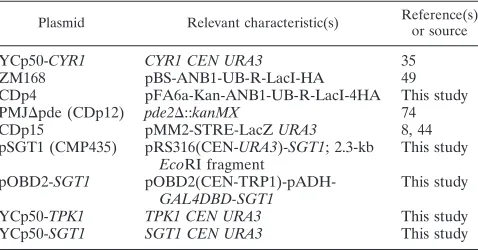
TABLE 2. Plasmids used in this study
Plasmid Relevant characteristic(s) Reference(s)or source
YCp50-CYR1 CYR1 CEN URA3 35
ZM168 pBS-ANB1-UB-R-LacI-HA 49
CDp4 pFA6a-Kan-ANB1-UB-R-LacI-4HA This study PMJ⌬pde (CDp12) pde2⌬::kanMX 74
CDp15 pMM2-STRE-LacZURA3 8, 44
pSGT1 (CMP435) pRS316(CEN-URA3)-SGT1; 2.3-kb
EcoRI fragment This study pOBD2-SGT1
pOBD2(CEN-TRP1)-pADH-GAL4DBD-SGT1 This study
YCp50-TPK1 TPK1 CEN URA3 This study
YCp50-SGT1 SGT1 CEN URA3 This study
on September 8, 2020 by guest
http://ec.asm.org/
G antibody coupled to horseradish peroxidase, and revealed by chemilumines-cence detection. For one of the experiments (see Fig. 3D), CDY116 was grown in YP-Gal at 24°C, whereas CDY116(pSGT1) was grown at 24°C on synthetic medium containing Casamino Acids, adenine, tryptophan, and 2% galactose but
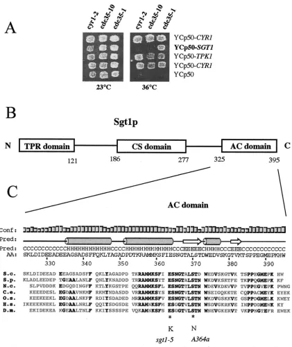
no uracil in order to maintain selection for plasmid pSGT1. Protein extracts and immunoprecipitates were prepared as described above, except that protein A-Sepharose beads containing covalently coupled 12CA5 anti-HA antibody were used. Immunoprecipitates used for anti-Sgt1p immunoblots were solubilized with FIG. 1. Isolation ofSGT1and structure of the encoded protein. (A)SGT1is an allele-specific suppressor ofcdc35-1.cdc35-1(CMY282),
cdc35-10(CMY391), andcyr1-2(CMY389) mutants were transformed with the indicated plasmids at 23°C, with selection for growth on synthetic medium without uracil. Patches of cells were then replica plated and incubated at 23 and 36°C to test for the suppression of thermosensitive growth. Ycp50-CYR1and Ycp50 are positive and negative controls, respectively, for complementation. (B) Schematic diagram of domains found within the Sgt1p sequence. The TPR and Sgt1 CHORD (CS domain) motifs were previously described (38, 63), and the highly conserved C-terminal sequence may be a domain that interacts with adenylyl cyclase (AC domain) in yeast cells. (C) Sequence and secondary structure prediction of the highly conserved C-terminal domain of Sgt1p from a series of eucaryotic organisms (S.c.,S.cerevisiae; S.p.,S. pombe; N.c.,N.crassa; C.e.,
Caenorhabditis elegans; O.s.,O.sativa, H.s.,H.sapiens, D.m.,Drosophila melanogaster) showing the positions of thesgt1-S371N(A364a) mutation and one of the twosgt1-5mutations. The secondary structure prediction (Pred) (H, alpha helix; E,strand; C, coil) was obtained with the Psi-Pred2 algorithm (34) (http://bioinf.cs.ucl.ac.uk/psipred/). The height of the bars is an estimate of the confidence (Conf) of the prediction. The region of Sgt1p between amino acids (AA) 338 and 365 is predicted with a high level of confidence to adopt a helix-turn-helix structure. Amino acid residues that are identical for at least five of the seven Sgt1p homologs are shown in bold.
on September 8, 2020 by guest
http://ec.asm.org/
sample buffer in the absence of -mercaptoethanol in order to reduce the liberation of immunoglobulin heavy chains from the protein A–anti-HA antibody beads that would otherwise obscure the Sgt1p signal on the immunoblots.
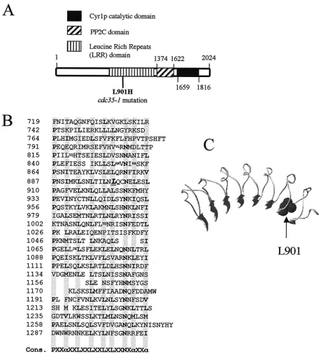
Sequence-structure threading and analysis.The sequence analysis of and the structure prediction for the different Sgt1p domains ofSaccharomyces cerevisiae were carried out by considering independently the following sequence regions:
positions 1 to 133, 168 to 219, and 220 to 395. The compatibility of the individual sequences and sequence alignments with known three-dimensional (3D) pro-tein structures was tested by using the sequence-structure threading methods FUGUE (61) (http://www-cryst.bioc.cam.ac.uk/⬃fugue/) and 3D-PSSM (36) (http: //www.bmm.icnet.uk/servers/3dpssm/). These algorithms recognize protein folds by using one-dimensional and 3D sequence profiles coupled with secondary FIG. 2. Thecdc35-1mutation affects a conserved leucine in the LRR domain of adenylyl cyclase. (A) Schematic diagram of the Cyr1p/Cdc35p sequence showing the position of the L901H substitution within the LRR domain of the cdc35-1mutant and the positions of the protein phosphatase 2C (PP2C) and adenylyl cyclase catalytic domains, as proposed by the Pfam database (3). (B) Sequence of the Cyr1p LRR domain showing the position of Leu-901 (underlined and in italic type). Note that this sequence corresponds to that of the wild-typeCYR1/CDC35gene in the A364a background, which differs at a number of positions from the published sequence of the gene in the S288C background (see Materials and Methods). Cons., consensus;␣, preferred hydrophobic residue. Shaded columns show the positions of preferred amino acids in the leucine-rich repeats. Small letters show sites of amino acid insertions within the repeats. (C) Structural model of the Cyr1p LRR domain in the environment of Leu-901.
on September 8, 2020 by guest
http://ec.asm.org/
FIG. 3. Sgt1p, but not Skp1p, coimmunoprecipitates with Cyr1p. (A) 3HA-Cyr1p overexpressed from a galactose-inducible promoter in strains CDY33 and CDY35 was immunoprecipitated with anti-HA antibody 12CA5, and the immunoprecipitates were analyzed by Western blotting to determine the efficiency of 3HA-Cyr1p IP and the fraction of coprecipitated Sgt1-13myc. Ten micrograms of the crude extract (E) and 10g of the supernatant (SN) are shown along with the total amount of the immunoprecipitated material (IP) from 1 mg of protein extract. (B) Sgt1-13myc expressed from its endogenous promoter in strains CDY33 and CDY34 was immunoprecipitated with anti-myc antibody 9E10, and the
on September 8, 2020 by guest
http://ec.asm.org/
structure and solvation potential information. They allow for the detection of remote sequence homology and for the optimization of sequence alignments.
For the region from positions 1 to 133, a preliminary multiple-sequence align-ment was calculated by using the Clustal W algorithm (33) withS.cerevisiae Sgt1p-homologous sequences from Schizosaccharomyces pombe, Neurospora crassa,Homo sapiens,Arabidopsis thaliana, andOryza sativa. The multiple-se-quence alignment was analyzed with the threading methods 3D-PSSM and FUGUE, both of which identified with high levels of confidence (over 95 and 99%, respectively) the tetratricopeptide repeat (TPR) domain of protein phos-phatase 5 as the most compatible fold (sequence identity, 13%; PDB no. 1A17). The scores for the prediction were anEvalue of 1.87⫻10⫺4and aZscore of 8.12 with the 3D-PSSM and the FUGUE methods, respectively. The 3D-PSSM method also identified the TPR domains of the Hop protein (PDB no. 1ELR and 1ELW) as highly probable folds for Sgt1 (positions 1 to 133), although their sequence identity with Sgt1p was rather low (15 and 16% for TPR1 and TPR2, respectively). The protein phosphatase 5 and Hop TPR domains bind the acidic C terminus of Hsp90 (56, 58). Interestingly, analysis of theS.cerevisiaeSgt1p sequence alone without the use of the multiple-sequence alignment did not allow any confident prediction.
For the second region (positions 168 to 219; CS domain), analysis of theS. cerevisiaeSgt1p sequence alone allowed the 3D-PSSM and FUGUE methods to identify with high levels of confidence (over 95 and 99%, respectively) the fold of the Hsp90 cochaperone p23 as the most probable fold (sequence identity, 18%; PDB no. 1EJF). The scores were anEvalue of 8.12⫻10⫺6and aZscore of 11.05 with the 3D-PSSM and the FUGUE methods, respectively. Similar values were obtained for the sequence ofH.sapiensSgt1 (21% sequence identity with p23) and the sequence of the Siah-interacting protein (SIP) (16% sequence identity with p23) (46).
Next, the sequence alignment was optimized and a structural model was generated by using the Centre de Bioinformatique Structurale server and the incremental threading optimization evaluation method (16, 39) (http://bioserv .infobiosud.univ-montp1.fr/bioserver/). Finally, regarding the 3D structure of the C-terminal region spanning the adenylyl cyclase-interacting domain (AC do-main), no prediction could be proposed with confidence. Nevertheless, the Psi-Pred2 method for secondary structure prediction (34) (http://bioinf.cs.ucl.ac.uk /psipred/) detected with a high level of confidence a helix-turn-helix motif in the region that is conserved in both the Sgt1p and the SIP families.
A structural model of the Cyr1p LRR domain was generated to analyze the location of mutated residue L901 in the LRR structure. The structure of the internalin B LRR domain (PDB no. 1D0B) was used as a template because it had the highest sequence identity with Cyr1p in the region of the mutation. The structural model was generated as described above.
RESULTS
SGT1suppresses the thermosensitivity of acdc35-1mutant.
cdc35-1is a temperature-sensitive allele of the adenylate cy-clase geneCYR1/CDC35that was isolated in the A364a genetic background (54). In genetic crosses with CYR1⫹/CDC35⫹
A364a strains, tetrads with 2TS⫹:2ts⫺ segregation were
ob-tained, as expected. However, in outcrosses with CYR1⫹/
CDC35⫹ strains in the S288C/YPH background, 3TS⫹:1ts⫺
segregation was observed, consistent with the presence of an unlinked suppressor locus in the S288C/YPH background. Fur-ther crosses showed that this suppressor acted in a dominant fashion. We thus sought to identify it by transforming the
cdc35-1A364a mutant with an S288C genomic DNA library in the YCp50 centromeric plasmid (55) and selecting for temper-ature-resistant growth. Three different genes were isolated in this manner (Fig. 1A):CYR1, encoding the wild-type adenylyl cyclase;TPK1/SRA3, encoding one of the catalytic subunits of PKA (9); andSGT1, encoding an Skp1p-interacting protein (38). These data suggested thatSGT1contributes to adenylyl cy-clase activity and that thesgt1allele found in the A364a strain background is deficient for this function. Sequencing of the sgt1allele in the A364a background showed that it encoded a protein that was identical to that encoded by theSGT1allele in the S288C background, except for an S371N substitution. Ser-371 is absolutely conserved in all known Sgt1p sequences and is found in a highly conserved C-terminal region of the protein (Fig. 1B and C). We found no phenotype associated with the sgt1-S371Nallele in the A364a background in the presence of a wild-typeCDC35/CYR1gene. Interestingly, one of the two amino acid substitutions found in thesgt1-5mutant (E364K) affects another highly conserved residue that maps close to the S371N substitution (Fig. 1C), and the sgt1-5 mutant has a thermosensitive G1 arrest phenotype that resembles that of
cdc35/cyr1mutants (38). Thus, thesgt1-5mutant may also have an adenylyl cyclase deficiency (see below).
Thecdc35-1allele has a point mutation in the LRR domain
of Cyr1p.YCp50-CYR1and YCp50-TPK1complemented two
other temperature-sensitive alleles for adenylyl cyclase,cyr1-2 and cdc35-10, whereas Ycp50-SGT1 could suppress only the thermosensitivity of cdc35-1(Fig. 1A) and is thus an allele-specific suppressor. Thecyr1-2mutation creates a UGA stop codon that is thought to lead to low levels of adenylyl cyclase by readthrough translation (50), and the cdc35-10mutation creates thermolabile adenylyl cyclase activity (6). In contrast, the cdc35-1-encoded adenylyl cyclase activity in membrane preparations was not thermosensistive and did not respond to G protein stimulation in vitro, suggesting that cdc35-1 may affect a Cyr1p regulatory site (10, 68). We sequenced the entire cdc35-1allele and found that it differs from the wild-type allele in the same genetic background by only a T2702A transversion that leads to an L901H substitution affecting a conserved leucine in the LRR domain of the protein (Fig. 2A and B). A structural model of this region of the Cyr1p LRR domain shows that L901 is likely to be part of the hydrophobic core of an LRR (Fig. 2C). The leucine-to-histidine mutation is pre-dicted to destabilize the structure of the hydrophobic core in this local region. The LRR domain has been implicated in Ras2 activation of Cyr1p (20, 62, 67). We thus tested whether the suppression ofcdc35-1bySGT1requiredRAS2. Acdc35-1 SGT1 strain grew at 37°C, whereas a cdc35-1 SGT1 ras2⌬
noprecipitates were analyzed by Western blotting to determine the efficiency of Sgt1-13myc IP and the fraction of coprecipitated overexpressed 3HA-Cyr1p. Ten micrograms of the crude extract (E) and 10g of the supernatant (SN) are shown along with the total amount of the immu-noprecipitated material (IP) from 4 mg of protein extract. (C) 3HA-Cyr1p overexpressed from a galactose-inducible promoter was immunopre-cipitated with anti-HA antibody 12CA5, and the immunoprecipitates were analyzed by Western blotting to determine the fraction of coprecipi-tating Sgt1-13myc and Skp1p. Forty micrograms of the crude extract (E) and 40g of the supernatant (SN) are shown along with the total amount of the immunoprecipitated material (IP) from 4 mg of protein extract. Rabbit anti-Skp1p antibodies were kindly provided by Wade Harper. (D) 3HA-Cdc35-1p overexpressed from a galactose-inducible promoter in strain CDY116 with or without pSGT1was immunoprecipitated from 1 mg of protein extract with protein A-Sepharose beads containing covalently coupled anti-HA antibody 12CA5. The immunoprecipitated material was then analyzed by immunoblotting with rabbit anti-Sgt1p polyclonal antibodies (upper panel) to test for coprecipitation of Sgt1-S371N or wild-type Sgt1p with Cdc35-1p or anti-HA antibodies to determine the level of 3HA-Cdc35-1p immunoprecipitated from each cellular extract (lower panels). In the upper panel, 20g each of crude extract (E) and supernatant (SN) was loaded next to the total amount of the immunoprecipitated material (IP).
on September 8, 2020 by guest
http://ec.asm.org/
strain showed thermosensitive growth similar to that of a cdc35-1 sgt1-S371N mutant (unpublished data). SGT1 and RAS2are thus both required to suppress the thermosensitive growth of thecdc35-1mutant.
Cyr1p and Sgt1p can be coimmunoprecipitated.The
local-ization of thecdc35-1mutation to the LRR domain and the allele specificity of cdc35-1 suppression by SGT1 suggested that Sgt1p and Cyr1p may physically interact. This notion was tested by immunoprecipitating tagged versions of each protein and determining whether the putative partner was coprecipi-tated. Sgt1-13myc was expressed from its normal chromosomal promoter. Attempts to visualize by immunoblotting Cyr1-3HA expressed from its normal chromosomal promoter were unsuc-cessful, so we overexpressed 3HA-Cyr1p from a galactose-inducible promoter. Under these conditions, we could readily demonstrate the presence of Sgt1-13myc in 3HA-Cyr1p immu-noprecipitations (Fig. 3A) and the presence of 3HA-Cyr1p in Sgt1-13myc immunoprecipitations (Fig. 3B). Approximately 1% of Sgt1p in the extracts was coprecipitated with overex-pressed 3HA-Cyr1p. This relatively low value may reflect the presence of Sgt1p in several different protein complexes (38) or a transient or labile interaction.
The coimmunoprecipitation of Sgt1-13myc and 3HA-Cyr1p indicates that they can form a complex and that they may interact directly. In contrast, we did not observe the coimmu-noprecipitation of Skp1p with 3HA-Cyr1p under conditions in which we readily observed the coimmunoprecipitation of Sgt1-13myc (Fig. 3C). This result suggests that Sgt1p forms exclusive complexes with Cyr1p and Skp1p.
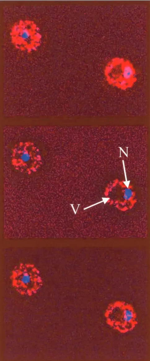
We also visualized the intracellular localization of Sgt1-13myc by indirect immunofluorescence with anti-myc antibod-ies. A stack of optical sections was acquired through fixed yeast cells expressing Sgt1-13myc and stained with DAPI to visualize nuclear DNA. The stack of images was then treated with a deconvolution program to remove out-of-plane fluorescence from each section. Figure 4 shows three consecutive images from az-section stack in which each image is separated by 150 nm. Sgt1-13myc was found throughout the cytosol as filiform or punctate aggregates. Sgt1-13myc was also observed in the nucleus as filiform or punctate aggregates, although its con-centration there might have been lower than that in the cy-tosol, and it was largely excluded from the vacuole. Similar images were also seen for the immunofluorescent localization of 6HA-Sgt1p (data not shown). No immunofluorescence sig-nal was detected under the same conditions from a congenic strain that did not express epitope-tagged Sgt1p. The observed nuclear and cytosolic localizations of Sgt1p are consistent with its proposed nuclear and cytosolic functions.
Interaction of Sgt1p with the LRR domain.The localization
of the cdc35-1 mutation to the LRR domain of Cyr1p sug-gested that Sgt1p may directly or indirectly bind this domain. We compared the ability of wild-type Sgt1p and Sgt1p-S371N to bind the Cdc35-1p mutant. 3HA-Cdc35-1p was overex-pressed from theGAL promoter at 24°C in cells expressing FIG. 4. Indirect immunofluorescence analysis of strain CDY34
done to visualize the intracellular localization of Sgt1-13myc with pu-rified mouse anti-myc monoclonal antibody 9E10. DNA was visualized with DAPI staining. Three consecutive optical sections separated by 150 nm are shown from az-section stack after deconvolution of the stack in order to remove out-of-plane fluorescence from each section. Sgt1-13myc fluorescence is shown in red, DNA is shown in blue, and
colocalization is shown in magenta. Under identical conditions, no immunofluorescence signal was observed for a congenic strain that did not express a myc-tagged protein. N, nucleus; V, vacuole. The yeast cells shown are about 5m in diameter.
on September 8, 2020 by guest
http://ec.asm.org/
only Sgt1p-S371N or in cells expressing Sgt1p-S371N and wild-type Sgt1p. 3HA-Cdc35-1p was then immunoprecipitated from cell extracts with anti-HA antibodies, and coprecipitation of Sgt1p was tested by immunoblotting with anti-Sgt1p rabbit polyclonal antibodies. Sgt1p coprecipitated with Cdc35-1p (Fig. 3D), as it did with wild-type Cdc35p/Cyr1p (Fig. 3A), but Sgt1p-S371N did not coprecipitate with Cdc35-1p (Fig. 3D). The defect in the physical interaction between Cdc35-1p and Sgt1p-S371N correlates with the thermosensitivity of strains containing both of the mutations and supports the idea that the C terminus of Sgt1p may interact with the LRR domain of Cdc35p.
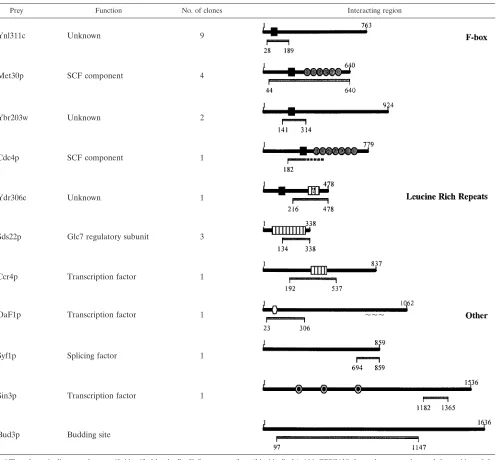
In order to further define its binding specificity, we used Sgt1p as bait in a two-hybrid screen with a library of coding fragments from the yeast genome as interacting prey. Three classes of interacting protein sequences were identified (Table 3). The first class was represented by protein fragments con-taining the F-box motif (52). Since Skp1p binds the F-box motif (60) and Sgt1p binds Skp1p (38), this class of proteins may indirectly interact with Sgt1p through Skp1p. The second pro-tein class contained well-defined (Sds22p and Ccr4p) or po-tential (Ydr306c) LRR domains. Sds22p is mainly composed of LRR sequences, and the Sgt1p-interacting region is almost entirely derived from the LRR domain. Thus, Sgt1p may gen-TABLE 3. Sgt1p-interacting protein fragments coded within the yeast genome and identified by a two-hybrid screena
Prey Function No. of clones Interacting region
Ynl311c Unknown 9
Met30p SCF component 4
Ybr203w Unknown 2
Cdc4p SCF component 1
Ydr306c Unknown 1
Sds22p Glc7 regulatory subunit 3
Ccr4p Transcription factor 1
OaF1p Transcription factor 1 ~~~
Syf1p Splicing factor 1
Sin3p Transcription factor 1
Bud3p Budding site
aThe schematic diagrams show motifs identified by the ProfileScan server (http://hits.isb-sib.ch/cgi-bin/PFSCAN) for each gene product and the position of the two-hybrid-interacting region. Ydr306c was predicted with a low level of confidence to contain an LRR motif (question mark). Symbols represent the interacting region (gray bar), F-box motif (black square), LRR (white bar), WD40 repeat (gray circle), Zn2Cys6 cluster domain (white hexagon), and paired amphipathic helix repeat (black circle within white circle).
on September 8, 2020 by guest
http://ec.asm.org/
on September 8, 2020 by guest
http://ec.asm.org/
erally bind LRR domains. Another intriguing observation is that Ydr306c is an F-box protein (52), but the interacting region identified in our screen contains the putative LRR se-quence and not the F-box motif. Thus, Sgt1p may interact both directly and indirectly with some F-box proteins. The third class of interacting proteins had no shared sequence motifs and is of uncertain functional significance.
Increased intracellular levels of cAMP modify the sgt1-5
phenotype.IfSGT1 is required for adenylyl cyclase function,
somesgt1mutants should show phenotypes similar to those of cdc35/cyr1mutants. BecauseSGT1is essential forS.cerevisiae viability, we studied conditional mutants. Thesgt1-5 thermo-sensitive mutant is affected in its SCF function and is unable to enter S phase at the restrictive temperature like other SCF mutants (38); however, rather than showing a multibud phe-notype like other SCF mutants, it instead shows arrested growth with unbudded cells similar to the arrest seen forcdc35/ cyr1mutants (45, 54). Moreover, iodine staining showed that thesgt1-5 strain accumulates glycogen at the restrictive tem-perature (Fig. 5A), as do cAMP pathway mutants, such as cdc35-1 (22). These phenotypes suggested that the cAMP pathway may be deficient in thesgt1-5mutant at the restrictive temperature.
We tested a possible contribution of deficient adenylyl cy-clase function to the sgt1-5phenotype by deleting the PDE2 gene, which encodes a high-affinity cAMP phosphodiesterase (57).pde2 mutants have increased intracellular cAMP levels and are phenotypically more sensitive to the addition of cAMP to the growth medium (47, 74). Deletion ofPDE2suppressed the glycogen accumulation of both thesgt1-5and thecdc35-1 mutants at 37°C (Fig. 5A). Furthermore, when ansgt1-5 pde2 double mutant was shifted to 37°C, it showed arrested growth after two or three cell divisions, whereas the parentalsgt1-5 single mutant showed arrested growth under the same condi-tions after one cell doubling (Fig. 5B). Deletion ofPDE2also partially suppressed the unbudded G1-phase arrest of the
sgt1-5mutant (Fig. 5C to E). Approximately half the cells of the sgt1-5 pde2 double mutant showed arrested division at 37°C, with large buds and a 2C DNA content. About 75% of large-bud cells contained an undivided nucleus, and the re-mainder were arrested in late nuclear division. These results suggested that inadequate cAMP synthesis partially accounts for the unbudded G1-phase arrest, but they also suggested that the sgt1-5 mutant is deficient in at least one other pathway required for growth and budding, as well as pathways required for nuclear division and cytokinesis. The defect in nuclear division is probably due to a kinetochore assembly problem (38).
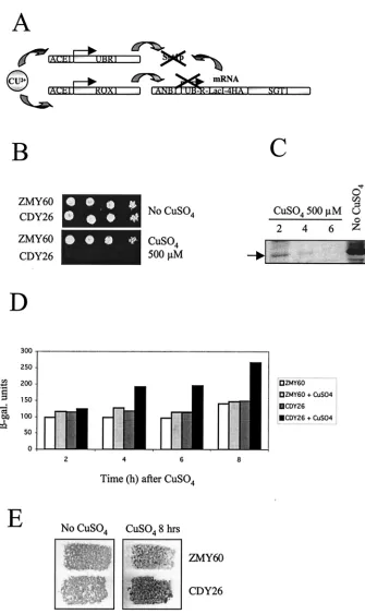
The cAMP pathway is affected in a conditional null mutant
for SGT1. The currently available thermosensitive alleles of SGT1may be deficient only in a subset of its functions (38). To further test the possibility that Sgt1p is an activator of adenylyl cyclase, we constructed a conditional null mutant forSGT1by using N-degron-Sgt1p expressed from a repressible promoter (Fig. 6A) and the system developed by Moqtaderi et al. (49). In this system, copper induces the expression of Ubr1p and Rox1p. Ubr1p is a ubiquitin ligase that binds proteins contain-ing an N-degron and facilitates their ubiquitination and sub-sequent destruction by the 26S proteasome. Rox1p represses the transcription of theANB1promoter controlling the expres-sion of N-degron-Sgt1p. Thus, the addition of copper both blocks the expression of N-degron-Sgt1p and triggers its pro-teolysis. As expected, the constructed strain was unable to grow on medium containing 500 M CuSO4 (Fig. 6B). The levels of N-degron-Sgt1p decreased rapidly after CuSO4 addi-tion and were markedly lower after 4 h of incubaaddi-tion (Fig. 6C). We used a stress response element (STRE)-LacZ reporter system to evaluate the activity of the cAMP pathway (44). STRE-mediated expression is controlled by the Msn2p and Msn4p transcription factors, whose activity is inversely propor-tional to the activity of the cAMP pathway (7, 24, 27, 65). Induced proteolysis of Sgt1p led to increased-galactosidase expression from STRE-LacZ (Fig. 6D) and to the accumula-tion of glycogen, as indicated by iodine staining of cells (Fig. 6E). Combined with our other data, these results suggested that depleting Sgt1p results in reduced cAMP levels, which activate Msn2p and Msn4p, and triggers glycogen accumula-tion.
Sequence analysis of the CS domain of Sgt1p suggests
fea-tures of a cochaperone.An amino-terminal region of Sgt1p was
suggested to adopt a TPR fold similar to that of Sti1/Hop (38), a cochaperone for Hsp70 and Hsp90 (11, 58). We obtained similar results by using the 3D-PSSM and FUGUE sequence threading methods (36, 61) to model this region (see Materials and Methods). The central region of Sgt1p contains a cysteine-and histidine-rich domain (CHORD) motif (CS domain) that is shared with the SIP family (46) and some other CHORD-containing proteins (63). The 3D-PSSM and FUGUE methods fitted with high levels of confidence (over 95 and 99%, respec-tively) the CS motif ofS. cerevisiaeSgt1p to the fold of the Hsp90 cochaperone p23 (sequence identity, 18%; PDB no., 1EJF). Similar results were obtained for the CS motifs ofH. sapiensSgt1p and SIP, an Sgt1-related protein that also binds Skp1p (46).
Figure 7A shows a sequence alignment of the CS motifs of Sgt1p, SIP, and p23. The positions of the hydrophobic residues buried in the structure of p23 (73) are highlighted in gray (solvent-accessible surface, below 20%). Figure 7B shows a
FIG. 5. Elevation of intracellular cAMP levels affectssgt1-5phenotypes. (A) Wild-type (WT; YPH499),sgt1-5(YKK57),cdc35-1(CMY282),
pde2⌬(CDY1),sgt1-5 pde2⌬(CDY3), andcdc35-1 pde2⌬(CDY5) strains were stained with iodine to visualize the accumulation of glycogen in patches of yeast colonies replicated on YPD plates at 24 and 37°C. The darkly stainingsgt1-5andcdc35-1patches of cells incubated at 37°C indicate a high level of glycogen in these cells. (B) Exponentially growingsgt1-5cells (open circles) andsgt1-5 pde2⌬cells (filled circles) on YPD plates at 24°C were transferred to 37°C, and the cell multiplication factor (number of cells at various times [Nt] divided by the number of cells at time zero
[N0]) was determined at the indicated times (T). (C) Fluorescence-activated cell sorting analysis of DNA content. Times (T) are given in hours.
1n, G1-phase DNA content; 2n, G2-phase DNA content. (D) Morphological analysis ofsgt1-5andsgt1-5 pde2⌬cells at 24°C (time zero) and after
transfer to 37°C for the indicated times (T, in hours). (E) Fluorescent phase-contrast images ofsgt1-5andsgt1-5 pde2⌬cells stained with propidium iodide to visualize nuclear DNA. Bar, 10m.
on September 8, 2020 by guest
http://ec.asm.org/
FIG. 6. Induced proteolysis of N-degron-Sgt1p leads to arrest of cell growth and activation of the expression of a STRE-LacZ reporter construction. (A) Schematic diagram of the N-degron system (49) used to create a conditional null mutant of Sgt1p by induced proteolysis. (B to E) The addition of 0.5 mM CuSO4to strain CDY26 (N-degron-Sgt1p) growing on synthetic complete medium at 30°C but not parental strain
ZMY60 blocks cell growth (B); induces the proteolysis of N-degron-4HA-Sgt1p after 2, 4, and 6 h of incubation with CuSO4, as determined by
immunoblotting with anti-HA antibodies (arrow, position of R-LacI-4HA-Sgt1p) (C); activates the expression of STRE-LacZ (-gal., -galacto-sidase) (D); and triggers glycogen accumulation specifically in strain CDY26 after 8 h of incubation, as shown by iodine staining of patches of cells replicated on agar medium plates (E).
on September 8, 2020 by guest
http://ec.asm.org/
ribbon representation of the structural model of the CS motif derived from the fold recognition methods. Mutation N213I in thesgt1-3 mutant appears to disrupt the binding of Sgt1p to Skp1p (the other mutations insgt1-3, L31P and F99L, are lo-cated in the TPR domain, which does not appear to be re-quired for Skp1p binding) (38, 46). From the structural model, we found close to conserved N213 several exposed residues that are conserved inS.cerevisiaeSgt1p,H.sapiensSgt1p, and SIP. Together with N213, they may take part in the binding of Sgt1p or SIP to the Skp1p N-terminal domain (60). Figure 7C shows the accessible surface of this set of residues colored with respect to sequence diversity. Their corresponding positions in the sequence alignment are highlighted. Figure 7D shows the accessible surface of the set of residues close to N213 with a
color code related to the amino acid type. This view suggests that the putative binding region may involve both hydrophobic and polar interactions. The remarkable structural similarity of the Sgt1p TPR and CS domains to Hsp90 cochaperones sug-gests that Sgt1p may have a similar function, although we have not succeeded in immunoprecipitating Hsp90 with Sgt1-13myc (unpublished data).
DISCUSSION
Sgt1p was initially described as an Skp1p-interacting protein involved in kinetochore assembly and SCF activity (38). We demonstrate here that Sgt1p also contributes to the activity of the cAMP pathway and physically interacts with yeast adenylyl FIG. 7. Sequence threading suggesting that the C5 domain of Sgt1p adopts a fold similar to that of the Hsp90 cochaperone p23. (A) Sequences of the CS domains ofS.cerevisiaeSgt1 (Sgt1_sc),H.sapiensSgt1 (Sgt1_hs), and SIP that align with that of the Hsp90 cochaperone p23 (PDB no. 1EJF), validated by using fold recognition methods. The positions of the hydrophobic residues buried in the structure of p23 (73) are highlighted in gray (solvent-accessible surface, below 20%). The arrows indicate the positions of the strands in the structure of p23, and their colors correspond to those of thestrands in panel B. The red asterisk indicates conserved N213. (B) The structural model derived from the fold recognition methods is shown as a ribbon representation. The colors of the strands indicate the order of the secondary structure elements from the N terminus (blue) to the C terminus (red). (C) The accessible surface of a set of residues close to N213 (implicated in binding Skp1p) is shown and colored with respect to the sequence diversity (blue for conserved, green for similar, and yellow for nonconserved positions). Their corresponding positions in the sequence alignment are indicated. (D) The accessible surface of the set of residues close to N213 is shown with a color code related to the amino acid type (gray for hydrophobic, yellow for polar, and blue for positively charged amino acids).
on September 8, 2020 by guest
http://ec.asm.org/
cyclase. An initial genetic interaction was based on the pres-ence of a defective allele ofSGT1 in the A364a strain back-ground. In an otherwise wild-type background, the A364asgt1 allele had no evident phenotypic effect. However, when com-bined with acdc35-1mutation, it led to thermosensitive growth due to the inactivation of adenylyl cyclase. Thecdc35-1 muta-tion itself had no evident phenotypic effect in anSGT1⫹
back-ground. This interaction was allele specific becauseSGT1⫹did
not suppress two other thermosensitive adenylyl cyclase muta-tions,cyr1-2andcdc35-10. Thecdc35-1mutation is an L901H substitution in the LRR region of adenylyl cyclase, and the defective sgt1 allele contains an S371N mutation affecting a highly conserved residue in the C-terminal region of the pro-tein. We further showed that Sgt1p and Cyr1p/Cdc35p, but not Skp1p and Cyr1p/Cdc35p, could be coimmunoprecipitated from yeast extracts. Furthermore, Sgt1p, but not the Sgt1p-S371N mutant, could be coimmunoprecipitated with the Cdc35-1p mutant. These results suggest that the C-terminal conserved domain of Sgt1 may interact with the LRR domain of adenylyl cyclase and that this interaction may exclude an interaction between Sgt1p and Skp1p. Supporting a possible interaction site on Sgt1p for LRR domains is our observation of two-hybrid interactions between Sgt1p and fragments of Sds22p and Ccr4p that contain LRR motifs.
How might Sgt1p contribute to adenylyl cyclase activity in yeast cells? Biochemical analyses of yeast and fungal adenylyl cyclases are not as advanced as the analysis of mammalian adenylyl cyclases. Like mammalian and fission yeast adenylyl cyclases, budding yeast Cyr1p can be activated by a G␣family protein called Gpa2p (71). However, Cyr1p activity also re-quires the small G protein Ras, and this property is not con-served in either fission yeast or mammals (4, 23). Immuno-affinity purification of overexpressed, epitope-tagged Cyr1p demonstrated that it was approximately stoichiometrically as-sociated with a 70-kDa protein called cyclase-asas-sociated tein (CAP)/Srv2p (18, 19, 26). Other Cyr1p-interacting pro-teins, including Sgt1p, Ras2p, and Gpa2p, were not identified. This result may mean that these regulators were present in substoichiometric quantities relative to the overexpressed Cyr1p or that their affinity for Cyr1p was lower than that of CAP. The N-terminal domain of CAP was shown to form a coiled-coil interaction with the C terminus of Cyr1p and to be required along with the Cyr1p LRR domain for Ras2p-medi-ated activation of adenylyl cyclase (20, 51, 67). Nevertheless, the precise mechanistic basis for the activation of Cyr1p by Ras2p and CAP or by Gpa2p is unknown. Adenylyl and gua-nylyl cyclases generally function as homo- or heterodimers (31). Studies of G␣activation of mammalian adenylyl cyclase suggest that it can both facilitate dimerization and induce a conformational change in the dimerized subunits that in-creases catalytic activity (70). The immunoaffinity-purified Cyr1p-CAP complex sediments as a dimer on glycerol gradi-ents (19), a finding which would suggest that Sgt1p, Ras2p, and Gpa2p are not required for dimerization. It remains possible that these proteins facilitate the dimerization of the complex in situ within cells or that they act as allosteric activators of the dimerized complex.
Sgt1p and, notably, the C-terminal region that we have im-plicated in a Cyr1p interaction, are highly conserved in euca-ryotes. Adenylyl cyclase catalytic domains are also conserved,
but sequences outside of the catalytic domains are not. Re-markably, the C-terminal region of Git7p, the Sgt1p homolog in fission yeast, has also been shown to be required for adenylyl cyclase activity in this yeast (57a). Our results suggest that Sgt1p interacts with the LRR domain of Cyr1p, a domain that is also found in fission yeast and other fungal adenylyl cyclases (42, 75, 76). It is thus likely that Sgt1p is generally required for adenylyl cyclase activity in yeasts and fungi. However, the ab-sence of an LRR domain in metazoan adenylyl cyclases may indicate that Sgt1p is not required for adenylyl cyclase activity in multicellular organisms, although it may well interact with other LRR-containing proteins in metazoans.
Given the role of Sgt1p in several different types of multi-meric protein complexes, it is possible that it acts as some sort of protein chaperone, assembly factor, or allosteric activator. This hypothesis is consistent with structure predictions suggest-ing that the Sgt1p N-terminal region is similar to the TPR regions of the Sti1/Hop cochaperone and that the central CS domain adopts a fold similar to that of the p23 cochaperone. Hop facilitates protein substrate transfer from Hsp70 to Hsp90, whereas p23 stimulates the ATPase activity of Hsp90 and the release of bound substrate (77). Several experimental observations are also consistent with a role for Sgt1p as a cochaperone or assembly factor. First, Sgt1p is required to functionally activate the Skp1p and Ctf13p subunits of the CBF3 kinetochore complex but is not itself a subunit of this complex (38). Second, Sgt1p may be present in substoichio-metric quantities in SCF (38) and Cyr1p (this work) complexes. Further work should elucidate the role of Sgt1p in the function of the multiple distinct complexes that are critically involved in the control of cell growth and cell division.
ACKNOWLEDGMENTS
We thank Lee Hartwell for supporting the initial stages of this work and Emmanuelle Boy-Marcotte, Michel Jacquet, Georges Renault, Wade Harper, Amit Banerjee, and Jeffrey Field for plasmids, antibod-ies, and advice. We are grateful to Anne Chevalier for help with the two-hybrid experiments and Charlie Hoffman for the stimulating elec-tronic exchange of information.
C.D. was supported by Allocation Couple´e de l’Ecole Normale Su-pe´rieure de Paris.
REFERENCES
1.Bai, C., P. Sen, K. Hofmann, L. Ma, M. Goebl, J. W. Harper, and S. J. Elledge.1996. SKP1 connects cell cycle regulators to the ubiquitin proteol-ysis machinery through a novel motif, the F-box. Cell86:263–274. 2.Baroni, M. D., P. Monti, and L. Alberghina.1994. Repression of
growth-regulated G1 cyclin expression by cyclic AMP in budding yeast. Nature371:
339–342.
3.Bateman, A., E. Birney, R. Durbin, S. R. Eddy, K. L. Howe, and E. L. Sonnhammer.2000. The Pfam protein families database. Nucleic Acids Res.
28:263–266.
4.Beckner, S. K., S. Hattori, and T. Y. Shih.1985. The ras oncogene product p21 is not a regulatory component of adenylate cyclase. Nature317:71–72. 5.Beebe, S. J.1994. The cAMP-dependent protein kinases and cAMP signal
transduction. Semin. Cancer Biol.5:285–294.
6.Boutelet, F., A. Petitjean, and F. Hilger.1985. Yeastcdc35mutants are defective in adenylate cyclase and are allelic with cyr1 mutants while CAS1, a new gene, is involved in the regulation of adenylate cyclase. EMBO J.
4:2635–2641.
7.Boy-Marcotte, E., G. Lagniel, M. Perrot, F. Bussereau, A. Boudsocq, M. Jacquet, and J. Labarre.1999. The heat shock response in yeast: differential regulations and contributions of the Msn2p/Msn4p and Hsf1p regulons. Mol. Microbiol.33:274–283.
8.Boy-Marcotte, E., M. Perrot, F. Bussereau, H. Boucherie, and M. Jacquet.
1998. Msn2p and Msn4p control a large number of genes induced at the diauxic transition which are repressed by cyclic AMP in Saccharomyces cerevisiae. J. Bacteriol.180:1044–1052.
on September 8, 2020 by guest
http://ec.asm.org/
9.Cannon, J. F., and K. Tatchell.1987. Characterization of Saccharomyces cerevisiaegenes encoding subunits of cyclic AMP-dependent protein kinase. Mol. Cell. Biol.7:2653–2663.
10.Casperson, G. F., N. Walker, and H. R. Bourne.1985. Isolation of the gene encoding adenylate cyclase inSaccharomyces cerevisiae. Proc. Natl. Acad. Sci. USA82:5060–5063.
11.Chang, H. C., D. F. Nathan, and S. Lindquist.1997. In vivo analysis of the Hsp90 cochaperone Sti1 (p60). Mol. Cell. Biol.17:318–325.
12.Chester, V. E.1968. Heritable glycogen-storage deficiency in yeast and its induction by ultra-violet light. J. Gen. Microbiol.51:49–56.
13.Ciechanover, A., A. Orian, and A. L. Schwartz.2000. Ubiquitin-mediated proteolysis: biological regulation via destruction. Bioessays22:442–451. 14.Connelly, C., and P. Hieter.1996. Budding yeastSKP1encodes an
evolu-tionarily conserved kinetochore protein required for cell cycle progression. Cell86:275–285.
15.Deshaies, R. J.1999. SCF and Cullin/ring H2-based ubiquitin ligases. Annu. Rev. Cell Dev. Biol.15:435–467.
16.Douguet, D., and G. Labesse.2001. Easier threading through web-based comparisons and cross-validations. Bioinformatics17:752–753.
17.D’Souza, C. A., and J. Heitman.2001. Conserved cAMP signaling cascades regulate fungal development and virulence. FEMS Microbiol. Rev.25:349– 364.
18.Fedor-Chaiken, M., R. J. Deschenes, and J. R. Broach.1990.SRV2, a gene required for RAS activation of adenylate cyclase in yeast. Cell61:329–340. 19.Field, J., J. Nikawa, D. Broek, B. MacDonald, L. Rodgers, I. A. Wilson, R. A. Lerner, and M. Wigler.1988. Purification of a RAS-responsive adenylyl cyclase complex fromSaccharomyces cerevisiaeby use of an epitope addition method. Mol. Cell. Biol.8:2159–2165.
20.Field, J., H. P. Xu, T. Michaeli, R. Ballester, P. Sass, M. Wigler, and J. Colicelli.1990. Mutations of the adenylyl cyclase gene that blockRAS func-tion inSaccharomyces cerevisiae. Science247:464–467.
21.Flores, A., J. F. Briand, O. Gadal, J. C. Andrau, L. Rubbi, V. Van Mullem, C. Boschiero, M. Goussot, C. Marck, C. Carles, P. Thuriaux, A. Sentenac, and M. Werner.1999. A protein-protein interaction map of yeast RNA polymerase III. Proc. Natl. Acad. Sci. USA96:7815–7820.
22.Francois, J., and J. L. Parrou.2001. Reserve carbohydrates metabolism in the yeastSaccharomyces cerevisiae. FEMS Microbiol. Rev.25:125–145. 23.Fukui, Y., T. Kozasa, Y. Kaziro, T. Takeda, and M. Yamamoto.1986. Role
of a ras homolog in the life cycle ofSchizosaccharomyces pombe. Cell44:
329–336.
24.Garreau, H., R. N. Hasan, G. Renault, F. Estruch, E. Boy-Marcotte, and M. Jacquet.2000. Hyperphosphorylation of Msn2p and Msn4p in response to heat shock and the diauxic shift is inhibited by cAMP inSaccharomyces cerevisiae. Microbiology146:2113–2120.
25.Gerisch, G.1987. Cyclic AMP and other signals controlling cell development and differentiation inDictyostelium. Annu. Rev. Biochem.56:853–879. 26.Gerst, J. E., K. Ferguson, A. Vojtek, M. Wigler, and J. Field.1991. CAP is a
bifunctional component of the Saccharomyces cerevisiaeadenylyl cyclase complex. Mol. Cell. Biol.11:1248–1257.
27.Gorner, W., E. Durchschlag, M. T. Martinez-Pastor, F. Estruch, G. Am-merer, B. Hamilton, H. Ruis, and C. Schuller.1998. Nuclear localization of the C2H2zinc finger protein Msn2p is regulated by stress and protein kinase A activity. Genes Dev.12:586–597.
28.Guarente, L., and M. Ptashne.1981. Fusion of Escherichia coli lacZ to the cytochrome c gene ofSaccharomyces cerevisiae. Proc. Natl. Acad. Sci. USA
78:2199–2203.
29.Hall, D. D., D. D. Markwardt, F. Parviz, and W. Heideman.1998. Regulation of the Cln3-Cdc28 kinase by cAMP inSaccharomyces cerevisiae. EMBO J.
17:4370–4378.
30.Hanoune, J., and N. Defer.2001. Regulation and role of adenylyl cyclase isoforms. Annu. Rev. Pharmacol. Toxicol.41:145–174.
31.Hurley, J. H.1998. The adenylyl and guanylyl cyclase superfamily. Curr. Opin. Struct. Biol.8:770–777.
32.Irniger, S., M. Baumer, and G. H. Braus.2000. Glucose and ras activity influence the ubiquitin ligases APC/C and SCF inSaccharomyces cerevisiae. Genetics154:1509–1521.
33.Jeanmougin, F., J. D. Thompson, M. Gouy, D. G. Higgins, and T. J. Gibson.
1998. Multiple sequence alignment with Clustal X. Trends Biochem. Sci.
23:403–405.
34.Jones, D. T.2000. A practical guide to protein structure prediction. Methods Mol. Biol.143:131–154.
35.Kataoka, T., D. Broek, and M. Wigler.1985. DNA sequence and character-ization of theS.cerevisiaegene encoding adenylate cyclase. Cell43:493–505. 36.Kelley, L. A., R. M. MacCallum, and M. J. Sternberg. 2000. Enhanced genome annotation using structural profiles in the program 3D-PSSM. J. Mol. Biol.299:499–520.
37.King, R. W., R. J. Deshaies, J. M. Peters, and M. W. Kirschner.1996. How proteolysis drives the cell cycle. Science274:1652–1659.
38.Kitagawa, K., D. Skowyra, S. J. Elledge, J. W. Harper, and P. Hieter.1999. SGT1encodes an essential component of the yeast kinetochore assembly pathway and a novel subunit of the SCF ubiquitin ligase complex. Mol. Cell
4:21–33.
39.Labesse, G., and J. Mornon. 1998. Incremental threading optimization (TITO) to help alignment and modelling of remote homologues. Bioinfor-matics14:206–211.
40.Lockwood, A. H., S. K. Murphy, S. Borislow, A. Lazarus, and M. Pendergast.
1987. Cellular signal transduction and the reversal of malignancy. J. Cell. Biochem.33:237–255.
41.Longtine, M. S., A. McKenzie III, D. J. Demarini, N. G. Shah, A. Wach, A. Brachat, P. Philippsen, and J. R. Pringle.1998. Additional modules for versatile and economical PCR-based gene deletion and modification in Sac-charomyces cerevisiae. Yeast14:953–961.
42.Mallet, L., G. Renault, and M. Jacquet.2000. Functional cloning of the adenylate cyclase gene of Candida albicans in Saccharomyces cerevisiae within a genomic fragment containing five other genes, including homo-logues ofCHS6andSAP185. Yeast16:959–966.
43.Mann, C., J. Y. Micouin, N. Chiannilkulchai, I. Treich, J. M. Buhler, and A. Sentenac.1992.RPC53encodes a subunit ofSaccharomyces cerevisiaeRNA polymerase C (III) whose inactivation leads to a predominantly G1arrest. Mol. Cell. Biol.12:4314–4326.
44.Martinez-Pastor, M. T., G. Marchler, C. Schuller, A. Marchler-Bauer, H. Ruis, and F. Estruch.1996. TheSaccharomyces cerevisiaezinc finger proteins Msn2p and Msn4p are required for transcriptional induction through the stress response element (STRE). EMBO J.15:2227–2235.
45.Matsumoto, K., I. Uno, and T. Ishikawa.1983. Control of cell division in Saccharomyces cerevisiae mutants defective in adenylate cyclase and cAMP-dependent protein kinase. Exp. Cell Res.146:151–161.
46.Matsuzawa, S. I., and J. C. Reed.2001. Siah-1, SIP, and Ebi collaborate in a novel pathway for beta-catenin degradation linked to p53 responses Mol. Cell7:915–926.
47.Mitsuzawa, H.1993. Responsiveness to exogenous cAMP of a Saccharomy-ces cerevisiaestrain conferred by naturally occurring alleles ofPDE1and PDE2. Genetics135:321–326.
48.Montminy, M.1997. Transcriptional regulation by cyclic AMP. Annu. Rev. Biochem.66:807–822.
49.Moqtaderi, Z., Y. Bai, D. Poon, P. A. Weil, and K. Struhl.1996. TBP-associated factors are not generally required for transcriptional activation in yeast. Nature383:188–191.
50.Morishita, T., A. Matsuura, and I. Uno.1993. Characterization of the cyr1–2 UGA mutation inSaccharomyces cerevisiae. Mol. Gen. Genet.237:463–466. 51.Nishida, Y., F. Shima, H. Sen, Y. Tanaka, C. Yanagihara, Y. Yamawaki-Kataoka, K. Kariya, and T. Kataoka.1998. Coiled-coil interaction of N-terminal 36 residues of cyclase-associated protein with adenylyl cyclase is sufficient for its function inSaccharomyces cerevisiaeras pathway. J. Biol. Chem.273:28019–28024.
52.Patton, E. E., A. R. Willems, and M. Tyers.1998. Combinatorial control in ubiquitin-dependent proteolysis: don’t Skp the F-box hypothesis. Trends Genet.14:236–243.
53.Pringle, J. R., A. E. Adams, D. G. Drubin, and B. K. Haarer.1991. Immu-nofluorescence methods for yeast. Methods Enzymol.194:565–602. 54.Pringle, J. R., and L. H. Hartwell.1981. TheSaccharomyces cerevisiaecell
cycle. Cold Spring Harbor Laboratory, Cold Spring Harbor, N.Y. 55.Rose, M. D., P. Novick, J. H. Thomas, D. Botstein, and G. R. Fink.1987. A
Saccharomyces cerevisiae genomic plasmid bank based on a centromere-containing shuttle vector. Gene60:237–243.
56.Russell, L. C., S. R. Whitt, M. S. Chen, and M. Chinkers.1999. Identification of conserved residues required for the binding of a tetratricopeptide repeat domain to heat shock protein 90. J. Biol. Chem.274:20060–20063. 57.Sass, P., J. Field, J. Nikawa, T. Toda, and M. Wigler.1986. Cloning and
characterization of the high-affinity cAMP phosphodiesterase of Saccharo-myces cerevisiae. Proc. Natl. Acad. Sci. USA83:9303–9307.
57a.Schadick, K., H. M. Fourcade, P. Boumenot, J. J. Seitz, J. L. Morrell, L. Chang, K. L. Gould, J. F. Partridge, R. C. Allshire, K. Kitagawa, P. Hieter, and C. S. Hofman.2002.Schizosaccharomyces pombeGit7p, a member of the Saccharomyces cerevisiae Sgt1p family, is required for glucose and cyclic AMP signaling, cell wall integrity, and septation. Eukaryot. Cell1:558–567. 58.Scheufler, C., A. Brinker, G. Bourenkov, S. Pegoraro, L. Moroder, H. Bar-tunik, F. U. Hartl, and I. Moarefi.2000. Structure of TPR domain-peptide complexes: critical elements in the assembly of the Hsp70-Hsp90 multichap-erone machine. Cell101:199–210.
59.Schmitt, A. P., and K. McEntee.1996. Msn2p, a zinc finger DNA-binding protein, is the transcriptional activator of the multistress response in Sac-charomyces cerevisiae. Proc. Natl. Acad. Sci. USA93:5777–5782. 60.Schulman, B. A., A. C. Carrano, P. D. Jeffrey, Z. Bowen, E. R. Kinnucan,
M. S. Finnin, S. J. Elledge, J. W. Harper, M. Pagano, and N. P. Pavletich.
2000. Insights into SCF ubiquitin ligases from the structure of the Skp1-Skp2 complex. Nature408:381–386.
61.Shi, J., T. L. Blundell, and K. Mizuguchi.2001. FUGUE: sequence-structure homology recognition using environment-specific substitution tables and structure-dependent gap penalties. J. Mol. Biol.310:243–257.
62.Shima, F., T. Okada, M. Kido, H. Sen, Y. Tanaka, M. Tamada, C. D. Hu, Y. Yamawaki-Kataoka, K. Kariya, and T. Kataoka.2000. Association of yeast adenylyl cyclase with cyclase-associated protein CAP forms a second
on September 8, 2020 by guest
http://ec.asm.org/
binding site which mediates its Ras-dependent activation. Mol. Cell. Biol.
20:26–33.
63.Shirasu, K., T. Lahaye, M. W. Tan, F. Zhou, C. Azevedo, and P. Schulze-Lefert.1999. A novel class of eukaryotic zinc-binding proteins is required for disease resistance signaling in barley and development inC.elegans. Cell
99:355–366.
64.Sikorski, R. S., and P. Hieter.1989. A system of shuttle vectors and yeast host strains designed for efficient manipulation of DNA inSaccharomyces cerevisiae. Genetics122:19–27.
65.Smith, A., M. P. Ward, and S. Garrett.1998. Yeast PKA represses Msn2p/ Msn4p-dependent gene expression to regulate growth, stress response and glycogen accumulation. EMBO J.17:3556–3564.
66.Stemmann, O., and J. Lechner.1996. TheSaccharomyces cerevisiae kineto-chore contains a cyclin-CDK complexing homologue, as identified by in vitro reconstitution. EMBO J.15:3611–3620.
67.Suzuki, N., H. R. Choe, Y. Nishida, Y. Yamawaki-Kataoka, S. Ohnishi, T. Tamaoki, and T. Kataoka.1990. Leucine-rich repeats and carboxyl terminus are required for interaction of yeast adenylate cyclase with RAS proteins. Proc. Natl. Acad. Sci. USA87:8711–8715.
68.Sy, J., and Y. Tamai.1986. An altered adenylate cyclase in cdc35–1 cell division cycle mutant of yeast. Biochem. Biophys. Res. Commun.140:723–727. 69.Tasken, K., B. S. Skalhegg, K. A. Tasken, R. Solberg, H. K. Knutsen, F. O. Levy, M. Sandberg, S. Orstavik, T. Larsen, A. K. Johansen, T. Vang, H. P. Schrader, N. T. Reinton, K. M. Torgersen, V. Hansson, and T. Jahnsen.
1997. Structure, function, and regulation of human cAMP-dependent pro-tein kinases. Adv. Second Messenger Phosphoprot. Res.31:191–204. 70.Tesmer, J. J., and S. R. Sprang.1998. The structure, catalytic mechanism
and regulation of adenylyl cyclase. Curr. Opin. Struct. Biol.8:713–719. 71.Thevelein, J. M., and J. H. de Winde.1999. Novel sensing mechanisms and
targets for the cAMP-protein kinase A pathway in the yeastSaccharomyces cerevisiae. Mol. Microbiol.33:904–918.
72.Tokiwa, G., M. Tyers, T. Volpe, and B. Futcher.1994. Inhibition of G1 cyclin activity by the Ras/cAMP pathway in yeast. Nature371:342–345. 73.Weaver, A. J., W. P. Sullivan, S. J. Felts, B. A. Owen, and D. O. Toft.2000.
Crystal structure and activity of human p23, a heat shock protein 90 co-chaperone. J. Biol. Chem.275:23045–23052.
74.Wilson, R. B., G. Renault, M. Jacquet, and K. Tatchell.1993. Thepde2gene ofSaccharomyces cerevisiaeis allelic torca1and encodes a phosphodiester-ase which protects the cell from extracellular cAMP. FEBS Lett.325:191– 195.
75.Yamawaki-Kataoka, Y., T. Tamaoki, H. R. Choe, H. Tanaka, and T. Kataoka.1989. Adenylate cyclases in yeast: a comparison of the genes from Schizosaccharomyces pombeandSaccharomyces cerevisiae. Proc. Natl. Acad. Sci. USA86:5693–5697.
76.Young, D., M. Riggs, J. Field, A. Vojtek, D. Broek, and M. Wigler.1989. The adenylyl cyclase gene fromSchizosaccharomyces pombe. Proc. Natl. Acad. Sci. USA86:7989–7993.
77.Young, J. C., I. Moarefi, and F. U. Hartl.2001. Hsp90: a specialized but essential protein-folding tool. J. Cell Biol.154:267–273.