Original Research Article
Clinical profile and geographical distribution of malaria patients
attending a tertiary care centre in Mangaluru, South India
Pavithra H.
1, Imaad Mohammed Ismail
1*, Ayisha Kahar
2, Sai Bhargav
2,
Samanth Bharadhwaj M. G.
2, M. Arun Kumar
2, Vishnu Rajeev
2INTRODUCTION
Malaria is a life threatening mosquito borne disease caused by Plasmodium parasite and transmitted by bite of female anopheles mosquito. Plasmodium has 4 species:
Plasmodium vivax, Plasmodium falciparum, Plasmodium malariae and Plasmodium ovale, of which P.falciparum
is the most fatal type. Apart from its medical effects such as fever, chills and body pain, it also causes increase in the premature mortality, causing an impediment to the economic development of the community affected.
Approximately 2,020 million people are exposed to the risk of contracting malaria, which accounts for 36% of the world population.1 According to World Malaria Report, in the year 2017, 219 million cases of malaria occurred worldwide accounting for 4,35,000 deaths. Nearly 80% of these deaths were contributed by 17 countries of WHO African region and India.2,3 India accounts for about 70% of malaria cases in South East Asia (SEA) followed by Indonesia and Myanmar.4 In the year 2017, 0.84 million people were diagnosed with malaria in India and 194 deaths were reported.4
ABSTRACT
Background: Around 0.84 million people contracted malaria in India in the year 2017 and it resulted in 194 deaths. This study was conducted to describe the demographic and clinical profile as well as the geographical distribution of malaria patients attending a tertiary care hospital in Mangaluru.
Methods: It was a record based descriptive study. All the malaria cases which were admitted from 1st May 2017 till 30th April 2018 were included in the study. QGIS software was used to plot the cases geographically.
Results: A total of 97 malaria cases were present, of which 74.2% were males. The mean age of the participants was 33.4 years. Fever and chills were the most common presenting complaints (94.8% and 85.6% respectively) and thrombocytopenia was the foremost complication. Diagnosis was done using blood smear in 45 cases and rapid diagnostic kit in 59 cases. 8 patients were detected positive by both the tests. Plasmodium vivax (69%) was the most common infection. QGIS plotting of address showed that 61.4% were from Mangaluru block. Rest of the cases were from other parts of Karnataka, Kerala and Bihar.
Conclusions: Malaria has a male preponderance and usually affects the economically productive age group. Thrombocytopenia was a common complication, thus necessitating close vigilance on platelet counts in malaria patients. Thick and thin blood smear is recommended to be performed to every case irrespective of their rapid diagnostic kit results. Mangaluru block accounted for 60 cases out of 97 cases which necessitates intensification of preventive measures in the block.
Keywords: Malaria, Clinical profile, QGIS, Geographic distribution
1
Department of Community Medicine, 1,2Yenepoya Medical College, Mangaluru, Karnataka, India
Received: 17 July 2019
Revised: 27 September 2019
Accepted: 30 September 2019
*Correspondence:
Dr. Imaad Mohammed Ismail, E-mail: [email protected]
Copyright: © the author(s), publisher and licensee Medip Academy. This is an open-access article distributed under the terms of the Creative Commons Attribution Non-Commercial License, which permits unrestricted non-commercial use, distribution, and reproduction in any medium, provided the original work is properly cited.
Malaria is highly endemic and persistent throughout the year in several parts of South Western regions of India, including a substantial portion of the Karnataka state. Dakshina Kannada district is a coastal district of Karnataka state and is endemic to malaria, as it receives high rainfall and has humid tropical environment. The construction sites, suburban slum areas and consistent showers provide with stagnant water and breeding places for the vector, resulting in high vector density creating malaria hotspots. A total of 4,741 cases were reported in Dakshina Kannada in the year 2017.5
Geographic information system (GIS) is a software that lets users to visualize, analyze and interpret geographical data and helps in understanding and solving the issues regarding the relationships and patterns. The advantages are that, it acts as a tool of instant retrieval of information and has the ability to pinpoint areas requiring immediate attention. Hence, it would be a brilliant tool to locate the clustering of malaria cases in the prone areas.6
As malaria continues as a major public health problem in Dakshina Kannada district, it is imperative to control the disease by identifying the pattern of the clinical profile of the patients and the endemic areas of malaria. Finding out the pattern of symptoms and complications will help us to make better diagnosis and provide efficient treatment. This study was thus conducted with the objective to describe the demographic and clinical profile as well as to describe the geographical distribution of malaria patients attending Yenepoya Medical College Hospital, Mangaluru.
METHODS
This was a record based descriptive study conducted at Yenepoya Medical College Hospital among patients admitted with malaria. The hospital is located in the Southern part of Mangaluru city, Dakshina Kannada District, Karnataka State. Malaria cases which were admitted from 1st May 2017 till 30th April 2018 were included in the study. The medical records department of the hospital was requested to provide case reports of these patients. All the laboratory confirmed cases were included in the study. Incomplete or missing records were excluded from the study. The details regarding ID number, age, sex, residence and occupation along with clinical profile including, signs and symptoms, method of diagnosis, treatment and complications were obtained.
Data was entered in predesigned Microsoft (MS) excel sheet. Data was analyzed using Statistical package for social sciences (SPSS) (23.0 IBM, New York, USA). Descriptive analysis was carried out and qualitative data is presented as frequencies and percentages whereas quantitative data is presented as mean and standard deviation. QGIS (2.18, QGIS Development Team) was used to plot the address of patients and the distribution of cases was analysed. Each taluk from which the patient
hailed from, was considered as a block of QGIS while plotting the address.
Institutional ethics committee approval was obtained for conducting the study. Permission to carry out the study was taken from hospital authorities. No personal identifiers of the patients were collected and confidentiality and anonymity of the data was maintained throughout the study.
RESULTS
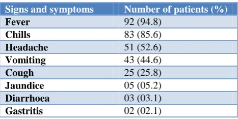
A total of 97 confirmed cases of malaria that were reported from the hospital from 1st May 2017 till 30th April 2018 were included for the study. No case was excluded as all the case files provided by MRD had adequate data. Among the malaria cases, 72 were males (74.2%) and 25 (25.8%) were females. The mean age of the participants was 33.4 years (±15.4 years) with minimum age being 3 years and maximum being 71 years. 94.8% study participants had fever and 85.6% had chills as the symptom of malaria. It was observed that 25.8% of the patients had cough along with the other symptoms of malaria (Table 1).
Table 1: Profile of symptoms of malaria among the admitted patients (n=97).
Signs and symptoms Number of patients (%)
Fever 92 (94.8)
Chills 83 (85.6)
Headache 51 (52.6)
Vomiting 43 (44.6)
Cough 25 (25.8)
Jaundice 05 (05.2)
Diarrhoea 03 (03.1)
Gastritis 02 (02.1)
Table 2: Peripheral smear results with species differentiation among the malaria cases (n=97)
Species of plasmodium Number of patients (%)
P. vivax 67 (69.0)
P. falciparum 17 (17.5)
Mixed infection 13 (13.5)
Table 3: Complications due to malaria among the admitted patients (n=97)
Complications Number of patients (%) Thrombocytopenia 18 (18.6)
Liver impairment 06 (06.2)
Renal impairment 01 (01.0)
both the tests. Among the patients, P. vivax was the most common infection followed by P. falciparum (Table 2). Regarding resistance, it was noted that only 1 case of P. vivax infection had chloroquine resistance. A total of 19
patients out of the 97 admitted with malaria reported with complications and thrombocytopenia was the most common being present in 18 patients (Table 3).
Figure 1: Distribution of malaria cases according to the QGIS blocks (n=97).
The distribution of malaria cases was displayed block-wise using QGIS (Figure 1). Out of total 97 patients that have reported to our tertiary care hospital during the study period, 60 were from Mangalore block, followed by 8 from Bantwal block, both these blocks belonging to Dakshina Kannada district of Karnataka state. Puttur and Belthangady blocks, also in to the same district, accounted for 4 cases and 3 cases respectively. We also identified 2 cases each from Davangere and Haveri blocks. Udupi, Byadagi, Hadagali, Shikaripura and Hirekerur blocks of Karnataka had 1 case each. From the neighbouring state Kerala, Kasargod block accounted for 3 cases, Manjeshwar block had 2 cases, Iritty, Taliparamba and Mallapurram block had 1 case each. Far off state like Bihar accounted for 5 cases where the migrant workers from Bihar got infected in Mangalore, rather than importing the disease from Bihar.
DISCUSSION
Among the 97 malarial cases studied, the mean age of the patients affected was about 33 years with 3 years and 71
years being the minimum and maximum age respectively. Thus malaria was seen to be affecting all the age groups. A similar study conducted in Mangalore city by Dayanand et al, found out that the mean age of the affected patients was around 36 years, which is comparable with our study.5 It is important to note that the people in the working age group are getting affected with the disease which will have a financial impact on the family, especially of the lower income class.
Out of all the cases we reviewed, 74.2% of the cases were males and 25.8% were females. This male preponderance observed can be co-related to the increased exposure to males related to their occupation. Similar trend is observed when these findings are compared with the study conducted by Dayanand et.al, which mentioned the male proportion as 73.3% and female as 26.7%.5
be very well compared with the findings obtained from a study done in KMC Attavar, Karnataka by Chowta et al, where all the malarial cases presented with fever followed by 51.55% with headache, 31.55% with vomiting, 20.37% with jaundice and 7.4% with cough.7 It is interesting to note that 1 in 4 malaria cases had cough as a symptom. Thus history of cough may be considered as one of the symptoms of malaria which will aid towards the disease diagnosis.
The symptoms can be attributed to the pathophysiology of malaria disease, which involves the lysis of the erythrocytes infected with plasmodium and further release of hemozoin pigment and other toxins including glycosylphosphatidyl inositol into the blood. This further leads to activation of macrophages and endothelial cells to release cytokines and inflammatory mediators such tumour necrosis factors, interferon gamma, interleukins, lymphotoxins and free radicals which are responsible for the various symptoms of malaria.9
The guidelines for malaria diagnosis mention that, RDTs can be used in diagnosis and treatment of malaria, in areas where microscopy results are not available in 24 hours. However, peripheral smear microscopy is still the gold standard test for the diagnosis.8 The sensitivity and specificity of rapid diagnostic tests with respect to the Leishman stain thick smear is 98.2% and 100%. The diagnosis of malaria in our study was done by thick and thin smears in 45 cases and by rapid diagnostic tests (RDTs) in 59 cases. Both tests were done in 8 patients.10 Thick and thin smears are done for the direct walk in cases in the study hospital. Patients who are already diagnosed by RDT and referred to the hospital may not be again investigated by thick and thin smear.
Out of the 97 cases studied, majority (69.0%) were infected by P. vivax. Further, 17.5% were infected by
P. falciparum and 13.5% had mixed infections. These findings are comparable with a retrospective study was conducted in Hiriadka, Udupi to determine the burden of Malaria and to assess the morbidity patterns. In the study it was found that 84.9% had P. vivax infection, 14.6% had
P. falciparum and 0.5% had mixed infections.11 A multi-centric study conducted in India by Siwal et al found that, 52% were P. vivax infections, 42% were P. falciparum
infections and 6% were mixed infections.12 P. vivax
infection has always been shown to have higher prevalence and has accounted for 98% of all malaria cases within urban malaria scheme in India in the year 2014.13
Complications are frequently associated with malaria. According to our data thrombocytopenia was the most common complication with incidence if 18.6%. This was followed by liver impairment among 6.2% of the total cases and renal impairment among 1% being the least. The other complications like cerebral malaria and pancreatitis were not found. As 1 in 5 malaria patients
had thrombocytopenia, it is advisable to keep a check on platelet count in all malaria cases.
In another study done in Mangaluru, liver impairment was found to be with highest frequency (9.3%) followed by thrombocytopenia (3.7%), renal impairment, pancreatitis and cerebral malaria with 1.9% each.7 A study done by Muddaiah et al in Dakshina Kannada district showed that thrombocytopenia accounted for 8.42% of the complications and liver impairment was found in 14.73% of the cases.14 The overall complications are found to be comparatively less in our study and may be attributed to the adequate healthcare measures at the tertiary care hospital.
On QGIS plotting, 3 clusters of malaria cases were found on the map. The first cluster consisted of those blocks which are in close proximity to the hospital (both Karnataka and Kerala blocks). Some cases from Kerala might have been diagnosed there and would have been referred to the study hospital for treatment purposes. In the other cases from Kerala, the symptomatic patients themselves would visit the study hospital for reasons such as close location of a tertiary care hospital and availability of Government benefit schemes (including some Kerala Government Schemes such as RSBY).
The second cluster had blocks from middle part of Karnataka like Davangere, Haveri, Shikaripura and Hirekerur. The reasons for patients coming from such long distances include the health camps conducted in these regions by the study hospital, provision of health facilities at subsidized rates and also the apt implementation of government schemes by the institution. The third cluster had cases from Bihar, who are immigrants for job, contracting the disease in Mangaluru and visiting the study hospital for health services.
The strengths of this study are that there is complete capture of the data of patients admitted with malaria and block wise plotting of data was done using GIS which has given representation of the geographical distribution of cases. The weakness of the study is that, the study results may not be generalisable as the study was conducted in a single tertiary care hospital.
CONCLUSION
Only about half of the malaria cases were diagnosed by examination of thick and thin blood smear. As it is the gold standard for diagnosis of malaria which also aids in identification of falciparum species, it is recommended to be performed to every case irrespective of their rapid diagnostic kit results.
Geographic mapping of the cases revealed that most cases were from in and around the study hospital. Mangaluru block accounted for 60 cases out of 97 cases which necessitates intensification of preventive measures in the block. Cases which were observed from other parts of India like Bihar can be attributed to the immigration of workers to Mangalore city.
ACKNOWLEDGEMENTS
The authors acknowledge the medical students of 2016 E batch for their contribution towards the study. The authors thank the management of Yenepoya Medical College and MRD for their constant support during this research work.
Funding: No funding sources Conflict of interest: None declared
Ethical approval: The study was approved by the Institutional Ethics Committee
REFERENCES
1. Dash AP. A profile of National Institute of Malaria Research. New Delhi: National Institute of malaria research; 2009: 92-99.
2. World Malaria Report 2018. Geneva: World Health Organization; 2018: xii.
3. Kumar A, Valecha N, Jain T, Dash AP. Burden of malaria in India: retrospective and prospective view. Am J Trop Med Hyg. 2007;77(6):69-78.
4. Malaria statistics of India 2018. Ministry of Health and Family Welfare. GOI. Available at http://www.nvbdcp.gov.in/. Accessed on 14 May 2019.
5. Dayanand KK, Punnath K, Chandrashekhar V, Achur RN, Kakkilaya SB, Gosh SK, et al. Malaria prevalence in Mangaluru city area in the south western coastal region of India. Malar J. 2017;16(1):492.
6. Nagpan BN, Saxena R, Shrivastava A. GIS decision support of control of vector borne disease in India. Available at http://ictpost.com/gis-in-decision-support-of-control-of-vector-borne-disease-in-india/. Accessed on 16 May 2018.
7. Chowta MN, Chowta N. Study of Clinical Profile of Malaria at KMC Hospital, Attavar. J Clin Diagn Res. 2007;1(3):110-5.
8. National Framework for Malaria Elimination in India (2016-2030). Directorate of National Vector Borne Disease Control Programme: Ministry of Health and Family Welfare, GOI; 2016.
9. Kasper et al. Harrison’s Principles of Internal Medicine. 19th edition. New York: McGraw Hill; 2018: 1371.
10. Ebrahim JJ, Mohammed IA, Maghram AA, Mandouh HA, Ramprasad N, Khan FR. Comparative study of thick smear, thin smear, QBC and antigen card test in diagnosis of malaria. Int J Pure Appl Sci Technol. 2013;17(1):54-9.
11. Kamath R, Gaitonde S, Tripathi P, Das D, Banerjee M, Shetty M, et al. Clinico-epidemiological profile of malaria: Analysis from a primary health centre in Karnataka, Southern India. GJMEDPH. 2012;1(6):1-6.
12. Siwal N, Singh US, Dash M, Kar S, Rani S, Rawal C, et al. Malaria diagnosis by PCR revealed differential distribution of mono and mixed species infections by Plasmodium falciparum and
Plasmodium vivax in India. PLoS One. 2018;13(3):e0193046.
13. Anvikar AR, Shah N, Dhariwal AC, Sonal GS, Pradhan MM, Ghosh SK, et al. Epidemiology of Plasmodium vivax malaria in India. Am J Trop Med Hyg. 2016;95(6):108-20.
14. Muddaiah M, Prakash PS. A study of clinical profile of malaria in a tertiary referral centre in South Canara. J Vector Borne Dis. 2006;43(1):29-33.