R E S E A R C H A R T I C L E
Open Access
Evaluation of ES-derived neural progenitors as a
potential source for cell replacement therapy in
the gut
Valentina Sasselli
1, Maria-Adelaide Micci
1, Kristen M Kahrig
1and Pankaj Jay Pasricha
1,2*Abstract
Background:Stem cell-based therapy has recently been explored for the treatment of disorders of the enteric nervous system (ENS). Pluripotent embryonic stem (ES) cells represent an attractive cell source; however, little or no information is currently available on how ES cells will respond to the gut environment. In this study, we
investigated the ability of ES cells to respond to environmental cues derived from the ENS and related tissues, both in vitroandin vivo.
Methods:Neurospheres were generated from mouse ES cells (ES-NS) and co-cultured with organotypic
preparations of gut tissue consisting of the longitudinal muscle layers with the adherent myenteric plexus (LM-MP). Results:LM-MP co-culture led to a significant increase in the expression of pan-neuronal markers (βIII-tubulin, PGP 9.5) as well as more specialized markers (peripherin, nNOS) in ES-NS, both at the transcriptional and protein level. The increased expression was not associated with increased proliferation, thus confirming a true neurogenic effect. LM-MP preparations exerted also a myogenic effect on ES-NS, although to a lesser extent. After transplantation in vivointo the mouse pylorus, grafted ES-NS failed to acquire a distinct phenotype al least 1 week following transplantation.
Conclusions:This is the first study reporting that the gut explants can induce neuronal differentiation of ES cells in vitroand induce the expression of nNOS, a key molecule in gastrointestinal motility regulation. The inability of ES-NS to adopt a neuronal phenotype after transplantation in the gastrointestinal tract is suggestive of the presence of local inhibitory influences that prevent ES-NS differentiationin vivo.
Keywords:Embryonic stem cells, Enteric nervous system, Gastrointestinal motility, Stem cell transplantation
Background
Several enteric neuronal disorders, such as achalasia, Hirschprung disease, congenital pyloric stenosis and pseudo-obstruction, are characterized by the loss or mal-function of critical neuronal subpopulations of the enteric nervous system (ENS) [1,2]. These conditions result in a severe impairment of digestive functions, for which current surgical and pharmacological therapeutic approaches are not very satisfactory, with the exception of pyloromyotomy for pyloric stenosis [3]. Novel approaches for the treatment of enteric neuropathies are therefore needed and in this
respect, stem cell-based replacement of dead or malfunc-tioning enteric neurons is a promising therapeutic tool [4,5].
Multipotent neural stem cells (NSC) isolated from the brain of embryonic mice can be transplanted in the gut of a transgenic model of gastroparesis and promote significant functional recovery as early as 1 week post-grafting [6-8]. However, the therapeutic use of CNS-derived NSC is lim-ited by the scarce availability of donor tissue and the poor ability of multipotent NSC to generate large number of cells for transplantationin vitro. Recent evidence has also shown that enteric neuronal progenitor cells (ENPC) can be derived by autologous biopsy and can be grafted in
organo-typic gut preparations in vitro [9]. Nevertheless the
* Correspondence:[email protected]
1Division of Gastroenterology and Hepatology, University of Texas Medical Branch, Galveston, TX, USA
2Johns Hopkins Center for Neurogastroenterology, Johns Hopkins University School of Medicine, Baltimore, MD, USA
consistency of this approach and the functional recovery after ENPC grafting need further investigation.
Embryonic stem (ES) cells can theoretically overcome many of these limitations and represent an alternative approach to neural precursors. ES cells are pluripotent cells derived from the inner cell mass of pre-implantation blastocysts [10]. ES cells can be propagated almost indefinitely in culture as undifferentiated cells but, under appropriate conditions, are capable of differ-entiating both in vitro and in vivo into a variety of cell types including neural cells [11,12]. Over the years, several protocols have been developed to efficiently de-rive specialized neural cell types such as dopaminergic [13-15], serotonergic [16], GABAergic and glutamatergic [17], telencephalic [18], cerebellar [19], motor [20] neu-rons from ES cells. Experimental approaches include ag-gregation into embryoid bodies followed by treatment with retinoic acid (RA), selective propagation of ES aggregates in defined media supplemented with growth factors, culture of ES cells on a monolayer or condi-tioned media of bone marrow-derived stromal cells (PA6) and genetic lineage selection [21,22]. Furthermore, several studies have also shown ES differentiation into neural crest cells, the developmental progenitors of the ENS, by using combination of defined factors in culture [23–26]. From a therapeutic perspective, therefore, it is of great interest to know whether gut environmental sig-nals can instruct ES cells to differentiate into locally ap-propriate neuronal types and whether these signals can be further analyzed to improve post-grafting success. Ac-cordingly, the aim of this study was to investigate the effects of putative differentiating cues derived from the
ENS and associated gut tissue on ES cells both in vitro
andin vivo.
Methods
All experimental animal protocols were approved by the Institutional Animal Care and Use Committee at the University of Texas Medical Branch (Galveston, Texas) in accordance with the guidelines provided by the Na-tional Institute of Health.
Mouse ES cells culture
Mouse ES cells from 129/Ola strain (a kind gift of Dr. Jie Du) were maintained for no more than 25 passages in feeder-free conditions on gelatin coated flasks (bovine gel-atin from Sigma-Aldrich, flasks from Nunc), in Glasgow’s modified Eagle’s medium (Sigma-Aldrich, St. Louis, MO) supplemented with 2 mM L-glutamine (Invitrogen Corp., Carlsbad, CA), 1 mM sodium pyruvate and 1X solution of non-essential amino acids (both from Sigma-Aldrich, St. Louis, MO), 10% fetal bovine serum (FBS; HyClone),
50μM 2-mercaptoethanol (Sigma-Aldrich, St. Louis, MO),
100μg/ml Primocin antibiotic (Amaxa Inc.) and 1000 U/ml
LIF (ESGRO, Millipore). ES cells were sub-cultured using 0.05% trypsin/0.02% EDTA (Invitrogen Corp., Carlsbad, CA) in phosphate buffered saline (PBS) and plated at 25,000-30,000 cells cm-2 in freshly gelatinized flasks.
Alkaline phosphatase assay was carried out using the Alkaline Phosphatase Detection Kit (Millipore) accord-ing to manufacturer’s instructions.
Induction of neurospheres from mouse ES cells
ES cells were trypsinized and plated at a density of 30,000 cells cm-2 onto non-coated flasks in neurobasal medium containing 2 mM L-glutamine, B27 supplement and 100 U/ml penicillin-streptomycin (NB27 medium; all reagents from Invitrogen Corp.), plus 1000 U/ml LIF, 10 ng/ml FGF2 and 50 ng/ml EGF (Promega). Cells were cultured for 7 days as floating multicellular aggregates termed ES-derived neurospheres or ES-NS.
Isolation and in vitro culture of mouse CNS-derived neural stem cells (CNS-NSC)
Cell culture reagents were obtained from Invitrogen Co. (Carlsbad, California) except where noted. Staged-pregnant female GFP mice (TgN(GFPU)5Nagy, Jackson Laboratory, Bar Harbor, Maine) at embryonic day 15 (E15) were anesthetized with sodium pentobarbital (70 mg/kg, i.p.) and a midline incision was made to expose the embryos. The brains of embryonic mice were removed and the subventri-cular zone (SVZ) dissected from each brain hemisphere. The tissues were washed in Ca2+- and Mg2+-free Hank’s Buffered Salt Solution (HBSS) and digested using a combin-ation of dispase type I (0.1%) and trypsin (0.005%) for 10 minutes at 37°C. After digestion, a cell suspension was obtained by gentle trituration, pelleted and resuspended in Neurobasal medium containing B27, 2 mM glutamine and penicillin-streptomycin (NB27), plus 20 ng/ml fibroblast growth factor (bFGF) and 20 ng/ml epidermal growth fac-tor (EGF) (Promega, Madison, Winsconsin).
Differentiation of mouse ES-NS and CNS-NSC
To assess differentiation, multicellular aggregates were dis-sociated by incubation with Accutase (Innovative Cell Technologies, Inc., San Diego, CA) at 37°C for 10 minutes. Single cell suspensions were plated onto poly-ornithine (Sigma-Aldrich, St. Louis, MO)/mouse laminin (Invitrogen Corp, Carlsbad, CA) coated plates (BD FalconTM, BD Biosciences, San Jose, CA) in NB27 medium without growth factors and cultured for 7 days.
Co-culture with muscularis externa-myenteric plexus
expression ubiquitously in all tissues. Mice were euthanized by cervical dislocation. The small intestine was dissected in pieces of 1 cm length and placed over a glass rod. A small incision was made and muscle layers with the adherent my-enteric plexus were peeled off using cotton swab. The ma-jority of preparations contained only longitudinal muscle and myenteric plexus and therefore we referred to them as LM-MP. LM-MPs were cultured at 37°C in 5% CO2 for 24 hrs in DMEM containing 10% FBS and 100U/ml penicillin-streptomycin (all reagents from Invitrogen Corp.). ES-NS were dissociated and plated onto poly-ornithine/laminin coated plates at a density of 2000 cells cm-2 in NB27 medium supplemented with 1000 U/ml LIF, 10 ng/ml FGF2 and 50 ng/ml EGF. Cells were cultured for
18 hrs before placing the LM-MPs into a 0.4 μm
micro-porous insert (Corning Inc., Corning, NY, USA) inside each well (5 pieces per 24 mm-diameter insert). Co-cultured ES-NS were maintained in NB27 medium with-out growth factors and cultured for 10 days. Freshly pre-pared LM-MPs were placed in the inserts after 5 days. Control ES-NS were cultured in the same conditions with-out LM-MPs.
Flow cytometry analysis
Cells were dissociated using Accutase digestion according to manufacturer’s instructions (Innovative Cell Technolo-gies, Inc., San Diego, CA) and collected by centrifugation. 5 x 10⁵–1 x 10⁶cells were fixed in ice-cold 4% parafor-maldehyde for 15 minutes at room temperature (RT). Cells were then blocked and permeabilized for 5 minutes in PBS containing 0.1% Triton X-100 and 2% normal goat serum (NGS). Incubation with Ki67 antibody diluted in PBS containing 2% NGS was carried out for 30 minutes at RT, followed by incubation with secondary antibody for another 30 minutes (see Table 1 for a list of antibodies
used). Cells were resuspended in 500μl of PBS
contain-ing 2% NGS and processed with the analytical Flow Cyt-ometer FACSCanto (Becton-Dickinson, Franklin Lakes, NJ USA).
Real time PCR analysis
Total RNA was extracted from cells using the TRIzol kit (Invitrogen Corp.) according to manufacturer’s instruc-tions. First-strand cDNA was then generated using Taq-Man Reverse Transcription Regents (Applied Biosystems, Foster City, CA, USA). Quantitative real-time polymerase chain reaction (PCR) was carried out in multiplex using the PTC 200 Peltier Thermal Cycler equipped with Chromo 4 Continuous Fluorescence Detector (MJ Re-search Waltham, MA, USA). PCR was performed using GeneAmp PCR kit and AmpliTaq Gold polymerase (Ap-plied Biosystems) with the following cycling conditions: 2 minutes at 50°C, 10 minutes at 95°C, followed by 40 cycles of 15 seconds at 95°C, 1 minute at 60°C and
fluorescence plate reading. The following sequence-specific primers and probes were designed using Primer Express software 2.0 (Applied Biosystems): β-III tubulin:
Fw GGTCTGGCGCCTTTGGA, Rv AGTTGTTGC
CAGCACCACTCT, probe Cy5-CCTATTCAGGCCCGA CAACTTTATCTTTGGT-IBRQ; PGP9.5: Fw GCTTCG CCGACGTGCTA, Rv TTGCTTTTTCCTGAAGTTTT
CATG, probe Cy5-CTGCTGCTCCTGTTTCCCCTC
ACG-IBRQ; peripherin: Fw GCATCTCAGTGCCGGTT
CAT, Rv TGGGACTCTGTCACCACCTTCT, probe
TexRed-TTGCCTCTCTAAGTTTAAAGACGACTG-IBRQ; nNOS: Fw GGGAAACTCTCGGAGGAGGA, Rv TGAGGGTGACCCCAAAGATG, probe 6FAM-CGTGG TACCGGTTGTCATCCCCTCAG-TAMRA; GFAP: Fw GAGGAGGAGATCCAGTTCTTAAGGA, Rv GCCTCG TATTGAGTGCGAATC, probe 6FAM-CCAGACCTCA
CAGCGGCCCTGA-TAMRA; α smooth muscle actin:
Fw GAGACTCTCTTCCAGCCATCTTTC, Rv TGAT GCTGTTATAGGTGGTTTCGT, probe 6FAM-ATGCC CGCTGACTCCATCCCAA-TAMRA. Ribosomial RNA 18 s (primers and VIC-conjugated probe from Applied Biosystems) was measured as a reference gene. Cycle Threshold (CT) values were quantified using Opticon Monitor 2 software (MJ research) and normalized calcu-lating the mRNA level of each probe (x) relative to the amount of 18 s mRNA (reference gene) as follow: mRNA
(x) = 2 CT (18 s)–CT (x) x 100. Mouse brain cDNA was
used as positive control for all primers and probes and NO-RT reactions were used as negative control to check for genomic DNA contaminations.
Western blot analysis
Cells were dissociated and collected in pellets, which, after incubation overnight at -80°C, were lysed in ice-cold buffer containing 2% sodium dodecyl sulphate
(SDS), protease cocktail inhibitor (Sigma-Aldrich),
intensity was quantified using Optiquant software (Pack-ard, Meriden, CT, USA) and data were normalized to the expression of the housekeeping protein GAPDH.
Immunofluorescence analysis
Cells were fixed in ice-cold 4% paraformaldehyde for 15 minutes at RT, blocked and permeabilized for 1 hr with PBS containing 0.3% Triton X-100 and 5% NGS. Sam-ples were incubated with primary antibodies (Table 1A) diluted in PBS containing 5% NGS overnight at 4°C and with secondary antibodies (Table 1B) for 1 hour at RT. Sections and slides were cover-slipped with mounting media containing the nuclear stain DAPI (Vector La-boratories, Burlingame, CA). LM-MP preparations were processed similarly with the following modifications: in-cubation in Pronase (Biomedia) for 5 minutes at RT after fixation, primary and secondary antibody incubation for 72 hrs and 1.5 hrs respectively at RT in PBS containing 1.5% NGS, 0.3% Triton-X100 and 0.01% Na Azide. Sam-ples were examined with an Olympus BX60 microscope (Olympus, Melville, NY, USA) equipped with fluores-cence and digital imaging with a cooled CCD camera (Photometrics CoolSNAP, Roper Scientific, Duluth, GA, USA) and Metaview software (Universal Imaging Corp., West Chester, PA, USA).
Cell counts were performed by counting the number of immunopositive cells for a given antibody and
expressing the results as number of positive cells versus total cells number (determined using the nuclear coun-terstain DAPI that forms fluorescent complexes with natural double-stranded DNA and it is used to label the nuclei in all the cells). An independent investigator who was blinded to the treatment counted three regions per slides. A total of 3 experiments were counted.
Intra-pyloric transplantation
To enable the detection of the cellsin vivo, dissociated ES-NS cells were labeled with CM-DiI (Molecular Probes,
Eugene, OR) according to manufacturer’s instructions.
After washing in PBS, the cells were resuspended at a con-centration of 50,000 cells/μl in PBS containing 500 μM caspase-1 inhibitor (Ac-YVAD-CMK, Calbiochem, La Jolla, CA, USA) and kept on ice. Adult male C57BL/6 J mice (Jackson laboratories) were anesthetized with 0.4 ml of 6% ketamine/4% xylazine intraperitoneally. A midabdominal incision was made and the pylorus identified. Two microli-ters of ES-NS suspension were injected bilaterally into the mid-pylorus immediately subserosal using a 22-gauge nee-dle attached to a 10μl Hamilton syringe.
One week following ES-NS transplantation, mice were deeply anesthetized with sodium pentobarbital (70 mg/kg intraperitoneally), transcardially perfused, and fixed with freshly prepared ice-cold 4% paraformaldehyde in 0.1 M PBS (pH 7.4). The pylorus was removed, post-fixed in 4% Table 1 List of primary (A) and secondary (B) antibodies used for Immunofluorescence (IF), Flow Cytometry (FC) and Western blot (WB) analysis
A.
Primary Ab Host Dilution Specificity Source
Ki67 Rabbit 1:100 FC Proliferating cells Abcam
Nestin Mouse 1:2000 WB, IF Neural progenitor cells BD
βIII-Tubulin Mouse 1:2000 IF; 1:5000 WB Neurons Promega
Chicken 1:2000 IF Neurons Aves Laboratories
PGP 9.5 Rabbit 1:2000 WB, IF Neurons Chemicon
Peripherin Rabbit 1:2000 WB, IF Neurons Chemicon
MAP-2 Chicken 1:2000 WB, IF Neurons Aves Laboratories
nNOS Mouse 1:2500 WB Neurons BD
Rabbit 1:200 IF Neurons Zymed
GFAP Rabbit 1:2000 IF 1:5000 WB Glial cells DAKO
α-SMA Rabbit 1:1000 IF 1:5000 WB Smooth muscle cells Abcam
GAPDH Mouse 1:10000 WB Loading control Abcam
B.
Secondary Ab Host Dilution Source
Alexa 633 anti rabbit Goat 1:200 FC Molecular Probes
Alexa 594 anti mouse or rabbit Goat 1:200 IF Molecular Probes
Alexa 488 anti mouse or rabbit Goat 1:200 IF Molecular Probes
paraformaldehyde, and cryoprotected by infiltration in 20% sucrose solution in PBS overnight at 4°C. Tissues were rap-idly frozen in O.C.T. embedding medium (Tissue Tek;
Sakura, Tokyo, Japan) over dry ice–chilled isopentane
(Sigma Chemical Co, St Louis, MO). Frozen serial sections
(16μm thick) were cut on a cryostat (TBS, Durham, NC),
placed on Superfrost Plus slides (VWR, West Chester, PA), and stored at -20°C until needed.
Statistical analysis
RT-PCR and WB data were expressed as mean ± SEM.
For comparisons between the groups One SampleTTest
was used. Statistical significance was assumed if P<0.05.
Results
Characterization of murine ES cells
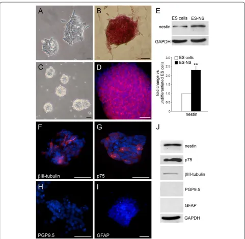
Characterization of the 129/Ola ES cell line was carried out by investigating common attributes of embryonic stem cells: colony morphology and alkaline phosphatase expres-sion. Murine ES cells grown in feeder-free conditions formed colonies with the typical round and compacted morphology (Figure 1A) and with a high expression of alka-line phosphatase, a marker expressed by embryonic stem cells but not by their differentiated derivatives (Figure 1B) [27].
Induction of neural progenitors from ES cells
In order to start with an enriched population of neural pre-cursors we have modified existing protocols for generating ES-derived neurospheres [14,15]. ES cells were grown for 7 days into non-coated tissue culture flasks in Neurobasal medium supplemented with the neurotrophic serum-free supplement B27 and the growth factors FGF-2, EGF and LIF. Under these conditions ES cells grow as floating multi-cellular aggregates, which morphologically resemble the neurospheres that are formed by neural stem cells (NSC) isolated from fetal and adult brain (Figure 1C). ES cells show expression of the neural stem cell marker nestin sug-gesting spontaneous neural differentiation under the cul-ture conditions used (data not shown). This is not unusual and has been previously reported by other investigators [28]. However, the expression of nestin was limited to few ES cells (data not shown) and was significantly increased after neural induction in ES-derived neurospheres (ES-NS) (Figure 1D), with a 2.3 fold increase in protein expression compared to undifferentiated ES cells cultures (P<0.01, n = 3; Figure 1E).
Some cells within the neurospheres also express the neural crest stem cell marker p75 and the neural
pro-genitor markerβIII-tubulin. On the other hand, the
ma-ture neuronal marker PGP9.5 and the glial marker GFAP were not detectable in ES-NS (Figure 1F-I and Figure 1J).
To further explore the neurogenic potential of ES-NS, neuronal differentiation of ES-NS was assayed after 1 week of culture in the absence of any growth factor and compared to that of uncommitted ES cells. Under these conditions ES-NS were able to generate neurons identified by a clear uni/bi-polar neuronal morphology (Figure 2A-B) and by the expression of the neuronal markerβIII-tubulin (1.8 fold increase as compared to ES
cells; P<0.01, n = 3. Figure 2C). Moreover, when we
compared differentiated ES-NS to cells differentiated from CNS-derived neurospheres (CNS-NS), we found
that the expression level of βIII-tubulin measured by
Western blot analysis was similar between the two groups (Figure 2D).
Interestingly, ES-NS did not generate astrocyte-like cells as indicated by the lack of expression of glial fibrillary acidic protein (GFAP), a marker of the glial lineage, as assessed by both Western blotting and RT-PCR (data not shown).
Small intestinal longitudinal muscle-myenteric plexus preparations
To investigate the effects of putative gut factors on the
differentiation of ES-NS we established an in vitro
co-culture protocol. The procedure involves the organotypic culture of strips of longitudinal muscle and the adherent myenteric plexus (LM-MP) derived from the small intes-tine of adult mice. LM-MP tissues were placed into the inside compartment of transwells separated from the
ES-NS by a microporous membrane (0.45 μm pore size)
that allows the exchange of soluble factors but not of cells. The maximum length of time for co-culture was defined as the approximate time needed for cells to reach confluency (10 days). To prove the effectiveness of this system we performed preliminary experiments in which LM-MP preparations were obtained from TgN (GFPU)5Nagy mice. No GFP positive cells were found in the compartment containing the ES-NS cells after 10 days of co-culture (data not shown), proving that only soluble molecules are exchanged between the two sides of the membrane. LM-MP preparations were also tested
by immunoreactivity for βIII-tubulin, GFAP and α
-smooth muscle actin (α-SMA) during the co-culturing
period, which showed integrity of both the neural plexus and smooth muscle for at least 5 days (Figure 3) and attested to their viability. Therefore, in the co-culture experiments, LM-MP strips were changed with fresh preparations at day 5 of the culturing period, in order to avoid tissue degeneration.
Neurogenic potentials of ES-NS in co-culture with LM-MP
to control ES-NS cultured without LM-MP (2.0, 1.3, 1.7 and 1.7 fold increases respectively, P<0.05, n = 7; Figure 4A). This was accompanied by a corresponding in-crease in protein expression as shown by Western blot analysis (2.4 fold forβIII-tubulin, 1.8 for PGP 9.5, 1.5 for
peripherin and 2.3 for nNOS, P<0.05, n = 6. Figure 4B). The expression of ChAT was not detected in differentiated ES-NS (data not shown). Interestingly, ES-NS did not gen-erate astrocyte-like cells as indicated by the lack of GFAP expression, both at the mRNA and protein level.
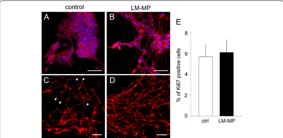
Immunofluorescence analysis corroborated these find-ings, revealing a significant increase of percentages of cells positive forβIII-tubulin, PGP9.5 and nNOS after exposure
to LM-MP compared to control cultures (βIII-tubulin
42.3 ± 7.0% vs 23.6 ± 6.3%, PGP9.5 39.9 ± 6.6% vs
21.6 ± 4.7%, nNOS 15.8 ± 3.5% vs 11.8 ± 4.6%, with 3.2, 2.3 and 1.6 fold increases respectively, P<0.05, n = 3. Figure 4C-D). Moreover, neurons showed a more devel-oped morphology after co-culture with LM-MP than in control cultures as highlighted by immunoreactivity for cytoskeleton proteins such as peripherin (Figure 5A-B) and βIII-tubulin (Figure 5C-D). It has to be noted that in the control group, the number of cells expressing peripherin in relation to the total number of cells in the culture was rela-tively low (as confirmed by the low signal in the WB
analysis shown in Figure 4B). However, the cells that did express peripherin were highly positive by immunofluores-cence analysis thus explaining the images that we are showing in Figure 5.
We also demonstrated that increased cell proliferation is unlikely to account for elevated levels of neuronal markers since the percentage of Ki67 expressing cells is similar in the co-culture and control groups (6.1 ± 1.2% and 5.6 ± 1.2% respectively, P = 0.81, n = 3; Figure 5E).
Immunofluorescence analysis also revealed the appear-ance of a small percentage of cells (less than 1% of the
total) positive for smooth muscle markerα-SMA in the
co-cultured ES-NS (Fig 6A-B). By Western blot analysis
we could detect α-SMA only after co-culture with
LM-MP (Fig 6D). This is most likely due to the signal being below the detection limit due to the low number of cells positive for this marker in the control culture. However, RT-PCR confirmed a 2.7 fold increase of SMA expres-sion in the co-cultured group as compared with control (P<0.01, n = 6; Figure 6C).
Intrapyloric transplantation of ES-derived neurospheres
To assess preliminary feasibility of ES-derived neuro-spheres transplantation, we used the experimental proto-col of intrapyloric transplantation previously described
for CNS-derived neural stem cells (CNS-NS) [6–9].
ES-NS were grafted in the pyloric muscle layer of adult male mice; survival and differentiation were assessed 1 week after transplantation. At the time point chosen grafted cells were found mainly in the muscle and the submuco-sal layers showing good survival (Figure 7A). On the other hand, immunohistochemistry failed to reveal
ex-pression of the neuronal markers βIII-tubulin,
periph-erin, MAP-2 and nNOS as well as the non-neuronal
markers GFAP and α-smooth muscle actin by the
engrafted ES-NS (Figure 7 B-D).
Discussion
Although ES cells can differentiate into neural precur-sors in the absence of any external influence, only a small percentage of neural cells survive and differentiate under these conditions [29,30]. In this report we have therefore developed a modified protocol to enrich our culture in ES-derived neural progenitors. Using this ap-proach we obtained multi-cellular aggregates that share many properties with neurospheres generated from CNS-derived neural stem cells, namely, sphere morph-ology, nestin expression and the ability to produce neu-rons. Moreover, ES-NS express p75, a marker found in neural crest-derived stem cells [31]. Having established a neuronally biased ES population, we next examined the effects of an enteric neuromuscular environment on its further differentiation. It has been shown that co-culture of progenitor cells with mature cells or tissues can drive Figure 2Neurogenic potential of ES-derived neurospheres.ES
their differentiation toward desired phenotypes [32,33].
We therefore established a controlled in vitro system
where ES-NS are co-cultured with strips of small intestine longitudinal muscle and the adherent myenteric plexus (LM-MP). Although the presence of the myenteric plexus in the LM-MP preparation might suppress ES-NS differ-entiation, our results show that intestinal LM-MP exert a considerable neurogenic effect on ES-NS, as evidenced by the enhanced expression of several neuronal markers, in-cluding peripherin, the intermediate filament which is a well established marker of peripheral neurons [34] and the enzyme neuronal nitric oxide synthase (nNOS), respon-sible for the production of the critical enteric neurotrans-mitter, nitric oxide [35]. On the other hand, ChAT was not expressed in ES-NS under either co-culture or control conditions. Although our data does not provide an explan-ation for the lack of ChAT expression, it is interesting to note that nNOS-expressing neurons appear early during ENS development while ChAT-expressing neurons are
only detected at later embryonic stages and post-natally [36–38].
The neurogenic effect observed after co-culture of ES-NS with LM-MP could be attributed to either increased proliferation and/or improved differentiation of neural progenitors within ES-NS. Our data show that the num-ber of neurons (PGP9.5 and nNOS positive) is increased after co-culture with LM-MP, but not the number of proliferating cells (Ki67 positive). Moreover, co-culture with LM-MP clearly affects the morphology of neurons produced, which possess longer axons and form a more extensive and organized network as compared to con-trols. This suggests that soluble factors released from the LM-MP promote survival and differentiation of neural progenitors but not their proliferation. Therefore the main effect in our ES-NS co-culturing protocol is to favor differentiation of neurons.
ES-NS. Previous reports have shown that CNS-derived NS can be induced to express smooth muscle markers in vitro [39], on the other hand, neural crest cells (NCC), which give rise to enteric neurons and glia dur-ing development, are also able to generate smooth muscle cells in different embryonic compartments [40].
Therefore our findings could be due to an in vitro
artifact or to a NCC-like potential of ES-NS.
ES-NS failed to express the glial marker GFAP under any of our culture conditions, unlike other studies using ES-derived neural precursors, which have reported the capability of these progenitors to differentiate into both the neuronal and glial lineagein vitro[11,14]. The discrep-ancy between these reports and our results may be due to differences in culture conditions used to produce ES-NS. Alternatively, the fact that neural progenitors have a
stage-dependent potential giving rise first to neurons and then to glia during neural commitment [41], may suggest that prolonged culture periods are necessary to generate glial cells. Therefore differentiation of ES-NS for longer periods of time (more than 10 days) and analysis of a more wide-spread panel of glial markers will need to be performed.
It is well known that the environment plays a key role on the differentiation of ES cells into defined pheno-types. We have developed a robust and informative in vitrosystem where soluble factors released by gut tis-sues are able to drive neuronal differentiation of ES-derivatives appropriate for the ENS lineage. The nature of these signals has yet to be determined, but may in-clude well-known soluble factors of the gut such as
GDNF, NO, TGFβ(respectively produced by glial,
Figure 5Differentiated neurons in LM-MP treated ES-NS exhibit a more complex and mature morphology than control neurons. Morphology of neurons is shown by immunoreactivity for the neuronal markers peripherin (red, A-B) andβIII-tubulin (red, C-D) in ES-NS co-cultured with LM-MP (B,D) or control (A,C). Neurons arising from ES-NS exposed to LM-MP show pronounced processes and form a network-like structure, whereas control neurons show either absence of neuritogenesis (A) or shorter blunt processes (C) white arrowheads. Nuclei are counterstained with DAPI (blue). Calibration bars = 25μm. (E) Analysis of the expression of the cell cycle marker Ki67 by flow cytometry does not reveal any significant change between the two groups. n = 3, P = 0.81, Student’sT-test.
analysis of the LM-MP conditioned medium will help to identify these factors.
Although transplantation of ES-NS in the pyloric muscle layer of wild type mice showed adequate survival of grafted cells at 1 week, no markers of terminal differentiation were noted. This contrasts with many reports that showed suc-cessful transplantation of ES-derived neural precursors in the CNS [13–15]. This may be due to the presence of in-hibitory factors, either secreted or contact-based that were
not captured in vitro using isolated LM-MP co-cultures.
This difference also probably reflects the vastly more com-plex environment of the gut wall, consisting of a mixture of epithelium, neurons, glia, interstitial cells of Cajal and smooth muscle, along with resident and circulation-derived immunocytes. These cellular elements may provide con-flicting cues to ES cells, resulting in inhibition or retard-ation of their differentiretard-ation. Further, non-neural factors such as neo-angiogenesis may be required for the optimal differentiation of these cells.
We have previously shown that CNS-derived NS grafted in the pylorus of mice generate neurons and glia and partially restore gastric function [7]. We here show that ES-derived neurospheres do not compare to CNS-NS after transplantation. Clearly, there is a lot more to be done in order to fully understand the mechanisms for this different behavior. It is possible that the lack of GFAP expression (and glia?) accounts for the poor out-comein vivo. Further work will need to be done to ad-dress this question.
In this regard, our study had several limitations. First, it should be noted that the time frame of these experiments was relatively short. Long-term survival and differentiation
of grafted cells needs to be investigated in further studies to fully assess the potential of ES-NS to form enteric neurons in vivo. It is also possible that“priming”of the ES, perhaps by exposure to LM-MP prior to the transplantation may re-sult in a better outcome. Finally, the inability to precisely localize the injection into the muscle layer may be a factor in preventing the appropriate signals from reaching the ES cells (in our study most of them were found in the sub-mucosa). Therefore, this study cannot confidently exclude the therapeutic potential of ES cell for repopulating the ENS. On the other hand, if they truly do not differentiate, they may retain the ability to proliferate (which we did not examine), which is of concern because of the risk of tumorigenesis.
Conclusions
A variety of cell sources are currently being explored for use in cell transplantation studies including CNS-derived NS, neural crest stem cells (NCCS), enteric neural progenitor cells (ENPC) and embryonic stem cells (ES) [5,42]. Among these sources, ES cells have the ad-vantage of being available in large numbers and of being capable to adopt a full spectrum of cell fates. In this study we have shown that ES-derived neurospheres (ES-NS) are able to generate specific phenotypes under the influence of diffusible factors in vitro, while they fail to differentiate after transplantation in the gut. The
inabil-ity of ES-NS to adopt a neuronal phenotype in vivo
might be due to local inhibitory influences that prevents their differentiation, and suggests that ES-NS may not be suitable candidates for ENS regeneration.
Figure 7Intrapyloric transplantation of ES-NS.(A) Grafted DiI-labeled ES-NS (red) after 7 days from transplantation were found mainly in the muscle and submucosal layers. (B-D) Immunofluorescence staining revealed a failure by the grafted ES-NS to express mature phenotype markers such as peripherin (green, B), GFAP (green, C) andα-smooth muscle actin (green, D). Nuclear staining with DAPI (blue). M, muscle; SM,
Competing interests
The author(s) declare that they have no competing interests.
Acknowledgements
The authors would like to thank Dr. Jie Du for providing the 129/Ola ES cells and Dr. Tor Savidge for valuable support with the RT-PCR assay.
This study was supported by a grant from the National Institute of Health (DK62834 and DK80920) and from the Moody Foundation, Galveston, TX.
Authors’contributions
VS carried out the experiments, performed the statistical analysis and drafted the manuscript. MAM participated in the design of the study, in the statistical analysis and in the drafting of the manuscript. KMK performed cell counting and participated in the cell transplantation experiments. PJP conceived the study, and participated in its design and coordination and helped to draft the manuscript. All authors read and approved the final manuscript.
Authors’information
The current affiliation of VS is Division of Molecular Neurobiology, MRC National Institute for Medical Research, The Ridgeway, Mill Hill London NW71AA, UK.
The current affiliation of MAM is Department of Anesthesiology, University of Texas Medical Branch, Galveston, TX, USA.
The current affiliation of PJP is Johns Hopkins Center for
Neurogastroenterology, Johns Hopkins University School of Medicine, Baltimore, MD, USA.
Received: 4 October 2010 Accepted: 26 June 2012 Published: 26 June 2012
References
1. Furness JB:The enteric nervous system: normal functions and enteric neuropathies.Neurogastroenterol Motil2008, 20(Suppl 1):32–38. 2. De Giorgio R, Guerrini S, Barbara G, Cremon C, Stanghellini V, Corinaldesi R:
New insights into human enteric neuropathies.Neurogastroenterol Motil
2004, 16(Suppl 1):143–147.
3. Di Nardo G, Blandizzi C, Volta U,et al:Review article: molecular, pathological and therapeutic features of human enteric neuropathies. Aliment Pharmacol Ther2008,28:25–42.
4. Thapar N:Future horizons in the treatment of enteric neuropathies. J Pediatr Gastroenterol Nutr2007, 45(Suppl 2):S110–114.
5. Schafer KH, Micci MA, Pasricha PJ:Neural stem cell transplantation in the enteric nervous system: roadmaps and roadblocks.Neurogastroenterol Motil2009,21:103–112.
6. Micci MA, Learish RD, Li H, Abraham BP, Pasricha PJ:Neural stem cells express RET, produce nitric oxide, and survive transplantation in the gastrointestinal tract.Gastroenterology2001,121:757–766.
7. Micci MA, Kahrig KM, Simmons RS, Sarna SK, Espejo-Navarro MR, Pasricha PJ:
Neural stem cell transplantation in the stomach rescues gastric function in neuronal nitric oxide synthase-deficient mice.Gastroenterology2005,
129:1817–1824.
8. Micci MA, Pattillo MT, Kahrig KM, Pasricha PJ:Caspase inhibition increases survival of neural stem cells in the gastrointestinal tract.
Neurogastroenterol Motil2005,17:557–564.
9. Metzger M, Caldwell C, Barlow AJ, Burns AJ, Thapar N:Enteric nervous system stem cells derived from human gut mucosa for the treatment of aganglionic gut disorders.Gastroenterology2009,136:2214-2225–e2211-2213.
10. Evans MJ, Kaufman MH:Establishment in culture of pluripotential cells from mouse embryos.Nature1981,292:154–156.
11. Okabe S, Forsberg-Nilsson K, Spiro AC, Segal M, McKay RD:Development of neuronal precursor cells and functional postmitotic neurons from embryonic stem cells in vitro.Mech Dev1996,59:89–102.
12. Ying QL, Stavridis M, Griffiths D, Li M, Smith A:Conversion of embryonic stem cells into neuroectodermal precursors in adherent monoculture. Nat Biotechnol2003,21:183–186.
13. Kim JH, Auerbach JM, Rodriguez-Gomez JA,et al:Dopamine neurons derived from embryonic stem cells function in an animal model of Parkinson's disease.Nature2002,418:50–56.
14. Takagi Y, Takahashi J, Saiki H,et al:Dopaminergic neurons generated from monkey embryonic stem cells function in a Parkinson primate model. J Clin Invest2005,115:102–109.
15. Morizane A, Takahashi J, Shinoyama M,et al:Generation of graftable dopaminergic neuron progenitors from mouse ES cells by a
combination of coculture and neurosphere methods.J Neurosci Res2006,
83:1015–1027.
16. Lee SH, Lumelsky N, Studer L, Auerbach JM, McKay RD:Efficient generation of midbrain and hindbrain neurons from mouse embryonic stem cells. Nat Biotechnol2000,18:675–679.
17. Bibel M, Richter J, Schrenk K,et al:Differentiation of mouse embryonic stem cells into a defined neuronal lineage.Nat Neurosci2004,7:1003–1009. 18. Watanabe K, Kamiya D, Nishiyama A,et al:Directed differentiation of
telencephalic precursors from embryonic stem cells.Nat Neurosci2005,
8:288–296.
19. Su HL, Muguruma K, Matsuo-Takasaki M, Kengaku M, Watanabe K, Sasai Y:
Generation of cerebellar neuron precursors from embryonic stem cells. Dev Biol2006,290:287–296.
20. Soundararajan P, Miles GB, Rubin LL, Brownstone RM, Rafuse VF:
Motoneurons derived from embryonic stem cells express transcription factors and develop phenotypes characteristic of medial motor column neurons.J Neurosci2006,26:3256–3268.
21. Stavridis MP, Smith AG:Neural differentiation of mouse embryonic stem cells.Biochem Soc Trans2003,31:45–49.
22. Du ZW, Zhang SC:Neural differentiation from embryonic stem cells: which way?Stem Cells Dev2004,13:372–381.
23. Mizuseki K, Sakamoto T, Watanabe K,et al:Generation of neural crest-derived peripheral neurons and floor plate cells from mouse and primate embryonic stem cells.Proc Natl Acad Sci U S A2003,100:5828–5833.
24. Pomp O, Brokhman I, Ben-Dor I, Reubinoff B, Goldstein RS:Generation of peripheral sensory and sympathetic neurons and neural crest cells from human embryonic stem cells.Stem Cells2005,23:923–930.
25. Hotta R, Pepdjonovic L, Anderson RB:et al. Stem Cells: Small Molecule Induction of Neural Crest-like Cells Derived from Human Neural Progenitors; 2009. 26. Takaki M, Nakayama S, Misawa H, Nakagawa H:In vitro formation of
enteric neural network structure in a gut-like organ differentiated from mouse embryonic stem cells.Stem Cells2006,24:1414–1422.
27. Pease S, Braghetta P, Gearing D, Grail D, Williams RL:Isolation of embryonic stem (ES) cells in media supplemented with recombinant leukemia inhibitory factor (LIF).Dev Biol1990,141:344–352.
28. Wiese C, Rolletschek A, Kania G, Blyszczuk P, Tarasov KV, Tarasova Y, Wersto RP, Boheler KR, Wobus AM:Nestin expression--a property of multi-lineage progenitor cells?Cell Mol Life Sci2004, 61(19-20):2510–22.
29. Tropepe V, Hitoshi S, Sirard C, Mak TW, Rossant J, van der Kooy D:Direct neural fate specification from embryonic stem cells: a primitive mammalian neural stem cell stage acquired through a default mechanism.Neuron2001,30:65–78.
30. Smukler SR, Runciman SB, Xu S, van der Kooy D:Embryonic stem cells assume a primitive neural stem cell fate in the absence of extrinsic influences.J Cell Biol2006,172:79–90.
31. Kruger GM, Mosher JT, Bixby S, Joseph N, Iwashita T, Morrison SJ:Neural crest stem cells persist in the adult gut but undergo changes in selfrenewal, neuronal subtype potential, and factor responsiveness. Neuron2002,35:657–69.
32. Van Vranken BE, Romanska HM, Polak JM, Rippon HJ, Shannon JM, Bishop AE:Coculture of embryonic stem cells with pulmonary mesenchyme: a microenvironment that promotes differentiation of pulmonary epithelium.Tissue Eng2005,11:1177–1187.
33. Troy TC, Turksen K:Directing epidermal fate selection by a novel co-culture system.Methods Mol Biol2006,330:105–111.
34. Troy CM, Brown K, Greene LA, Shelanski ML:Ontogeny of the neuronal intermediate filament protein, peripherin, in the mouse embryo. Neuroscience1990,36:217–237.
35. Mashimo H, Goyal RK:Lessons from genetically engineered animal models. IV. Nitric oxide synthase gene knockout mice.Am J Physiol1999,
277:G745–750.
36. Young HM:Functional development of the enteric nervous system–from migration to motility.Neurogastroenterol Motil2008, 20(Suppl 1):20–31. 37. Young HM, Jones BR,et al:The projections of early enteric neurons are
38. Vannucchi MG, Faussone-Pellegrini MS:Differentiation of cholinergic cells in the rat gut during pre- and postnatal life.Neurosci Lett1996, 206(2–3):105–8. 39. Oishi K, Ogawa Y, Gamoh S, Uchida MK:Contractile responses of smooth
muscle cells differentiated from rat neural stem cells.J Physiol2002,
540:139–152.
40. Le Douarin NM, Dupin E:Cell lineage analysis in neural crest ontogeny. J Neurobiol1993,24:146–161.
41. Temple S:The development of neural stem cells.Nature2001,414:112–117. 42. Micci MA, Pasricha PJ:Neural stem cells for the treatment of disorders of
the enteric nervous system: strategies and challenges.Dev Dyn2007,
236:33–43.
doi:10.1186/1471-230X-12-81
Cite this article as:Sasselliet al.:Evaluation of ES-derived neural progenitors as a potential source for cell replacement therapy in the gut.BMC Gastroenterology201212:81.
Submit your next manuscript to BioMed Central and take full advantage of:
• Convenient online submission
• Thorough peer review
• No space constraints or color figure charges
• Immediate publication on acceptance
• Inclusion in PubMed, CAS, Scopus and Google Scholar
• Research which is freely available for redistribution