ISSN 0015–5659 www.fm.viamedica.pl
The sciatic nerve in human cadavers
— high division or low formation?
A.A. Khan
1, M.A. Asari
2, M.A. Pasha
31Department of Clinical and Neuroanatomy, Universiti Sains, Malaysia 2Department of Neurohistology, Universiti Sains. Malaysia
3Department of Surgery and Clinical Anatomy, Universiti Sains. Malaysia
[Received: 28 October 2015; Accepted: 13 November 2015]
Variations of the sciatic nerve have been extensively studied in the past including its relationship with the piriformis muscle and associated clinical conditions like piriformis syndrome and sciatica. In the present study we noticed some interest-ing variations of the sciatic nerve, which were slightly different from the cases described earlier. In the previous studies most of the authors described the higher division of sciatic nerve and none of them discussed its formation. In this study we tried to look its formation from the sacral plexus and its divisions in the thigh. We noticed that in one cadaver the two components of the sciatic nerve originated directly from the sacral plexus and coursed down without merging in the thigh. Should this be called a higher division or non formation of the sciatic nerve? On the other hand in two other cadavers, the two divisions after emerging separately from the sacral plexus, united in the gluteal region and in the thigh respectively. Should we call this as higher division or low formation of the sciatic nerve? In two other cadavers the sciatic nerve emerged from the greater sciatic foramen below the piriformis and divided in the gluteal region itself. Ideally this should be called as higher division of sciatic nerve. (Folia Morphol 2016; 75, 3: 306–310)
Key words: sciatic nerve, common fibular nerve, tibial nerve, piriformis muscle, greater sciatic foramen
INTRODUCTION
The sciatic nerve (SN) is the largest branch of the sacral plexus. It has two components, the tibial nerve
(TN) and common fibular nerves (CFN) enclosed in
a common fascial sheath. In the majority of cases, SN leaves the pelvic cavity by passing through the greater sciatic foramen below the piriformis muscle and nor-mally it divides into two separate trunks outside the pelvis, usually at the level of the upper angle of the popliteal fossa [22].
Variations in the level of division of SN are not un-common phenomena. In most cases of high division, the SN separates after emerging from the greater
sci-atic foramen but quite infrequently it separates within the pelvic cavity. The incidence of high division of SN, that is, the division of SN above its usual site varies
considerably. For instance, the reported incidences of
SN division before its exit in the gluteal region vary from 4.0% [23], to 20.9% [17] to 48.0% [7].
Regarding the relationship between SN to piri-formis, various studies reported a variety of different anatomic relations between the SN or its terminal branches and the piriformis [2, 5, 6, 10, 19]. There are two important aspects with regard to SN
varia-tions, first, its position in relation to piriformis muscle
and secondly, the location where it divides into two
terminal trunks. In some cases SN may emerge above the piriformis or through the muscle. While in other
variants its branches, CFN and TN may lie on either
side of the muscle, or one branch either above or below or one branch passing through the piriformis (the most common variant). These varied relation-ships of the SN and its branches with the piriformis muscle, while passing through the greater sciatic fora-men may result in non-discogenic sciatica or piriform syndrome [16, 20, 21]. The piriformis syndrome is a group of symptoms that include low back or buttock pain referred to the leg. It can be described as a nerve entrapment syndrome characterised by the entrap-ment of the SN by piriformis muscle hypertrophy,
inflammation, or irritation [3, 15]. Further Güvençer
et al.[7] stated that each anatomical variation may
reflect a different and a case-specific clinical presen -tation. In addition, higher division of the SN may be the cause of an incomplete block of the SN during popliteal block anaesthesia [18].
While most studies describe the division of SN none have described its low formation. Noting the importance of high division or low formation of SN and the incidence of piriformis syndrome due to anomalous relationship between piriformis muscle and SN and its terminal branches, we undertook this study with the aim to highlight interesting cases of high division of SN and its relation with the piriformis muscle as well as low formation of SN. We hope our
findings will be helpful to the clinician seeking to
explain causes of sciatica and piriformis syndrome.
MATERIALS AND METHODS
We dissected 24 lower limbs of formalin fixed male
cadavers available in the Department of Anatomy,
Health Campus, Universiti Sains Malaysia to see the
variations of the division of the SN. After careful dissec-tion SN was exposed in the thigh and gluteal region. We noted the division of the SN in all the specimens. In specimens, who were having high division of SN, we
tried to define the exact location of the division. In three specimens, in whom CFN and TN nerves were coming
separately through the greater sciatic foramen, we tried to dissect the sacral plexus in the pelvis to see the origin of these two branches from the sacral plexus. All the details were recorded and photographs were taken.
RESULTS
Out of 24 limbs dissected, in 6 limbs we found interesting variation of SN. In three limbs (No. 1, 2
and 3), TN and CFN were seen emerging through the
greater sciatic foramen separately. In specimen 4, 5 and 6, SN was dividing below the piriformis in the thigh. In specimen 4 and 5 it was dividing high up in the thigh, while in specimen 6, it was dividing quite low in the popliteal fossa and sural nerve and sural
communicating nerve were arising from the CFN. In the other 18 limbs we did not find any such variations.
Out of 3 limbs with so called higher division of
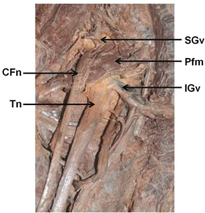
SN, in case 1 (Fig. 1) CFN exited the pelvis above the
piriformis (SP) and TN emerged below the piriformis
(IP). Following sacral plexus exploration, we noticed
that the lumbosacral trunk was coursing down and
receiving contribution from first sacral primary ra -mus and then dividing into two roots just above the piriformis muscle; one root was continuing down
through SP as CFN and the other root joined the TN just behind the piriformis muscle (Fig. 2). Thus, in this
limb TN had formed quite low just before emerging through the greater sciatic foramen. By looking at this arrangement we surmise that SN is not formed at all
in this limb and CFN and TN are direct branches from
the sacral plexus. Interestingly in this specimen we also noticed that inferior gluteal vessels were passing through the TN and peroneal communicating nerve
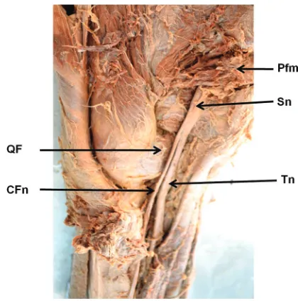
was arising in the middle of the thigh (Fig. 2). In case 2 (Fig. 3), CFN was seen passing through
the lower part of the piriformis muscle along with the inferior gluteal vessels and TN emerged through
the IP of the greater sciatic foramen. In the gluteal region both the nerves were running down close to each other and at the lower border of the quadratus femoris muscle these two divisions were seen to be enclosed in a common facial sheath and then pursue a normal course in the thigh as the SN.
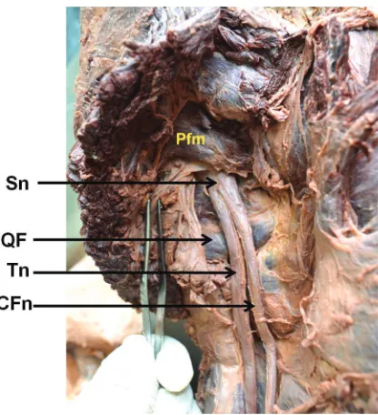
In case 3 (Fig. 4), TN and CFN emerged separately from the greater sciatic foramen. CFN was passing
above and TN had passed below the piriformis. Inter-estingly these two divisions united in the upper part of the thigh to form the SN.
In case 4 (Fig. 5), SN was seen dividing just at the
upper border of the quadratus femoris muscle in the
gluteal region and in case 5 (Fig. 6), it was dividing
just after emerging through the greater sciatic fora-men below the piriformis muscle.
Figure 2. The left gluteal region showing the region of greater sci-atic foramen after reflecting piriformis muscle;LST — lumbo-sacral trunk; SP — lumbo-sacral plexus; CFn — common fibular nerve; Tn — tibial nerve; IGv — inferior gluteal vessels; Pfm — piriformis muscle.
Figure 3. The left gluteal region after reflecting gluteus maximus muscle; CFn — common fibular nerve; Tn — tibial nerve; IGv — inferior gluteal vessels; Pfm — piriformis muscle; GM — gluteus medius; QF — quadratus femoris muscle; GMx — gluteus maxi -mus -muscle; Sn — sciatic nerve.
Figure 4. The right gluteal region after reflecting gluteus maximus muscle; CFn — common fibular nerve; Tn — tibial nerve; Pfm — piriformis muscle; QF — quadratus femoris muscle; Sn — sciatic nerve.
DISCUSSION
According to previous studies, higher division of SN in the thigh is not a very uncommon phenom-enon but its division higher up in the pelvis or before emerging through the greater sciatic foramen and its anomalous relation with the piriformis is not so common.
Many previous studies on the varied relationship between the piriformis and SN, have found variations in 15–30% of cases [17, 20].
Beaton and Anson [4] have classified these varia -tions into six categories:
— Type 1: Undivided nerve below undivided piri -formis muscle;
— Type 2: Divisions of nerve between and below undivided muscle;
— Type 3: Divisions above and below undivided muscle;
— Type 4: Undivided nerve between heads;
— Type 5: Divisions between and above heads;
— Type 6: Undivided nerve above undivided muscle.
Out of these variations, type “1” is considered as normal relationship between the piriformis and the SN and type 2 to 6 are labelled as an abnormal
rela-tionship. The type 6 variation defined hypothetically
by Beaton and Anson [4] was later reported by Ozaki et al. [14] and Sayson et al. [20]. Similarly Babinski et al. [2] and Mas et al. [11] reported a rare variation,
in which CFN and TN emerged as separate branches
through the IP but TN was passing anterior to the superior gemellus muscle. Though not reported by Beaton and Anson [4], Güvençer et al.[7] have re-ferred to this variation as Beaton and Anson type 7.
On the other, Machado et al. [10] reported three
types of variation, including type 1 where the CFN
penetrated the piriformis and the TN passed under the
piriformis (16%), type 2 where the CFN passed above
the piriformis and the TN passed under the piriformis and type 3 where the SN penetrated the piriformis.
Pokorny et al. [17] customised the Beaton and
Anson classification and stated that the type 1 vari -ation, undivided nerve below undivided muscle, was seen in 79.1% of their specimens [16]. In our study we also noted that in 19 specimens the SN was dividing in the popliteal fossa.
In the present study we also found one specimen
(case 2) with Type 2 variation in which CFN was pen -etrating the piriformis and TN was passing below the piriformis. Similar variant was reported in one case by
Arifoğlu et al. [1], and by Kırıcı and Ozan [8]. Natsis
et al. [13] dissected Caucasian cadavers (294 limbs) and only in 12 limbs (4.1%) he found the CFN passed
through and the TN below a double piriformis, while
Güvençer et al. [7], found this variation in 14% of their
cases. While two specimens (1 and 3) from our study appeared to be Beaton and Anson type 3 variation (Fig. 4), in which CFN was passing through the SP and
the TN through the IP, these variations were reported
by Ugrenovic et al. [23] in 1.5% of their cases. In our
study, we noted that the two divisions were joining together and forming SN in the upper part of the thigh. No other study mentioned this type of SN for-mation. These cases cannot be called as a typical case of higher division of SN. In case 1 SN had not formed
at all. The CFN and TN had originated separately from
the sacral plexus and coursed down the thigh as two nerves. Other interesting feature which we found in this case is that the Inferior gluteal vessels were pass-ing through the TN, which is not reported in any of
the previous studies. Moreover, in this case first sacral
nerve root was joining lumbosacral trunk quite low. Matejcik [12]also reported low level of connection
between truncus lumbosacralis and first sacral nerve root in 10 cases. Contrarily in our cases 2 and 3, CFN
and TN after emerging through the greater sciatic foramen as separate branches, joined together in the upper part of the thigh forming the SN. We suggest that this arrangement be termed as low formation of SN rather than a higher division of SN.
We did not find Beaton and Anson type 4 varia -tions as this variation is extremely rare and is reported
only by Chen [5], and by Kosukegawa et al. [9]. Case 4 and 5 are typically called as higher division
of the SN, as in these cases SN was dividing after emerging from the IP of the greater sciatic foramen high up in the thigh. This type of variation is not de-scribed by Beaton and Anson [4], Prakash et al. [18] reported this type of division inthree (2 male and 1 female) out of 86 (3.5%) extremities, they studied.
Further they also stated that neurological sign and
symptoms may depend on the level of SN division.
CONCLUSIONS
In three limbs the TN and CFN components of the
SN had emerged separately with varied relationship with the piriformis. Out of these, in two cases, these two components had united in the thigh to form the SN, while in the third case they had a separate course in the thigh. We do not consider this as a higher division of the SN. On the other hand we believe they should be termed as either low formation, if the two components joined together after emerging through the pelvis or failure of formation of the SN, if these two components do not join at all. In the present study only in two limbs the SN was dividing in the gluteal region and we ap-propriately consider these cases as high division of the SN. It is hoped our conclusions from this study will add a new perspective to the formation and division of the SN, and help the clinician to better manage patients with sciatica and piriformis syndrome.
REFERENCES
1. Arifoğlu Y, Sürücü HS, Sargon MF, Tanyeli E, Yazar F (1997)
Double superior gemellus together with double piriformis and high division of the sciatic nerve. Surg Radiol Anat, 19: 407–408.
2. Babinski MA, Machado FA, Costa WS (2003) A rare varia -tion in the high division of the sciatic nerve surrounding
the superiorgemellus muscle. Eur J Morphol, 41: 41–42.
3. Barton PM (1991) Piriformis syndrome: a rational approach to management. Pain, 47: 345–352.
4. Beaton LE, Anson BJ (1937) The relation of the sciatic nerve
and its subdivisions to the piriformis muscle. Anat Rec, 70: 1–5. 5. Chen WS (1994) Bipartite piriformis muscle: an unusual
case of sciatic nerve entrapement. Pain, 58: 269–272. 6. Chiba S (1992) Multiple positional relationships of nerves
arising from the sacral plexus to the piriformis muscle in humans. Kaibogaku Zasshi, 67: 691–724.
7. Güvençer M, Iyem C, Akyer P, Tetik S, Naderi S (2009)
Variations in the high division of the sciatic nerve and relationship between the sciatic nerve and the piriformis. Turkish Neurosurg, 19: 139–144.
8. Kırıcı Y, Ozan H (1999) Double gluteus maximus muscle
with associated variations in the gluteal region. Surg Radiol Anat, 21: 397–400.
9. Kosukegawa I, Yoshimoto M, Isogai S, Nonaka S, Yamash -ita T (2006) Piriformis syndrome resulting from a rare anatomic variation. Spine, 31: 664–666.
10. Machado FA, Babinski MA, Brasil FB, Favorito LA, Abidu-Figureiedo M, Costa MG (2003) Anatomical variations
between sciatic nerve and priform muscle during fetal
period in human. Int J Morphol, 21: 29–35.
11. Mas N, Ozekşi P, Ozdemir B, Kapakin S, Sargon MF, Celik HH, Yener N (2003) A case of bilateral high division of the
sciatic nerves, together with a unilateral unusual course of the tibial nerve. Neuroanatomy, 2: 13–15.
12. Matejcík V (2010) Anatomical variations of lumbosacral plexus. Surg Radiol Anat, 32: 409–414.
13. Natsis K, Totlis T, Konstantinidis GA, Paraskevas G, Piagkou M, Koebke J (2014) Anatomical variations between the sciatic
nerve and the piriformis muscle: a contribution to surgi-cal anatomy in piriformis syndrome. Surg Radiol Anat, 36: 273–280.
14. Ozaki S, Hamabe T, Muro T (1999) Piriformis syndrome re -sulting from an anomalous relationship between the sciatic nerve and piriformis muscle. Orthopedics, 22: 771–772. 15. Papadopoulos EC, Khan SN (2004) Piriformis syndrome
and low back pain: a new classification and review of the literature. Orthop Clin Am, 35: 65–71.
16. Pecina M (1979) Contribution to the etiological explanation
of the piriformis syndrome. Acta Anat (Basel), 105: 181–187. 17. Pokorny D, Jahoda D, Veigl D, Pinskerova V, Sonsa A (2006)
Topographic variations of the relationship of the sciatic nerve and the piriformis muscle and its relevance to palsy after total hip arthroplasty. Surg Radiol Anat, 28: 88–91. 18. Prakash, Bhardwaj AK, Devi MN, Sridevi NS, Rao PK, Singh G
(2010) Sciatic nerve division: a cadaver study in the Indian
population and review of them literature. Singapore Med J,
51: 721.
19. Robinson DR (1997) Pyriformis syndrome. The relation to
sciatic pain. Am J Surg,73: 355–380.
20. Sayson SC, Ducey JP, Maybrey JB, Wesley RL, Vermilion D
(1994) Sciatic entrapment neuropathy associated with an anomalous piriformis muscle. Pain, 59: 149–152. 21. Smoll NR (ALFABET!!) (2010) Variations of the piriformis
and sciatic nerve with clinical consequence: a review. Clin
Anat, 23: 8–17.
22. Standring S (2005) Gray’s anatomy. The anatomical basis of clinicalpractice. 39 Ed. Elsevier Churchill Livingstone,
Spain 1403, 1404, 1446.
23. Ugrenovic S, Jovanovic I, Krstic V, Stojanovic V, Vasovic L,