R E S E A R C H
Open Access
Formation and optimization of
three-dimensional organoids generated from
urine-derived stem cells for renal function
in vitro
Guoliang Sun
1, Beichen Ding
2, Meimei Wan
3, Liang Chen
1,3*, John Jackson
3and Anthony Atala
3Abstract
Background:Organoids play an important role in basic research, drug screening, and regenerative medicine. Here, we aimed to develop a novel kind of three-dimensional (3D) organoids generated from urine-derived stem cells (USCs) and to explore whether kidney-specific extracellular matrix (kECM) could enable such organoids for renal function in vitro.
Methods:USCs were isolated from human urine samples and cultured with kECM extraction to generate 3D organoids in vitro. Eight densities from 1000 to 8000 cells per organoids were prepared, and both ATP assay and Live/Dead staining were used to determine the optimal USC density in forming organoids and kECM additive concentration. The morphology and histology of as-made organoids were evaluated by hematoxylin and eosin (H.E.) staining, immunofluorescence staining and whole mount staining. Additionally, RT-qPCR was implemented to detect renal-related gene expression. Drug toxicity test was conducted to evaluate the potential application for drug screening. The renal organoids generated from whole adult kidney cells were used as a positive control in multiple assessments.
Results:The optimized cell density to generate ideal USC-derived organoids (USC-organoids) was 5000 cells/well, which was set as applying density in the following experiments. Besides, the optimal concentration of kECM was revealed to be 10%. On this condition, Live/Dead staining showed that USC-organoids were well self-organized without significant cell death. Moreover, H.E. staining showed that compact and viable organoids were generated without obvious necrosis inside organoids, which were very close to renal organoids morphologically. Furthermore, specific proximal tubule marker Aquaporin-1 (AQP1), kidney endocrine product erythropoietin (EPO), kidney glomerular markers Podocin and Synaptopodin were detected positively in USC-organoids with kECM.
Nephrotoxicity testing showed that aspirin, penicillin G, and cisplatin could exert drug-induced toxicity on USC-organoids with kECM.
(Continued on next page)
© The Author(s). 2020Open AccessThis article is licensed under a Creative Commons Attribution 4.0 International License, which permits use, sharing, adaptation, distribution and reproduction in any medium or format, as long as you give appropriate credit to the original author(s) and the source, provide a link to the Creative Commons licence, and indicate if changes were made. The images or other third party material in this article are included in the article's Creative Commons licence, unless indicated otherwise in a credit line to the material. If material is not included in the article's Creative Commons licence and your intended use is not permitted by statutory regulation or exceeds the permitted use, you will need to obtain permission directly from the copyright holder. To view a copy of this licence, visithttp://creativecommons.org/licenses/by/4.0/. The Creative Commons Public Domain Dedication waiver (http://creativecommons.org/publicdomain/zero/1.0/) applies to the data made available in this article, unless otherwise stated in a credit line to the data.
* Correspondence:[email protected]
1Department of Urology, Tongji Hospital, Tongji Medical College of Huazhong University of Science and Technology, 1095 Jiefang Avenue, Qiaokou, Wuhan 430030, HB, China
3Wake Forest Institute for Regenerative Medicine, Wake Forest University, Winston Salem, NC, USA
(Continued from previous page)
Conclusions:USC-organoids could be developed from USCs via an optimal procedure. Combining culture with kECM, USC-organoid properties including morphology, histology, and specific gene expression were identified to be similar with real renal organoids. Additionally, USC-organoids posed kECM in vitro showed the potential to be a drug screening tool which might take the place of renal organoids to some extent in the future.
Keywords:Urine-derived stem cells, Organoids, Extracellular matrix, Drug screening, Renal function
Background
Kidney is considered as an extremely essential and com-plex organ, which is responsible for several functions in-cluding drug metabolism [1,2]. Because of the ubiquitous drug nephrotoxicity, it is imperative to find appropriate drug screening models [3–5]. Currently, much medical re-search still relies on traditional two-dimensional (2D) in vitro cell culture or animal models for drug testing and toxicity assays [6]. However, these two models have insur-mountable disadvantages. Although 2D cell culture sys-tems are derived from human tissues, their differences from in vivo architecture and physiology inevitably result in the failing in drug clinical transformation [6–9]. They also cannot represent the disease heterogeneity, as they are originally derived from homogeneous cell lines [6]. Otherwise, animal models unavoidably face many prob-lems, such as high cost, ethical issues, and heterogeneity compared to human systems [6].
With the rapid development of regenerative medicine, organoid becomes a valuable research platform for drug development, which has the potential to overcome defi-ciencies originated from traditional models [1,6]. Orga-noids are complex three-dimensional (3D) structures developed from stem cells or organ-specific progenitors through a self-organization process [10]. It is thought to closely recapitulate human in vivo environment because it consists of several cell types and contains multicellular organ structures, displaying architectures and functional-ities similar to in vivo organs [11]. So far, organoids of various organs [12–16] have been generated including different renal-like cell types and self-organizing renal organoids [17–23]. They have been verified to be excel-lent high-throughput drug screening models [6].
Generally, adult somatic stem cells and human pluri-potent stem cells (hPSCs) including human embryonic stem cells (hESCs) and human-induced pluripotent stem cells (hiPSCs) are two major sources for organoid gener-ation [24]. However, these approaches face many chal-lenges. Adult somatic stem cells are usually isolated from surgical or puncture specimen, which is invasive and inconvenient [25]. Meanwhile, intrinsic tumorigen-icity properties are identified in undifferentiated hiPSCs, and hESCs cannot be applied widely because of medical ethical issues [26]. Thus, we urgently need a simple, safe, and non-invasive method to generate renal organoids.
Urine-derived stem cells (USCs) have been proved to possess the desired regenerative properties, including ro-bust proliferative potential, multipotential differentiation, and paracrine effects [27]. Currently, USCs have been successfully induced to differentiate into urothelium [28]. Moreover, USCs have been applied for personalized modeling of genetic kidney disorders and motor neuron disease [29]. Compared to adult somatic stem cells and hPSCs, USCs can be obtained noninvasively at a less cost and simpler method [27]. This indicates the possibility of organoid generation from USCs and subsequent application in drug screening field.
Renal function including glomerular filtration, renal tubu-lar reabsorption, and endocrine are important evaluation indicators for renal organoids. So far, renal organoids can-not completely recapitulate kidney complex organization, but they enable in vitro modeling of renal function to some extent [30]. Typically, protein Aquaporin-1 (AQP1) which forms a water-specific channel for high permeability to water in kidney proximal tubules and erythropoietin (EPO) which is a hormone mainly secreted by the kidney to stimulate the production of red blood cells are both fre-quently used as specific markers for renal characteristic and function [31]. Besides, Podocin is a slit diaphragm-associated protein, and Synaptopodin is a cytoskeleton-associated pedicel protein, which both are kidney glomeru-lar markers [32]. Because animal-derived kidney-specific extracellular matrix (kECM) can provide an optimal micro-environment for renal cells, it can affect cell differentiation and contribute much to renal function in 3D culture [30,
33]. O’Neill et al. and Peloso et al. had ever prepared kECM through kidney decellularization and digestion and used it to regulate kidney cell growth and metabolism [34, 35]. Thus, kECM was used in our study as well as AQP1, EPO, Podocin, and Synaptopodin were regarded as specific detection indicators for renal characteristic and function.
Methods
Cell isolation and culture
The USCs were isolated and cultured according to the protocol reported previously [36]. In brief, 14 urine sam-ples (80–400 ml) freshly collected from 3 healthy male individuals were centrifuged at 500gfor 5 min. After dis-carding urine supernatant, cell pellets were gently sus-pended in USC culture medium consisting of embryo fibroblast medium (EFM, Lonza, Basel, Switzerland) and keratinocyte serum-free medium (KSFM, Gibco, Wal-tham, MA, USA) mixed at a ratio of 1:1 with 10% fetal bovine serum (FBS, Gibco, Waltham, MA, USA). The cells were then cultured in 24-well plates at 37 °C in a 20% O2/5% CO2incubator. After 3–5 days, USC individ-ual clones appeared, which was considered as passage 0 (p0). Each clone was trypsinized, and culture medium was changed. When reaching a confluence of 60–70%, they were passaged into 6-well plates (p1). Finally, USCs were expanded in a 150-mm culture dish (p2). USCs at p3–4 were used in all experiments.
Human kidney cells (HKCs) were previously isolated from donor human kidneys not used for transplantation and frozen in liquid nitrogen at passage 0 [37]. They were recovered and cultured in kidney culture medium (KCM) containing equal amount of the following two media: one is KSFM supplemented with 2.5% FBS, 1% penicillin-streptomycin, 0.4% insulin transferrin selenium (Sigma-Aldrich, St. Louis, MO, USA), 0.25% epidermal growth factor and bovine pituitary extract (manufacture’s supple-ments for KSFM); the other is high glucose Dulbecco’s modified Eagle’s medium (DMEM, Sigma-Aldrich, St. Louis, MO, USA) supplemented with 10% FBS and 1% penicillin-streptomycin. The p1–2 HKCs were applied in the subsequent steps.
3D organoids formation
The Gravity PLUS™ 3D culture and assay platform (InSphero, Brunswick, ME, USA) were utilized to gener-ate 3D organoids. During organoid formation, the KCM was applied to culture USCs for renal lineage differenti-ation. The trypsinized and resuspended USCs in 40μl KCM were seeded into each well of GravityPLUS plate gently and vertically by 8-channel pipette. All plates were incubated at 37 °C in 20% O2/5% CO2. Four days later, the 3D organoids were harvested, transferred from GravityPLUS plate into GravityTRAP plate by adding 70μl KCM into each well with a subtle pipetting pres-sure. Immediately, the residual medium was aspirated out carefully, and 70μl fresh KCM was added slowly. For long-term and robust culture, 70μl KCM per well was refreshed every day. We defined the obtained organoids derived from USCs as USC-organoids.
The HKCs were formed into renal organoids using the same protocol.
USC-organoids optimization
Seeding density optimization by ATP assay
To optimize the initial USC concentration to generate organoids, the individual USCs were calculated and seeded at eight densities from 1000 to 8000 cells (in in-tervals of 1000 cells) per well respectively in the above procedure. Each 96-well GravityTRAP plate was pre-pared for each density and incubated for 7 days. Then, the ATP measurement of CellTiter-Glo 3D Cell Viability Assay (Promega, Madison, WI, USA) was utilized to evaluate organoid proliferative ability according to the manufacturer’s instruction. Briefly, 40μl mixture of CellTiter-Glo 3D reagent and KCM were added into a sample plate and shaken at room temperature (RT) for 30 min. The content in the wells was then moved into assay plate, and their bioluminescence activity was appraised by plate reader. Later, ATP concentration was computed according to the standard curve.
kECM concentration optimization by Live/Dead staining
To explore the optimal kECM concentration for USC-organoid viability, we cultured USC-USC-organoids in the mix-ture of KCM and different ratios of porcine-derived kECM, and then evaluated cytotoxicity by Live/Dead staining.
To prepare for kECM extract, the discarded porcine kidneys in other experiments in our laboratory [31] were obtained and utilized. The production method was modified from previously reported protocol of liver extracellular matrix (ECM) extract [38], which was dem-onstrated in Additional file 1 (Supplementary Figure1). Briefly, the kidney decellularization procedure for kECM included complete pre-rinsing through the blood vessels, tissue cutting into blocks followed by slicing into pieces after flash freezing at −80 °C, reduplicative shaking in distilled water for 3 days, repeated treating with 2% Tri-ton X-100 for 4 days, and finally washing for 2 days for clean removal of Triton X-100. The obtained compo-nents were lyophilized and grounded into powder with a freezer mill. ECM powder was mixed with pepsin (Fisher Scientific, Pittsburgh, PA, USA) at a weight ratio of 10:1 and sterilized by gamma irradiation (1 Mrad). Under sterile condition, the mixture was centrifuged at 3000 rpm for 15 min after dissolving in 0.1 mol/L hydrochloric acid. The pellet was discarded repeatedly until the super-natant was clear. The decellularized ECM extracts were further purified by filtering through a 0.2-μm syringe fil-ter (Thermo Fisher Scientific, Rochesfil-ter, NY, USA) and neutralized by 1 mol/L NaOH to pH 7.0 for ready use.
USC-organoids in conditioned medium with the above three kECM concentrations. Simply, the organoid sam-ples were carefully transferred to Eppendorf tubes with top-cut 1 mL pipette tips and incubated with a reagent working solution consisting of 0.5μL calcein AM and 2μL Ethidium homodimer-1 in 1 mL KCM for 30–45 min at RT. Washed twice by phosphate-buffered saline (PBS), the samples were observed under an Olympus FV10i confocal microscope. ImageJ was used to quantify the average fluorescence brightness.
Histology evaluation for 3D organoids
We prepared three groups of samples for the following assessments: USC-organoids (optimized seeding density from the “Seeding density optimization by ATP assay” section), USC-organoids with kECM (optimized seeding density from the section “Seeding density optimization
by ATP assay” section and optimized kECM concentra-tion from the “kECM concentration optimization by
Live/Dead staining” section), and renal organoids (opti-mized HKCs seeding density).
Hematoxylin and eosin staining
On the 7th day of culture, 3D organoid samples were washed triply by PBS for 15 s and fixed with 4% parafor-maldehyde for 1 h at RT. After 3 times wash by PBS again, all samples were immersed into preheated and dissolved HistoGel (Thermo Fisher Scientific, Waltham, MA, USA), thrown into 60% EtOH, and treated by dehy-dration and paraffin embedding. Followed with deparaf-finization and hematoxylin and eosin (H.E.) staining process, sample slides were viewed using a Zeiss Axio-vert 200M microscope.
Immunofluorescence staining
The paraffin slides from the “Hematoxylin and eosin
(H.E.) staining” section without H.E. staining were chosen for immunofluorescence staining. All slides were incubated at 60 °C for 2 h for deparaffinization, washed by ddH2O, and transferred to microwave antigen re-trieval in citrate buffer (pH = 6.0) at 95 °C for 10 min. Let the slides cool at RT for 1.5 h and block them with protein blocking buffer (Dako, Carpinteria, CA, USA) for 1 h at RT. After incubation with 1:100 diluted spe-cific rabbit anti-human primary antibodies for AQP1 (#Ab15080, Abcam, Cambridge, MA, USA), EPO (#Ab126876, Abcam), Podocin (H-130, Santa Cruz Bio-technology, Santa Cruz, CA, USA), and Synaptopodin (P-19, Santa Cruz Biotechnology) overnight at 4 °C, corresponding secondary antibodies were carried out to treat washed samples for 1 h in dark at RT. The immunofluorescence signals from paraffin sections were imaged by Zeiss Axiovert 200M microscope. ImageJ was used to measure the average fluorescence brightness.
Whole mount staining
The 3D organoids were transferred to chamber slides (Thermo Fisher Scientific, Waltham, MA, USA) and washed in PBS repeatedly. Followed with fixation in 4% paraformaldehyde for 1 h, the 3D organoids were perme-abilized in 0.2% Triton X-100 for 30 min and washed by PBS again. Sequentially, organoids were incubated in pro-tein blocking buffer for 1 h at RT, in which the primary antibodies for AQP1 and EPO were diluted. On the next day, corresponding secondary antibody was applied. The same primary and secondary antibodies as mentioned above were used except that the dilution of the secondary antibody was 1:500. The fluorescence signals were visual-ized by Olympus FV10i confocal microscope. ImageJ was used to evaluate the average fluorescence brightness.
RT-qPCR
Total mRNA was extracted from 3D organoids by a RNeasy Plus Mini Kit (Qiagen, Valencia, CA, USA). The cDNA was synthesized by QuantiTect Rev. Transcription Kit (Qiagen), and the quantitative PCR was then per-formed by a QuantiTect SYBR Green PCR Kit (Qiagen) using the 7300 Real-Time PCR system (Applied Biosys-tems, Foster City, CA, USA). The primer sequences were as follows (5′-3′): GAPDH (forward GTCATCATCT CCGCCCCTTCTGC, reverse GATGCCTGCTTCACCA CCTTCTTG); AQP1 (forward CTGGGCATCGAGAT CATCGG, reverse ATCCCACAGCCAGTGTAGTCA); EPO (forward GGAGGCCGAGAATATCACGAC, re-verse CCCTGCCAGACTTCTACGG); Podocin (forward ACCAAATCCTCCGGCTTAGG, reverse CAACCTTT ACGCAGAACCAGA); Synaptopodin (forward CCCAAG GTGACCCCGAAT, reverse CTGCCGCCGCTTCTCA) (Sangon, Shanghai, China).
Function assay
Drug toxicity test
The 3D organoids from three groups were treated with three drugs respectively, aspirin, penicillin G, and cis-platin, for nephrotoxicity test. Aspirin, penicillin G, and cisplatin (all from Sigma-Aldrich, St. Louis, MO, USA) were chosen for drug toxicity test because they were proved to be excreted by glomerulus and renal tubules, respectively [39]. The working concentration of these three drugs was all 200μM, and the drug treatment time was 48 h [39]. Organoids were then homogenized and tested by γ-glutamyltransferase (GGT) Activity Colori-metric Assay Kit (Sigma-Aldrich).
Dose-response curves and the half maximal inhibitory concentration estimation
6.4 mM. Twenty-four hours later, the relative cell sur-vival was evaluated by MTS Kit (Promega, Madison, WI, USA) according to the manufacturer’s protocol. Relative cell survival was defined as the ratio of the number of vi-able cells in the containing medium and the drug-free medium. “Relative cell survival” values and drug concentrations were fitted to dose-response curves to estimate the half maximal inhibitory concentration (IC50) values.
Statistical analysis
In ATP assay, six replicated wells for each cell density were examined. In Live/Dead staining, histology evalu-ation, and RT-qPCR, three samples for each group were evaluated. In GGT activity measurement and drug screening experiment, five and three replicated samples for each group were used respectively.
The data were presented as the mean ± standard devi-ation (SD). Comparisons between two groups were per-formed using a two-tailed Student’s t test. A value of
P< 0.05 was considered as statistically significant. P< 0.05,P< 0.01, andP< 0.001 were marked as“*”,“**”, and
“***”respectively.
Results
Morphology of 2D culture cells
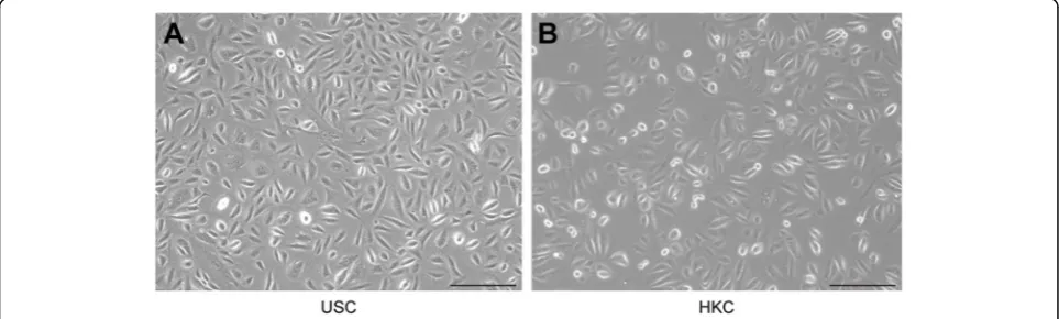
The morphology of 2D-cultured USCs and HKCs are shown in Fig. 1. Seen from Fig. 1a, the USCs were plump, compact, and bright, showing a rice grain-like appearance after initial seeding. The HKCs looked more rounded than USCs in Fig. 1b. The other properties of USCs have been reported in a previous article [40].
Morphology and optimization of USC-organoids
Morphology observation
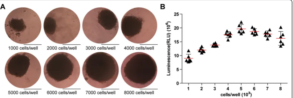
USCs were seeded at a density of 1000, 2000, 3000, 4000, 5000, 6000, 7000, 8000 cells/well, respectively. Organoids formed at day 3 and were observed at day 7.
Under the microscope, USCs were organized into a spheroid in a 3D pattern. Generally, with the USC amount increasing, the organoids became larger and more compact. Theoretically, organoid function will be better and closer to normal tissues if they carry more cells. However, as the volume increases, it is harder for nutrient solution to penetrate inside, so the cells in the sphere center might die due to lack of nutrition. On the basis of our previous results [39], the relatively ideal 3D organoid is about 200–300μm at diameter, which corre-sponds to USC-organoids with the seeding density of 3000–6000 cells/well in this experiment. In Fig. 2a, the size of the organoids grew larger ranging from about 160μm (1000 cells/well) to almost 400μm (8000 cells/ well) with the increasing initial cell density. HKC orga-noid morphology displayed the same pattern [39].
Proliferation assay for cell density optimization
Additionally, ATP activity test was designed to deter-mine USC-organoid proliferative ability. On day 14, these organoids were harvested and the ATP assays were conducted. The results clearly showed that the USC-organoid proliferative ability increased with the ascent of initial USC concentration and reached a peak at 5000 cells/well, after which the proliferative ability began to descend, as shown in Fig. 2b. These results demon-strated that the optimal initial USC concentration for organoid formation was 5000 cells/well, whose corre-sponding organoid diameter was about 250μm. Com-bined with the above morphological results, we concluded that 5000 cells/well was the optimal seeding density for USCs to form organoids and it was applied for further experiments.
Viability assay for kECM concentration optimization
Live/Dead staining was employed to evaluate organoid viability. According to the above data in cell density optimization, USCs were seeded at the density of
5000 cells/well to generate 3D organoids. The KCM was supplemented with 5%, 10%, or 15% solubilized kECM. At day 14, organoids were harvested for Live/Dead stain-ing. The results showed that the organoid cultured with 10% kECM were well self-organized and had the most green fluorescence (living cells) and least red fluores-cence (dead cells), indicating the best viability, seen from Fig. 3. These findings revealed that 10% kECM was the optimal concentration for organoid formation.
The above data indicated the optimal cell seeding density was 5000 cells/well and optimal kECM additive concentra-tion was 10% in our experimental system. As-formed USC-organoids were employed in the next assessments.
Histology evaluation for 3D organoids
H.E. staining, immunofluorescence staining, and whole mount staining were applied for comparative observa-tion for three groups of 3D organoids, USC-organoids, USC-organoids with kECM, and renal organoids. All organoids cultured for 14 days were submitted to the following staining assays.
Approximately, three groups of organoids looked uni-form in cellular aggregation and compact in structure organization in H.E. staining. In Fig.4a, the cell nucleus was blue stained, cytoplasm was red stained, and there was no large necrosis and voids in the organoids. In terms of cytoplasm staining, USC-organoids with kECM was closer to renal organoids, with more cytoplasm and deeper red staining.
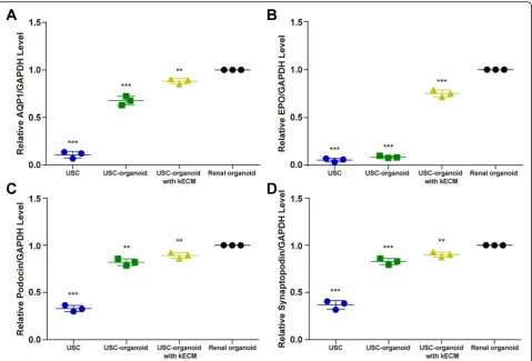
Additionally, marker proteins for specific proximal tu-bule AQP1, kidney endocrine product EPO, and kidney glomerular Podocin and Synaptopodin were marked as red on sectioned organoids by immunofluorescence
staining while DAPI as blue. In Fig. 4b, we found that three groups of 3D organoids showed similar patterns in terms of AQP1, indicating that they all possessed water metabolism function. The AQP1 dyeing intensity of USC-organoids with kECM was closer to renal orga-noids. Besides, USC-organoids with kECM had similar EPO expression level to renal organoids, but there was almost none EPO fluorescence in USC-organoids with-out kECM obviously. Furthermore, the Podocin and Synaptopodin dyeing intensity of USC-organoids with kECM was closer to renal organoids.
Moreover, the whole mount staining was performed to label the fluorescence of AQP1 and EPO in three-dimensional distribution. Figure 4c showed that USC-organoids with kECM were analogous to renal USC-organoids to a large extent.
Expression of renal-specific markers
0.034, USC-organoid 0.822 ± 0.036, USC-organoid with 10% kECM 0.894 ± 0.031]; Synaptopodin [USC 0.369 ± 0.047, USC-organoid 0.830 ± 0.033, USC-organoid with 10% kECM 0.901 ± 0.027]). The AQP1, EPO, Podocin, and Synaptopodin expression levels of USCs, USC-organoids, and USC-organoids with kECM were all significantly lower than renal organoids.
Evaluation of USC-organoids as a nephrotoxicity screening model
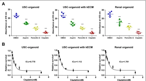
Drug toxicity test was carried out to assess the potential role of USC-organoids for nephrotoxicity screening. Similar trends were observed between USC-organoids with kECM and renal organoids in Fig.6a. Significantly, the decreased absorbance at 418 nm was observed in as-pirin, penicillin G, and cisplatin group than the control group, demonstrating that these drugs at normal drug concentration could exert similar drug-induced toxicity and the USC-organoids with kECM had similar drug screening ability to renal organoids.
The “relative cell survival” values and drug concentra-tions were fitted to dose-response curves to estimate the IC50values of cisplatin. Respectively, the IC50of cisplatin in USC-organoids, USC-organoids with kECM, and renal
organoids were 0.778 mM, 1.112 mM, and 1.781 mM, marked in Fig.6b.
Discussion
This study successfully sketched the optimized process of USC-organoid generation with kECM and proved that USC-organoids had similar morphology, histology, gene expression, and nephrotoxicity screening ability to renal organoids. Firstly, the USCs were collected, and porcine-derived kECM was prepared to imitate the in vivo envir-onment. Next, the optimal cell density and kECM con-centration were determined by examining organoids with proliferative ability and viability. Finally, we evalu-ated the characteristics of USC-organoids and compared it with renal organoids by multiple assays. Briefly, our study developed a novel type of organoid, namely USC-organoids, and validated its identity and potential in nephrotoxicity screening to take the place of renal organoids or renal tissue models.
The functional renal cells are in great need in both clinic and laboratory, which have insufficient and un-stable source, even worse the renal organoids for re-search. The urine-derived stem cells have many advantages compared to adult somatic stem cells and Fig. 3kECM concentration optimization via viability assay. Live/Dead staining was employed to evaluate the viability of USC-organoids in
hPSCs. They can be collected in a noninvasive manner from an easily accessible source [41]. In addition, there are less ethical restrictions about urine cells [26]. On one hand, there were sparse studies implying the in-volvement of USCs in organoid generation. Usui et al. elucidated that prostate cancer organoids were success-fully generated using dog urine cancer stem cells, and these organoids might become a useful tool for the re-search of cancer pathogenesis and treatment [42]. Be-sides, Wan et al. illustrated that differentiated
in vitro and in vivo [41]. Moreover, Yi et al. elaborated that they differentiated U-hiPSCs into motor neurons, and U-hiPSCs were promising source for motor neuron disease modeling [29].
Here, we systematically generated USC-organoids with the help of kECM and verified its renal function prelim-inarily. Currently, several categories of ECM including animal-derived ECM (general or tissue-specific) and syn-thetic matrices have been developed for 3D culture [44]. Frans et al. indicated that animal tissue-derived ECM could improve the expansion, differentiation, and passa-ging of renal organoids [30]. They also reviewed that synthetic matrices might not only contribute to renal organoid culture but also enable the clinical grade renal organoids’formation for cell therapy [30]. So far, animal tissue-derived ECM matrix is the only matrix reported to be used for organoid culture [30]. Further studies are
needed to verify the kECM contribution to renal func-tion of USC-organoids.
Several types of function can be assessed to identify renal characteristics including tissue-specific barrier and transport functions [30]. Takasato et al. demonstrated that renal organoid cells with a proximal tubular pheno-type had the cubilin-mediated endocytosis function [19]. Freedman et al. elaborated that methotrexate and dex-tran could be dex-transported into the lumen of renal orga-noids, and this was mediated by endocytosis [22]. In this study, we used EPO test to validate the renal function. EPO is an important glycoprotein hormone produced from renal fibroblasts, and it can stimulate red cell production [45, 46]. We proved that USC-organoids cultured with kECM had similar EPO expression pattern with renal organoids, indicating the endocrine function of USC-organoids.
Newly developed drugs including antibiotics and im-munosuppressants usually have potential in nephrotox-icity, and it is critical to predicting drug nephrotoxicity before they are used clinically [3,4]. Thus, novel and ac-curate models for screening drug nephrotoxic activity are urgently needed [5]. To date, isolated human pri-mary renal cells and immortalized cell lines had been utilized for drug screening, but they showed low specifi-city, reproducibility, and accuracy [5]. Besides, ESCs and hiPSCs are unable to simulate distinct renal cells, kECM, vascularization, etc. [47]. Furthermore, rodent models are poor predictors of nephrotoxicity because of the spe-cies differences in gene expression [5]. Presently, renal organoids containing several types of renal cells are thought to be an effective approach for predictive tox-icity [47]. So far, many studies reported that kidney organoids from hPSCs could be applied in drug toxicity testing [3, 48–50]. In our study, we demonstrated that USC-organoid was a promising drug nephrotoxicity screening tool. Aspirin and penicillin G are excreted by the kidneys and can exert an adverse effect on renal function [51]. Cisplatin may cause acute proximal tubu-lar necrosis [52]. GGT is expressed in proximal tubule, and it is essential for theγ-glutamyl cycle, which plays a critical role in the detoxification of xenobiotics [18]. Its
activity can be used to represent the renal cell function [18]. The GGT assay showed that renal function of USC-organoids and renal USC-organoids both were impaired by as-pirin, penicillin G, and cisplatin in a dose-dependent way.
Conclusions
This study develops USC-organoids successfully and uses kECM to guide their renal function. We verify USC-organoid properties including morphology, hist-ology, gene expression, and their potential application value for drug screening.
Supplementary information
Supplementary informationaccompanies this paper athttps://doi.org/10. 1186/s13287-020-01822-4.
Additional file 1.The preparation process of kECM.
Abbreviations
3D:Three-dimensional; USCs: Urine-derived stem cells; kECM: Kidney-specific extracellular matrix; H.E.: Hematoxylin and eosin; USC-organoids: USCs-derived organoids; AQP1: Aquaporin-1; EPO: Erythropoietin; hPSCs: Human pluripotent stem cells; hESCs: Human embryonic stem cells; hiPSCs: Human-induced pluripotent stem cells; EFM: Embryo fibroblast medium;
KSFM: Keratinocyte serum-free medium; FBS: Fetal bovine serum; p0: Passage 0; HKCs: Human kidney cells; KCM: Kidney culture medium;
DMEM: Dulbecco’s modified Eagle’s medium; GGT: Glutamyltransferase;
Fig. 6Application of USC-organoids in renal function and drug screening.aGGT activity measurement for renal function and drug screening in 3D organoids. Percentage decrease in the absorbance: USC-organoids (aspirin 38.2%, penicillin G 63.0%, cisplatin 84.6%), USC-organoids with kECM (aspirin 42.8%, penicillin G 63.1%, cisplatin 86.8%), renal organoid (aspirin 38.8%, penicillin G 60.4%, cisplatin 86.4%) (five replicated samples for each group, data presented as mean ± SD).bDose-response curves of cisplatin in in 3D organoids. The IC50of cisplatin in USC-organoids,
IC50: Half maximal inhibitory concentration; SD: Standard deviation; RT: Room
temperature; U-hiPSCs: Urine-derived human-induced pluripotent
Acknowledgements
We would like to thank Tongji Hospital for its help.
Authors’contributions
LC, GS, and BD contributed to the conception. LC and GS performed the whole experimental work. GS and BD contributed to data acquisition and analysis. GS and LC drafted the work. MW, JJ, and AA supervised the work. All authors read and approved the final manuscript.
Funding
This work was supported by the Natural Science Foundation of Hubei Province [grant number 2019CFB772].
Availability of data and materials
Not applicable.
Ethics approval and consent to participate
This study was approved by the Ethics Committee of Affiliated Hospital of Tongji Medical College of Huazhong University of Science and Technology. Informed consent was obtained from participants.
Consent for publication
Not applicable.
Competing interests
The authors declare that they have no competing interests.
Author details
1Department of Urology, Tongji Hospital, Tongji Medical College of Huazhong University of Science and Technology, 1095 Jiefang Avenue, Qiaokou, Wuhan 430030, HB, China.2Department of Urology, First Affiliated Hospital of Harbin Medical University, Harbin, HLJ, China.3Wake Forest Institute for Regenerative Medicine, Wake Forest University, Winston Salem, NC, USA.
Received: 11 March 2020 Revised: 25 June 2020 Accepted: 10 July 2020
References
1. Miyoshi T, Hiratsuka K, Saiz EG, Morizane R. Kidney organoids in translational medicine: disease modeling and regenerative medicine. Dev Dyn. 2019; 249(1):34–45.
2. Davies J. Engineered renal tissue as a potential platform for
pharmacokinetic and nephrotoxicity testing. Drug Discov Today. 2014;19: 725–9.
3. Perazella MA. Renal vulnerability to drug toxicity. Clin J Am Soc Nephrol. 2009;4:1275–83.
4. Tiong HY, Huang P, Xiong S, Li Y, Vathsala A, Zink D. Drug-induced nephrotoxicity: clinical impact and preclinical in vitro models. Mol Pharm. 2014;11:1933–48.
5. Little MH, Hale LJ, Howden SE, Kumar SV. Generating kidney from stem cells. Annu Rev Physiol. 2019;81:335–57.
6. O'Connell L, Winter D. Organoids - past learning and future perspectives. Stem Cells Dev. 2019;29(5):281–289.
7. Kamb A. What's wrong with our cancer models? Nat Rev Drug Discov. 2005; 4:161–5.
8. DiMasi JA, Grabowski HG. Economics of new oncology drug development. J Clin Oncol. 2007;25:209–16.
9. Yang L, Yang S, Li X, Li B, Li Y, Zhang X, Ma Y, Peng X, Jin H, Fan Q, et al. Tumor organoids: from inception to future in cancer research. Cancer Lett. 2019;454:120–33.
10. Rossi G, Manfrin A, Lutolf MP. Progress and potential in organoid research. Nat Rev Genet. 2018;19:671–87.
11. Wang S, Gao D, Chen Y. The potential of organoids in urological cancer research. Nat Rev Urol. 2017;14:401–14.
12. Takebe T, Sekine K, Enomura M, Koike H, Kimura M, Ogaeri T, Zhang RR, Ueno Y, Zheng YW, Koike N, et al. Vascularized and functional human liver from an iPSC-derived organ bud transplant. Nature. 2013;499:481–4.
13. Huang L, Holtzinger A, Jagan I, BeGora M, Lohse I, Ngai N, Nostro C, Wang R, Muthuswamy LB, Crawford HC, et al. Ductal pancreatic cancer modeling and drug screening using human pluripotent stem cell- and patient-derived tumor organoids. Nat Med. 2015;21:1364–71.
14. Huang SX, Islam MN, O'Neill J, Hu Z, Yang YG, Chen YW, Mumau M, Green MD, Vunjak-Novakovic G, Bhattacharya J, Snoeck HW. Efficient generation of lung and airway epithelial cells from human pluripotent stem cells. Nat Biotechnol. 2014;32:84–91.
15. Lancaster MA, Renner M, Martin CA, Wenzel D, Bicknell LS, Hurles ME, Homfray T, Penninger JM, Jackson AP, Knoblich JA. Cerebral organoids model human brain development and microcephaly. Nature. 2013;501:373– 9.
16. Spence JR, Mayhew CN, Rankin SA, Kuhar MF, Vallance JE, Tolle K, Hoskins EE, Kalinichenko VV, Wells SI, Zorn AM, et al. Directed differentiation of human pluripotent stem cells into intestinal tissue in vitro. Nature. 2011;470: 105–9.
17. Morizane R, Lam AQ, Freedman BS, Kishi S, Valerius MT, Bonventre JV. Nephron organoids derived from human pluripotent stem cells model kidney development and injury. Nat Biotechnol. 2015;33:1193–200. 18. Yamaguchi S, Morizane R, Homma K, Monkawa T, Suzuki S, Fujii S, Koda M,
Hiratsuka K, Yamashita M, Yoshida T, et al. Generation of kidney tubular organoids from human pluripotent stem cells. Sci Rep. 2016;6:38353. 19. Takasato M, Er PX, Chiu HS, Maier B, Baillie GJ, Ferguson C, Parton RG,
Wolvetang EJ, Roost MS, Chuva de Sousa Lopes SM, Little MH. Kidney organoids from human iPS cells contain multiple lineages and model human nephrogenesis. Nature. 2015;526:564–8.
20. Takasato M, Er PX, Chiu HS, Little MH. Generation of kidney organoids from human pluripotent stem cells. Nat Protoc. 2016;11:1681–92.
21. Hiratsuka K, Monkawa T, Akiyama T, Nakatake Y, Oda M, Goparaju SK, Kimura H, Chikazawa-Nohtomi N, Sato S, Ishiguro K, et al. Induction of human pluripotent stem cells into kidney tissues by synthetic mRNAs encoding transcription factors. Sci Rep. 2019;9:913.
22. Freedman BS, Brooks CR, Lam AQ, Fu H, Morizane R, Agrawal V, Saad AF, Li MK, Hughes MR, Werff RV, et al. Modelling kidney disease with CRISPR-mutant kidney organoids derived from human pluripotent epiblast spheroids. Nat Commun. 2015;6:8715.
23. Taguchi A, Kaku Y, Ohmori T, Sharmin S, Ogawa M, Sasaki H, Nishinakamura R. Redefining the in vivo origin of metanephric nephron progenitors enables generation of complex kidney structures from pluripotent stem cells. Cell Stem Cell. 2014;14:53–67.
24. Fatehullah A, Tan SH, Barker N. Organoids as an in vitro model of human development and disease. Nat Cell Biol. 2016;18:246–54.
25. Schutgens F, Rookmaaker MB, Margaritis T, Rios A, Ammerlaan C, Jansen J, Gijzen L, Vormann M, Vonk A, Viveen M, et al. Tubuloids derived from human adult kidney and urine for personalized disease modeling. Nat Biotechnol. 2019;37:303–13.
26. Yasuda S, Kusakawa S, Kuroda T, Miura T, Tano K, Takada N, Matsuyama S, Matsuyama A, Nasu M, Umezawa A, et al. Tumorigenicity-associated characteristics of human iPS cell lines. PLoS One. 2018;13:e0205022. 27. Liu G, Wu R, Yang B, Deng C, Lu X, Walker SJ, Ma PX, Mou S, Atala A, Zhang Y.
Human urine-derived stem cell differentiation to endothelial cells with barrier function and nitric oxide production. Stem Cells Transl Med. 2018;7:686–98. 28. Wan Q, Xiong G, Liu G, Shupe TD, Wei G, Zhang D, Liang D, Lu X, Atala A,
Zhang Y. Urothelium with barrier function differentiated from human urine-derived stem cells for potential use in urinary tract reconstruction. Stem Cell Res Ther. 2018;9:304.
29. Yi H, Xie B, Liu B, Wang X, Xu L, Liu J, Li M, Zhong X, Peng F. Derivation and identification of motor neurons from human urine-derived induced pluripotent stem cells. Stem Cells Int. 2018;2018:3628578.
30. Schutgens F, Verhaar MC, Rookmaaker MB. Pluripotent stem cell-derived kidney organoids: an in vivo-like in vitro technology. Eur J Pharmacol. 2016; 790:12–20.
31. Abolbashari M, Agcaoili SM, Lee MK, Ko IK, Aboushwareb T, Jackson JD, Yoo JJ, Atala A. Repopulation of porcine kidney scaffold using porcine primary renal cells. Acta Biomater. 2016;29:52–61.
32. Bariety J, Mandet C, Hill GS, Bruneval P. Parietal podocytes in normal human glomeruli. J Am Soc Nephrol. 2006;17:2770–80.
34. O'Neill JD, Freytes DO, Anandappa AJ, Oliver JA, Vunjak-Novakovic GV. The regulation of growth and metabolism of kidney stem cells with regional specificity using extracellular matrix derived from kidney. Biomaterials. 2013; 34:9830–41.
35. Peloso A, Ferrario J, Maiga B, Benzoni I, Bianco C, Citro A, Currao M, Malara A, Gaspari A, Balduini A, et al. Creation and implantation of acellular rat renal ECM-based scaffolds. Organogenesis. 2015;11:58–74.
36. Zhang Y, McNeill E, Tian H, Soker S, Andersson KE, Yoo JJ, Atala A. Urine derived cells are a potential source for urological tissue reconstruction. J Urol. 2008;180:2226–33.
37. George SK, Abolbashari M, Jackson JD, Aboushwareb T, Atala A, Yoo JJ. Potential use of autologous renal cells from diseased kidneys for the treatment of renal failure. PLoS One. 2016;11:e0164997.
38. Skardal A, Smith L, Bharadwaj S, Atala A, Soker S, Zhang Y. Tissue specific synthetic ECM hydrogels for 3-D in vitro maintenance of hepatocyte function. Biomaterials. 2012;33:4565–75.
39. Ding B, Sun G, Liu S, Peng E, Wan M, Chen L, Jackson J, Atala A. Three-dimensional renal organoids from whole kidney cells: generation, optimization, and potential application in nephrotoxicology in vitro. Cell Transplant. 2020;29:963689719897066.
40. Bharadwaj S, Liu G, Shi Y, Wu R, Yang B, He T, Fan Y, Lu X, Zhou X, Liu H, et al. Multipotential differentiation of human urine-derived stem cells: potential for therapeutic applications in urology. Stem Cells. 2013;31:1840–56.
41. Jiang YF, Chen M, Zhang NN, Yang HJ, Rui Q, Zhou YF. In vitro and in vivo differentiation of induced pluripotent stem cells generated from urine-derived cells into cardiomyocytes. Biol Open. 2018;7(1):bio029157. 42. Usui T, Sakurai M, Nishikawa S, Umata K, Nemoto Y, Haraguchi T, Itamoto K,
Mizuno T, Noguchi S, Mori T, et al. Establishment of a dog primary prostate cancer organoid using the urine cancer stem cells. Cancer Sci. 2017;108: 2383–92.
43. Li G, Xie B, He L, Zhou T, Gao G, Liu S, Pan G, Ge J, Peng F, Zhong X. Generation of retinal organoids with mature rods and cones from urine-derived human induced pluripotent stem cells. Stem Cells Int. 2018;2018: 4968658.
44. Tibbitt MW, Anseth KS. Hydrogels as extracellular matrix mimics for 3D cell culture. Biotechnol Bioeng. 2009;103:655–63.
45. Souma T, Suzuki N, Yamamoto M. Renal erythropoietin-producing cells in health and disease. Front Physiol. 2015;6:167.
46. Semenza GL. Oxygen sensing, homeostasis, and disease. N Engl J Med. 2011;365:537–47.
47. Kim YK, Nam SA, Yang CW. Applications of kidney organoids derived from human pluripotent stem cells. Korean J Intern Med. 2018;33:649–59. 48. Narayanan K, Schumacher KM, Tasnim F, Kandasamy K, Schumacher A, Ni M,
Gao S, Gopalan B, Zink D, Ying JY. Human embryonic stem cells differentiate into functional renal proximal tubular-like cells. Kidney Int. 2013;83:593–603.
49. Murphy SV, Atala A. 3D bioprinting of tissues and organs. Nat Biotechnol. 2014;32:773–85.
50. Chang JW, Park SA, Park JK, Choi JW, Kim YS, Shin YS, Kim CH. Tissue-engineered tracheal reconstruction using three-dimensionally printed artificial tracheal graft: preliminary report. Artif Organs. 2014;38:E95–e105. 51. Curiel RV, Katz JD. Mitigating the cardiovascular and renal effects of NSAIDs.
Pain Med. 2013;14(Suppl 1):S23–8.
52. Goldstein RS, Smith PF, Tarloff JB, Contardi L, Rush GF, Hook JB. Biochemical mechanisms of cephaloridine nephrotoxicity. Life Sci. 1988;42:1809–16.
Publisher’s Note