S H O R T R E P O R T
Open Access
Novel Protein kinase C
θ
: Coronin 1A complex in
T lymphocytes
Kerstin Siegmund
†, Nikolaus Thuille
†, Nina Posch, Friedrich Fresser and Gottfried Baier
*Abstract
Background:
Protein kinase C-
θ
(PKC
θ
) plays an important role in signal transduction down-stream of the T cell
receptor and T cells deficient of
PKC
θ
show impaired NF-
κ
B as well as NFAT/AP-1 activation resulting in strongly
decreased IL-2 expression and proliferation. However, it is not yet entirely clear, how the function of PKC
θ
- upon
T cell activation - is regulated on a molecular level.
Findings:
Employing a yeast two-hybrid screen and co-immunoprecipitation analyses, we here identify coronin 1A
(Coro1A) as a novel PKC
θ
-interacting protein. We show that the NH
2-terminal WD40 domains of Coro1A and the
C2-like domain of PKC
θ
are sufficient for the interaction. Furthermore, we confirm a physical interaction by GST-Coro1A
mediated pull-down of endogenous PKC
θ
protein. Functionally, wild-type but not Coro1A lacking its actin-binding
domain negatively interferes with PKC
θ
-dependent NF-
κ
B, Cyclin D1 and IL-2 transactivation when analysed with
luciferase promoter activation assays in Jurkat T cells. This could be phenocopied by pharmacological inhibitors of actin
polymerization and PKC, respectively. Mechanistically, Coro1A overexpression attenuates both lipid raft and plasma
membrane recruitment of PKC
θ
in CD3/CD28-activated T cells.
Using primary CD3
+T cells, we observed that (opposite to PKC
θ
) Coro1A does not localize preferentially to the
immunological synapse. In addition, we show that CD3
+T cells isolated from
Coro1A
-deficient mice show impaired
IKK/NF-
κ
B transactivation.
Conclusions:
Together, these findings both in Jurkat T cells as well as in primary T cells indicate a regulatory role of
Coro1A on PKC
θ
recruitment and function downstream of the TCR leading to NF-
κ
B transactivation.
Keywords:
T lymphocyte signaling, Transcriptional regulation, Protein kinase C
θ
(PKC
θ
), Coronin 1A (Coro1A), NF-
κ
B,
IL-2
Findings
An efficient adaptive immune response depends on the
activation of T lymphocytes by antigen-presenting cells
(APC) and the acquisition of appropriate effector T cell
function. Activation of T lymphocytes occurs upon
en-gagement of T cell receptors (TCR) and the
correspond-ing antigen-MHC complexes together with the ligation
of the co-receptor CD28 by B7 molecules
–
leading to
the formation of the immunological synapse (IS)
be-tween T cell and APC. PKC
θ
, a member of the protein
kinase C (PKC) family of serine/threonine kinases, is
rapidly, within seconds after TCR engagement, recruited
to the peripheral supra-molecular activation cluster
(pSMAC) of the immunological synapse (IS) [1,2]. It has
been shown that, the activation of the transcription
fac-tors NF-
κ
B, NFAT and AP-1 downstream of the TCR
critically depend on PKC
θ
[3-5], linking PKC
θ
function
to IL-2 transcription, whose promoter activation
de-pends on these transcription factors [6].
Activation of all PKC family members is controlled by
a so-called pseudo-substrate (PS) domain in the NH
2-terminus that resembles PKC substrates and forms an
auto-inhibitory loop to keep the enzyme in an inactive
con-formation. PKC
θ
is released from the auto-inhibition after
recruitment to the plasma membrane, where it binds to
di-acylglycerol (reviewed in [7]). Noteworthy, PKC mutations
that disrupt this intra-molecular interaction generate
* Correspondence:[email protected]†Equal contributors
Department for Pharmacology and Genetics, Division of Translational Cell Genetics, Medical University Innsbruck, Peter Mayr Str. 1a, A-6020 Innsbruck, Austria
constitutively active forms of PKC, which are useful tools
for analysing PKC functions. Another domain involved in
modulating PKC
θ
activity is its C2-like domain, which
rep-resents a major protein:protein interaction domain. Binding
of WD40 domain-containing receptor for activated PKC
proteins
–
so called RACK proteins - to the C2-like domain
of activated PKC forms another level of regulating its
en-zymatic function [8]. However, so far no RACK
physiolo-gically interacting with PKC
θ
has been identified. In the
present study, designed to discover PKC
θ
interacting
part-ners, we identified coronin 1A (Coro1A) as a functional
regulator of PKC
θ
activation.
Identification of Coro1A as physical interaction
partner of PKC
θ
Little is known about proteins that interact with PKC
θ
and regulate its function in T lymphocytes and thereby
modulate activation of this immune cell subset. To
con-tribute to this issue, we have employed a yeast
two-hybrid (Y2H) screen using the regulatory domain of
PKC
θ
(PKC
θ
-NH
2) fused to the DNA-binding domain
as
“
bait
”
. With this approach we identify a group of
clones that interact strongly with PKC
θ
. A detailed
description of all methods used is provided in the
Additional file 1 (Supplementary Methods). DNA
se-quencing reveals the interacting
“
prey
”
protein as the
NH
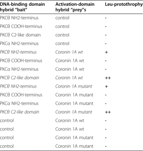
2-terminal domain of human Coro1A (Table 1).
Coro1A, a member of the evolutionary conserved
WD-repeat family of coronin proteins, is highly expressed
in all leukocytes. Originally, Coro1A has been isolated
as an actin/myosin binding protein and implicated in
F-actin dynamics by negatively regulating the function of
the nucleation-promoting Arp2/3 complex (reviewed in
[9]). In mice and human, genetic inactivation of Coro1A
results in immune deficiencies that are linked to a strong
reduction of naive T cell numbers in peripheral organs
[10-15]. Of note, Coro1A has been implicated in calcium
mobilization after TCR triggering in naive T cells as well
as TGF-
β
signaling in Th17 cells [11,14].
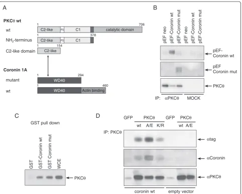
Using truncated versions of PKC
θ
and Coro1A
(Figure 1A), we demonstrate that the N-terminal WD40
domains of Coro1A and the C2-like domain of PKC
θ
are
sufficient for the interaction. Co-immunoprecipitation
(Co-IP) analysis in Jurkat T cells transfected with an
epitope-tagged Coro1A expression vector confirmed a
complex formation between PKC
θ
and Coro1A in T cells
(Figure 1B). Reversely, GST-Coro1A pull-downs revealed
interaction with endogenous PKC
θ
in mouse T cells
(Figure 1C). This PKC
θ
:Coro1A interaction was observed
both with and without CD3/CD28 stimulation of the cells
and thus being constitutive in resting cells. Of note, the
Co-IP experiments show strongly increased physical
asso-ciation of Coro1A with the constitutively active mutant
PKC
θ
A149E, while the binding to the dominant-negative
PKC
θ
K409R mutant remained unaltered when compared
to wild-type PKC
θ
(Figure 1D). This suggests that Coro1A
might function as a RACK protein regulating PKC kinase
activity. Of note, based on experiments using phorbol
ester as pleiotropic PKC activator, or serine/threonine
pro-tein phosphatase inhibitors, PKCs have been described as
kinases phosphorylating Coro1A and thereby
down-regulating its binding to actin [16,17]. Itho
et al.
identified
PKC
α
and PKC
δ
as the PKC isotypes responsible for
Coro1A phosphorylation [18].
Coro1A modulates PKC
θ
-mediated functions
After having observed a complex formation between
PKC
θ
and Coro1A, we next asked the question about the
functional relevance of this interaction. Therefore, it was
analysed whether Coro1A does influence the
transcrip-tional activation of genes that are established downstream
targets of PKC
θ
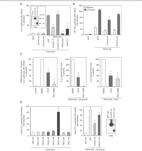
such as IL-2 and Cyclin D1. In functional
analyses using IL-2 promoter luciferase reporter assays,
overexpression of wild-type Coro1A but not the
COOH-deletion mutant, lacking the actin-binding domain,
nega-tively interferes with PKC
θ
-dependent IL-2 transactivation
in Jurkat T cells (Figure 2A). Thus, even though the
actin-binding function of Coro1A is not necessary for its
Table 1 Specific interaction between PKC
θ
and Coronin
1A in the GAL4 two-hybrid system
DNA-binding domain hybrid“bait”
Activation-domain hybrid“prey”s
Leu-protothrophy
PKCθNH2-terminus control
-PKCθCOOH-terminus control
-PKCθC2-like domain control
-PKCαNH2-terminus control
-PKCθNH2-terminus Coronin 1A wt +
PKCθCOOH-terminus Coronin 1A wt
-PKCαNH2-terminus Coronin 1A wt
-PKCθC2-like domain Coronin 1A wt ++
PKCθNH2-terminus Coronin 1A mutant +
PKCθCOOH-terminus Coronin 1A mutant
-PKCαNH2-terminus Coronin 1A mutant
-PKCθC2-like domain Coronin 1A mutant ++
control Coronin 1A wt
-control Coronin 1A wt
-control Coronin 1A mutant
-control Coronin 1A mutant
-PKCθand Coro1A were identified as interaction partners applying the GAL4 Two-Hybrid System. Saccharomyces cerevisiae reporter strain EGY48 was co-transformed with the bait construct encoding the NH2-terminal regulatory
domain of PKCθfused to a GAL4 DNA-binding domain and the human lymphocyte Matchmaker cDNA library. In re-transformation analysis, the NH2
interaction with PKC
θ
(Figure 1), it appears to be of
rele-vance for Coro1A modulating PKC
θ
function. In these
experiments, Jurkat T cells co-transfected with the
constitu-tively active mutant PKC
θ
A149E and wild-type or
truncated Coro1A, were stimulated with the calcium
iono-phore, ionomycin. Co-transfection with the
dominant-negative PKC
θ
K409R mutant or the dominant-negative
mutant of Rac1, Rac1 N17, which leads to inhibition of
IL-2 reporter transcription via actin polymerization defects
served as positive controls. Those findings suggest that
actin is part of a functional PKC
θ
:Coro1A axis identified in
the Jurkat T cell line. In addition, wild-type but not the
de-letion mutant of Coro1A repressed the induction of an
NF-κ
B-dependent promoter luciferase reporter (Figure 2B).
This effect could be phenocopied both by cell-permeable
pharmacological inhibitors of actin polymerisation and
PKC function, respectively (Figure 2C). Similarly, Cyclin D1
promoter reporter activation (that was PKC
isotype-selectively dependent on PKC
θ
function) was attenuated by
wild-type Coro1A co-expression (Figure 2D).
WD40
WD40
GST
GST
-Coronin wt
GST
Coronin mut
WCE
PKC
θ
GST pull down
A
D
C
GFP
PKC
θ
GFP
PKC
θ
wt
A/E K/R
wt A/E
IP: PKC
θ
α
tag
α
Coronin
α
PKC
θ
coronin wt
empty vector
Actin binding
C2-like PS C1 catalytic domain
1 706
294
460
C2-like
154 1
1
1
wt
PKC
θ
wt
NH
2-terminus
C2-like domain
Coronin 1A
wt
mutant
378 1
C2-like PS C1
pEF neo
pEF-Coronin wt
pEF-Coronin mut
pEF neo
pEF-Coronin wt
pEF-Coronin mut
pEF-Coronin wt
pEF
Coronin mut
PKC
θ
α
PKC
θ
MOCK
IP:
B
Figure 1PKCθphysically interacts with Coro1A. (A)The cartoon depicts interactions identified between deletion mutants of PKCθand Coro1A by Y2H as well as Co-IP experiments. It could be shown by deletion assays that the WD40 domain of Coro1A and the C2-like domain of
PKCθare sufficient for their interaction.(B)Co-immunoprecipitation analysis of Jurkat T cells transiently transfected with empty vector pEF-neo or
RGSH6-tagged Coro1A constructs (wild-type or deletion mutant). Twenty-four hours after transfection, PKCθ-specific immunoprecipitates were
immunostained with antibodies recognizing the tag, PKCθor Coro1A. To control for the specificity of the interaction immunoprecipitation with
control IgG (MOCK) was performed.(C)GST-Coro1A pull-down of endogenous PKCθfrom mouse CD3+T cells. WCE = whole cell extract as
positive control was used in parallel.(D)Jurkat T cells were transiently transfected with empty vector or RGSH6-tagged wild-type Coro1A together
with GFP or PKCθallelic versions (wild-type, A/E–constitutively active and K/R–dominant negative). Twenty-four hours after transfection,
PKCθ-specific antibodies were used for immunoprecipitation and the pulled-down proteins were analysed by immunoblotting the fusion-tag as
GFP
Coronin wt
Coronin mut PKC
θ K/R Rac N17 150 100 50 0
IL-2 Luciferase (Rlu1/Rlu2)
% activation
PMA/ionomycin - induced IL-2 Luciferase
(Rlu1/Rlu2)
% activation
IL-2 Luciferase (Rlu1/Rlu2)
% activation
IL-2 Luciferase (Rlu1/Rlu2)
% activation + Ionomycin * * * * * * * * * * * *
PKCθA/E + Ionomycin PKCθA/E + CaN
Coronin wt Coronin mut GFP 50 0 100 150 200 250 GFP GFP Coronin wt Coronin mut NF-κ
B Luciferase (Rlu1/Rlu2)
fold induction
PKCθA/E PKCθA/E +
Medium Ionomycin 100 80 60 40 20 0
control DMSO Cyt.D
PKC LMWI 100 80 60 40 20 0 DMSO Cyt.D PKC LMWI 100 80 60 40 20 0 DMSO Cyt.D PKC LMWI
PKCθA/E + Ionomycin 100 80 60 40 20 0
Cyclin D1 Luciferase (Rlu1/Rlu2)
fold induction
pEF neo
GFP
Coronin wt
Coronin mut
Cyclin D1 Luciferase (Rlu1/Rlu2)
fold induction 100 80 60 40 20 0
pEF neo CK
P α E/ A PKC δ A/E PKC ε A/E PKC η A/E PKC θ A/E PKC ζ A/E PKC ι A/E + Ionomycin
A
C
D
B
control Coronin wt Coronin mut
αtag wt
mut Coronin
pEF neo Coronin wt Coronin mut
αtag
Figure 2Coro1A modulates PKCθ-mediated effector function. (A)IL-2 promoter luciferase reporter assay performed with Jurkat T cells
transfected with the constitutively active mutant PKCθA/E and wild-type or truncated Coro1A–as indicated. Transfected cells were stimulated
with the calcium ionophore ionomycin overnight. The insertion in the upper left corner shows expression of recombinant Coro1A in Jurkat T cells
analysed by an anti-tag immunoblot. GFP-expressing plasmid was used as an inert protein overexpression control.(B)NF-κB-dependent promoter
luciferase reporter of transfected Jurkat T cells either stimulated with ionomycin or left untreated.(C)IL-2 promoter-dependent luciferase reporter
of Jurkat cells stimulated with phorbol ester/ionomycin, transfected with constitutively active mutant PKCθA/E and stimulated with ionomycin, or
alternatively, transfected with constitutively active mutants of both PKCθand Calcineurin (CaN) and/or treated by cytochalasin (Cyt) D and PKC
LMWI AEB071/Sotratstaurin as indicated.(D)Cyclin D1 promoter-dependent luciferase reporter of Jurkat cells stimulated with ionomycin and
transfected with constitutively active mutants of several PKC family members and with PKCθA/E and wild-type or truncated Coro1A, respectively.
The mean ± SE of three independent experiments is shown. Statistical significance was defined with p < 0.05 (Student’s t-test) and marked with
one asterisk (*). A/E: constitutively active and K/R: dominant negative mutant of PKCθ; Rac1 N17: dominant-negative mutant of Rac1; Rlu - relative
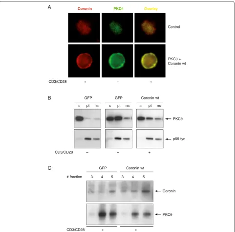
Mechanistically, in transient Jurkat transfection assays,
PKC
θ
and Coro1A co-localized in intact Jurkat T cells
(Figure 3A), and Coro1A overexpression inhibited both
plasma membrane and lipid raft recruitment of PKC
θ
in CD3/CD28-activated cells (Figure 3B/C). While we
cannot exclude additional Coro1A functions affecting
GFP Coronin wt
3 4 5 3 4 5
CD3/CD28 + +
CD3/CD28 + + +
Coronin # fraction
PKCθ Control
PKCθ + Coronin wt
Coronin PKCθ Overlay
A
C
s pt ns s pt ns s pt ns GFP GFP Coronin wt
CD3/CD28 – + +
PKCθ
p59 fyn
B
Figure 3Coro1A modulates PKCθ-mediated subcellular location in activated T cells.Jurkat cells were transfected with GFP inert protein
control, PKCθor Coro1A wild-type cDNA expression plasmids, respectively.(A)Co-localization of transfected PKCθand Coro1A occurred at a rate
of approximately 76% in intact cells as measured by immunofluorescence. Jurkat T cells were stimulated for 20 min with anti-CD3/anti-CD28
antibodies and analysed by subsequent staining with protein-specific antibodies. A representative image is shown.(B)Translocation of PKCθto
the plasma membrane is inhibited by overexpression of Coro1A. Jurkat cells were transfected with Coro1A or GFP, respectively. After 21 hrs cells
were stimulated with anti-CD3/anti-CD28 antibodies for 20 min or left unstimulated, as indicated and subcellular distribution of endogenous PKCθ
was determined by immunoblotting. The cell fractions are defined as the soluble (s) fraction, the particulate (pt) fraction and the Triton-X100 non-soluble (ns) fraction, which were prepared as described in the Additional file 1 (Supplementary Methods). The p59 fyn protein was detected
to control for cell fractionation.(C)Lipid rafts were prepared by fractionation of sucrose gradients and immunostained for PKCθand Coro1A. As a
w/o
peptide
6396p5.1.2
50 µM
KS140
10 µM
APC
(p)Tyr
PKC
θ
w/o
peptide
6396p5.1.2
50 µM
6396p5.1.2
50 µM
KS140
10 µM
APC
(p)Tyr
Coronin
A
C
D
B
stim.
–
5’
15’
5’
15’
–
5’
15’ 5’
15’
stim.
α
CD3/
CD28
α
CD3/
CD28
α
CD3
α
CD28
α
CD3
α
CD28
–
–
(p)I
κ
B
α
Actin
Coro1A
PDBu/
Iono
PDBu/
Iono
wt
coro1a–/–
wt
coro1a–/–
PDBu/
Iono
PDBu/
Iono
0
5‘
15‘
30‘
20
%
40
60
80
100
stimulation time
n=59
Statistics
Coronin1a in IS
unchanged
excluded
enriched
NF-
κ
B activation independent of PKC
θ
, based on the
experiments
described
above,
we
conclude
that
Coro1A, which is in a complex with PKC
θ
, modulates
PKC
θ
functionally.
Taken together, Coro1A likely may act as a safeguard
for stochastic membrane recruitment/IS translocation of
PKCtheta upon transient T cell activation signals, e.g. by
low affinity antigens.
Coro1A is involved in NF-
κ
B signaling in primary
T lymphocytes
Next, we investigated the subcellular localization of
Coro1A and PKC
θ
upon T cell activation. For this
pur-pose human T cell blasts from immunized donors were
incubated with APCs loaded or not with the
correspond-ing peptide and analysed by confocal microscopy for the
localization of PKC
θ
and Coro1A with regard to the IS
(stained by antibodies against (p)tyrosine). Of note, while
as already published, activation-induced PKC
θ
recruit-ment to the IS was consistently observed by confocal
microscopy [19], Coro1A was not recruited to the IS.
Coro1A was rather excluded from the IS in
approxi-mately 65% of antigen:APC-stimulated T cell blasts
(Figure 4A/B), suggesting a role as negative regulator in
TCR signaling.
Results on the molecular mechanism of Coro1A in T
cell signaling are controversial in part due to the
di-verse results obtained with the different conventional
knockout mice strains established in several
laborator-ies [10,11,20]. In particular, Mueller
et al. described a
physical interaction between Coro1A and PLC-
γ
1
pro-moting calcium mobilization from intracellular stores
upon activation of naive T cells [11], while no defect in
other pathways downstream of the TCR was detected.
In contrast, Föger
et al. did not observe any
impair-ment of T cell activation at all when analysing T cell
function using their knockout strain [10]. Using our
Coro1a
knockout mice [14], we addressed the potential
involvement of Coro1A in the NF-
κ
B signaling
path-way, known to be regulated by PKC
θ
, in primary
mouse T cells. The results revealed reduced levels of
phosphorylated inhibitor of NF-
κ
B (I-
κ
B
α
) in T cells
isolated from
Coro1a-deficient mice upon stimulation
with anti-CD3 and anti-CD28 (Figure 4C).
Further-more, NF-
κ
B:DNA binding upon anti-CD3/CD28
treatment was strongly reduced in
Coro1a-deficient T
cells when analysed by electrophoretic mobility shift
assay (EMSA) (Figure 4D).
Using a combination of phorbol ester and ionomycin,
which bypass early activation events downstream of
the TCR by directly activating PKC isotypes and
indu-cing calcium influx, respectively, only partially restored
I-
κ
B
α
phosphorylation
and
NF-
κ
B:DNA
binding,
pointing to an important role of Coro1A for PKC
acti-vation processes.
Taken together, the present results provide evidence
that Coro1A is a functional interaction partner of PKC
θ
in the established PKC
θ
/IKK/NF-
κ
B/IL-2 transactivation
pathway in CD3
+T cells.
Additional file
Additional file 1:Supplementary Methods.
Abbreviations
APC:Antigen presenting cell; AP-1: Activating protein 1; CaN: Calcineurin;
Coro1A: Coronin 1A; Co-IP: Co-immunoprecipitation; CA: Constitutively active; CycD1: Cyclin D1; CytD: Cytochalasin D; DN: Dominant negative; EMSA: Electrophoretic mobility shift assay; GFP: Green fluorescence protein; IL-2: Interleukin-2; IS: Immunological synapse; NFAT: Nuclear factor of
activation in T cells; NF-κB: Nuclear factorκB; PKC: Protein kinase C;
PDBu: Phorbol 12,13-dibutyrate; ko: Knockout; PKC LMWI: PKC low molecular weight inhibitor; TCR: T cell receptor; Y2H: Yeast two-hybrid.
Competing interests
The authors declare that they have no competing interests.
Authors’contributions
GB coordinated the project and analysed the research. GB and NT conceived and designed the experiments. NT, FF, KS and NP conducted the research. GB and KS wrote the manuscript. All authors read and approved the final manuscript.
Acknowledgements
This work was supported by grants from the FWF Austrian Science Fund (25044-B21 to GB, M1636-B23 to KS) and by the intramural funding program of the Medical University Innsbruck for young scientists MUI-START, Project 2013042002 (to KS).
(See figure on previous page.)
Figure 4Coro1A is not recruited into the IS and its gene ablation strongly reduces NF-κB responses. (A, B)Confocal microscopy of
Coro1A and PKCθin primary human T cells. A T cell clone (KS140) specific for the tetanus toxin peptide (TT830–843; QYIKANSKFIGITE) and a T cell
clone (6396p5.1.2) specific for the measles virus fusion protein peptide (F254–268; GDLLGILESRGIKAR) were used with autologous Epstein–Barr
virus (EBV)-transformed B cells as APC. Quantification of Coro1A subcellular localization on 59 synapses is shown as bar graph.(C)CD3+T cells
were isolated from either wild-type orCoro1aknockout mice. After 2 hour restingex vivothe cells were stimulated with soluble anti-CD3/CD28
and cross-linking anti-hamster IgG antibodies or PDBu for 5 and 15 minutes. Whole cell lysates (supplemented with phosphatase inhibitor) were
subjected to SDS-Page and immunoblotting against phosphorylated IκBα, actin and Coro1A.(D)CD3+T cells were isolated and stimulated as
described in(C), but the stimulation time was increased to 8 hours. Nuclear extracts were prepared and analysed by electromobility shift assays
Received: 3 November 2014 Accepted: 13 March 2015
References
1. Sims TN, Soos TJ, Xenias HS, Dubin-Thaler B, Hofman JM, Waite JC, et al.
Opposing effects of PKCθand WASp on symmetry breaking and relocation
of the immunological synapse. Cell. 2007;129:773–85.
2. Quann EJ, Liu X, Altan-Bonnet G, Huse M. A cascade of protein kinase C
isozymes promotes cytoskeletal polarization in T cells. Nat Immunol.
2011;12:647–54.
3. Baier-Bitterlich G, Uberall F, Bauer B, Fresser F, Wachter H, Grunicke H, et al.
Protein kinase C-θisoenzyme selective stimulation of the transcription factor
complex AP-1 in T lymphocytes. Mol Cell Biol. 1996;16:1842–50.
4. Sun Z, Arendt CW, Ellmeier W, Schaeffer EM, Sunshine MJ, Gandhi L, et al.
PKCθis required for TCR-induced NF-κB activation in mature but not
immature T lymphocytes. Nature. 2000;404:402–7.
5. Pfeifhofer C, Kofler K, Gruber T, Tabrizi NG, Lutz C, Maly K, et al. Protein
kinase Cθaffects Ca2+ mobilization and NFAT cell activation in primary
mouse T cells. J Exp Med. 2003;197:1525–35.
6. Isakov N, Altman A. Protein kinase C(θ) in T cell activation. Annu Rev
Immunol. 2002;20:761–94.
7. Steinberg SF. Structural basis of protein kinase C isoform function.
Physiol Rev. 2008;88:1341–78.
8. Mochly-Rosen D. Localization of protein kinases by anchoring proteins: a
theme in signal transduction. Science. 1995;268:247–51.
9. Uetrecht AC, Bear JE. Coronins: the return of the crown. Trends Cell Biol.
2006;16:421–6.
10. Foger N, Rangell L, Danilenko DM, Chan AC. Requirement for coronin 1 in T
lymphocyte trafficking and cellular homeostasis. Science. 2006;313:839–42.
11. Mueller P, Massner J, Jayachandran R, Combaluzier B, Albrecht I, Gatfield J,
et al. Regulation of T cell survival through coronin-1-mediated generation of inositol-1,4,5-trisphosphate and calcium mobilization after T cell receptor
triggering. Nat Immunol. 2008;9:424–31.
12. Shiow LR, Paris K, Akana MC, Cyster JG, Sorensen RU, Puck JM. Severe
combined immunodeficiency (SCID) and attention deficit hyperactivity disorder (ADHD) associated with a Coronin-1A mutation and a chromosome
16p11.2 deletion. Clin Immunol. 2009;131:24–30.
13. Haraldsson MK, Louis-Dit-Sully CA, Lawson BR, Sternik G, Santiago-Raber ML,
Gascoigne NR, et al. The lupus-related Lmb3 locus contains a
disease-suppressing Coronin-1A gene mutation. Immunity. 2008;28:40–51.
14. Kaminski S, Hermann-Kleiter N, Meisel M, Thuille N, Cronin S, Hara H, et al.
Coronin 1A is an essential regulator of the TGFβreceptor/SMAD3 signaling
pathway in Th17 CD4(+) T cells. J Autoimmun. 2011;37:198–208.
15. Siegmund K, Zeis T, Kunz G, Rolink T, Schaeren-Wiemers N, Pieters J. Coronin
1-mediated naive T cell survival is essential for the development of
autoimmune encephalomyelitis. J Immunol. 2011;186:3452–61.
16. Oku T, Kaneko Y, Murofushi K, Seyama Y, Toyoshima S, Tsuji T. Phorbol
ester-dependent phosphorylation regulates the association of p57/coronin-1
with the actin cytoskeleton. J Biol Chem. 2008;283:28918–25.
17. Oku T, Nakano M, Kaneko Y, Ando Y, Kenmotsu H, Itoh S, et al. Constitutive
Turnover of Phosphorylation at Thr-412 of Human p57/Coronin-1 Regulates
the Interaction with Actin. J Biol Chem. 2012;287:42910–20.
18. Itoh S, Suzuki K, Nishihata J, Iwasa M, Oku T, Nakajin S, et al. The role of
protein kinase C in the transient association of p57, a coronin family
actin-binding protein, with phagosomes. Biol Pharm Bull. 2002;25:837–44.
19. Monks CR, Kupfer H, Tamir I, Barlow A, Kupfer A. Selective modulation of
PKCθduring T cell activation. Nature. 1997;385:83–6.
20. Shiow LR, Roadcap DW, Paris K, Watson SR, Grigorova IL, Lebet T, et al. The
actin regulator coronin 1A is mutant in a thymic egress-deficient mouse strain and in a patient with severe combined immunodeficiency.
Nat Immunol. 2008;9:1307–15.
Submit your next manuscript to BioMed Central
and take full advantage of:
• Convenient online submission
• Thorough peer review
• No space constraints or color figure charges
• Immediate publication on acceptance
• Inclusion in PubMed, CAS, Scopus and Google Scholar
• Research which is freely available for redistribution