Assessment of Anatomical Variations of Paranasal Sinus Region: A Computed Tomography Study IJOCR
Assessment of Anatomical Variations of Paranasal
Sinus Region: A Computed Tomography Study
1Ahanthem Nandita, 2Sowbhagya Malligere Basavaraju, 3Balaji Pachipulusu, 4GU Pravin
IJOCR
ORIGINAL ARTICLE
10.5005/jp-journals-10051-0092
cavity, wherein there is absence of inflammatory signs, hyperplastic mucosa, purulent discharge, and sinonasal polyps or masses.1 This headache may also result from pressure on the nasal mucosa due to anatomical variations. Anatomical disparities of nose have been reported to pre-dispose to sinusitis.2 Chronic rhinosinusitis is one of the most common illnesses, and it has been known to nega-tively impact health-related quality of life.3 Common ana-tomical variations include deviated nasal septum, concha bullosa, air cells – Agger nasi, Haller cell, and Onodi cell.2 These variants may determinate contact points between nasal structures, thereby, stimulating “trigger points” and determining facial pain crisis. Facial pain may be localized to periorbital, frontal, or temporozygomatic region, which might be unilateral or bilateral.4 Three-dimensional (3D) imaging of paranasal sinuses is mandatory for diagnosis and treatment of the underlying anatomical variations, as these variations could be a cause for sinonasal symptoms, facial pain symptoms, and headache.5-7 Endoscopic sinus surgery is the treatment of choice for refractory sinusitis.7 Hence, understanding the complex anatomy of skull base is crucial for safe endoscopic sinus surgery to prevent from complication. The aim of the study is to assess the anatomical variation of paranasal sinus region and deter-mine the relationship of these variations with respect to age, side, and gender of an individual.
AIMS AND OBJECTIVES Aims
• To assess the prevalence of anatomical variations of paranasal sinus region.
• To determine the relationship of these variations with age, side, and gender of an individual.
Objectives
• To aid in diagnosis of facial pain and headache. • To ascertain the frequency and corelationship of these
variants to age and sex.
• To facilitate surgical planning in patients undergoing endonasal procedure.
MATERIALS AND METHODS
This study was carried out at the Department of Radio-diagnosis, RajaRajeswari Medical College & Hospital,
1Postgraduate Student, 2Reader, 3,4Professor and Head 1-4Department of Oral Medicine & Radiology, RajaRajeswari
Dental College & Hospital, Bengaluru, Karnataka, India
Corresponding Author: Ahanthem Nandita, Postgraduate Student, Department of Oral Medicine & Radiology, RajaRajeswari Dental College & Hospital, Bengaluru, Karnataka, India, e-mail: [email protected]
ABSTRACT
Introduction: Facial pain syndrome or rhinogenic headache is a headache secondary to mucosal contact points in the
sinonasal cavities, wherein there is absence of inflammatory
signs, hyperplastic mucosa, purulent discharge, and sinonasal polyps or masses. This headache may also result from pressure on the nasal mucosa due to anatomical variations. Anatomical disparities of nose have been reported to predispose to sinusitis. These variants may determinate contact points between nasal structures, stimulating “trigger” points and determining facial pain crisis. Facial pain is localized to the periorbital, frontal, temporozygomatic region, which may be unilateral or bilateral. The aim of this study is to assess the prevalence of anatomical variations – concha bullosa, nasal septum deviation, and air cells (Haller cells, Agger nasi cells, Onodi cells).
Materials and methods: This retrospective study was con-ducted at the Department of Radiodiagnosis, RajaRajeswari Medical College & Hospital, Bengaluru, India. Data comprised paranasal sinus computed tomography images of 50 patients (25 males and 25 females) that were retrieved from archives and analyzed for presence of anatomical variations, such as devia-tion of nasal septum, concha bullosa, and air cells – Agger nasi cell, Haller cell, and Onodi cell. Data obtained were analyzed with Chi-square test and Mann–Whitney test.
Results: Deviated nasal septum was seen in 88% of the cases followed by Agger nasi cells (66%), concha bullosa (64%), Haller cells (56%), and Onodi cells (38%). We found no statistical
significance when comparing the relationship of anatomical
variations with age, side, and gender.
Keywords: Agger nasi, Anatomical variations, Concha bullosa, Facial pain, Haller cell, Onodi cell.
How to cite this article: Nandita A, Basavaraju SM, Pachipu-lusu B, Pravin GU. Assessment of Anatomical Variations of Paranasal Sinus Region: A Computed Tomography Study. Int J Oral Care Res 2017;5(2):97-101.
Source of support: Nil Conflict of interest: None
INTRODUCTION
variations Incidence (%) Unilateral (%) Bilateral (%) n % n % χ2 value p-value
CB 64 56 16 50 16 50 0 1.00
DNS 88 22 50 22 50 0 1.00
ANC 66 28 38 14 42.4 19 57.6 2.228 0.14 HC 56 40 16 12 42.9 16 57.1 1.299 0.25 OC 38 38 0 11 57.9 8 42.1 0.764 0.38 CB: Concha bullosa; DNS: Deviated nasal septum; ANC: Agger nasi cell; HC: Haller cell; OC: Onodi cell
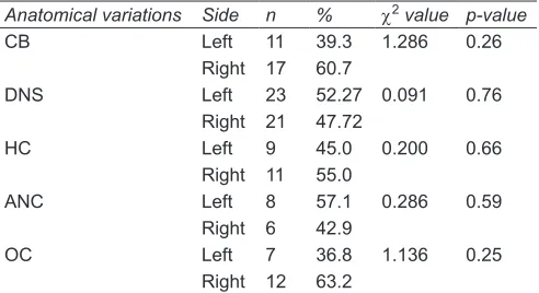
Table 2: Unilateral distribution of anatomical variations of
paranasal air sinuses using Chi-square goodness of fit test
Anatomical variations Side n % χ2 value p-value
CB Left 11 39.3 1.286 0.26 Right 17 60.7
DNS Left 23 52.27 0.091 0.76 Right 21 47.72
HC Left 9 45.0 0.200 0.66 Right 11 55.0
ANC Left 8 57.1 0.286 0.59 Right 6 42.9
OC Left 7 36.8 1.136 0.25 Right 12 63.2
CB: Concha bullosa; DNS: Deviated nasal septum; ANC: Agger nasi cell; HC: Haller cell; OC: Onodi cell
Bengaluru, India. The sources of data for our study were retrospectively selected paranasal sinus computed tomography (CT) images of 50 patients (25 males and 25 females) randomly collected from CT archives in the age group of 18 to 70 years.
Inclusion Criteria
• The CT paranasal sinus images in axial, coronal, and saggital planes.
• Age group 18 to 70 years.
Exclusion Criteria
• The CT images with alteration of paranasal sinus anatomy due to surgery, tumor, or facial trauma. • Patients less than 18 years.
All the patients underwent CT scan of the paranasal sinus region with Siemens Somatom Perspective 128 with syngo CT 2012 A software. The sections were taken with slice thickness of 5 mm. The exposure settings used were 130 kVp and 35 mA and reviewed for the presence of deviated nasal septum, concha bullosa, air cells – agger nasi, Haller cell, and Onodi cell in axial, coronal, and sagittal views. The data collected were subjected for statistical analysis.
RESULTS
The most common anatomical variation observed was deviated nasal septum, 44 (88%), followed by Agger nasi cells, 33 (66%), concha bullosa, 32 (64%), Haller cells, 28 (56%), and Onodi cells, 19 (38%).
Septal deviation was the most common variation seen. Out of 50 scans, nasal septum accounts for 44 (88%), among which 17 (47.72%) deviated toward left side and 23 (52.27%) deviated toward right side.
As per our study, out of 50 scans analyzed, Agger nasi cells were observed in 33 (66%), out of which 14 (28%) were present unilaterally and 19 (38%) bilaterally. Also, 8 (57.1%) were found on left side and 6 (42.9%) on right side. We revealed no statistical significance between gender and presence of Agger nasi cells (42.4% male, 57.6% female; p = 0.14).
Out of the 50 scans analyzed, concha bullosa was perceived in 32 (64%), out of which 28 (56%) were
present unilaterally and 4 (8%) present bilaterally. Also, 17 (60.7%) were noted on right side and 11 (39.3%) on left side. We noticed no statistical difference between gender and presence of concha bullosa (50% male, 50% female; p = 1).
Of the 50 scans analyzed, Haller cells were seen in 28 (56%), out of which 20 (40%) presented unilaterally and 8 (16%) bilaterally. Also, 11 (55.5%) were noted on right side and 9 (45.05%) on left side. We found no statistically difference between gender and the presence of Haller cells (42.9% male, 57.1% female; p = 0.25).
Of the 50 scans, Onodi cell were observed in 19 (38%), noted only unilaterally, out of which 12 (63.2%) were noted on right side and 7 (36.75) on left side. We revealed no statistically significance between gender and presence of Onodi cells (57.9% male, 42.1% female; p = 0.3) (Tables 1 and 2).
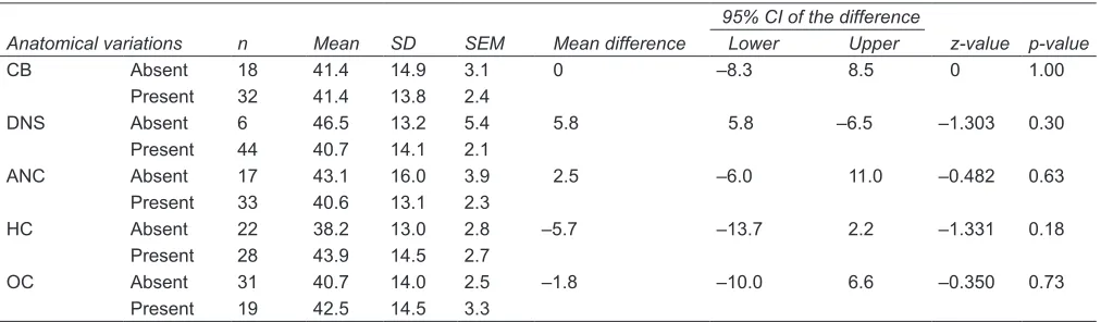
According to our study, we found no statistical sig-nificance between the mean age group and presence of anatomical variations (concha bullosa p = 1, Agger nasi p = 0.63, Haller cells p = 0.18, Onodi cells p = 0.73, and deviated nasal septum p = 0.30) (Table 3).
DISCUSSION
Assessment of Anatomical Variations of Paranasal Sinus Region: A Computed Tomography Study IJOCR
variations of the paranasal sinus region is mainly pre-disposed to recurrent sinusitis and, in selected cases, headache.9 Also, these variants may determine contact points between nasal structures leading to facial pain crisis.4 These variants are important in two distinct view-points: Firstly, their relationship to disturbing drainage and ventilation system and secondly, the potential impact on operative technique and surgical safety.9,10
Deviated Nasal Septum
As per literature, deviated nasal septum is the most frequent anatomical variation found. It represents a divergence of septum from the midline.11 Deviated nasal septum can be cartilaginous, bony type, or a combination of both. Deviation of septum causes lateral decompres-sion and displacement of the middle turbinate leading to nasal obstruction.12 The incidence of nasal septum varies from 14.1 to 80%.7 According to Earwaker (44%),11 Nar-endrakumar and Subramanian (76%),12 Pérez-Piñas et al (80%),13 Sarika et al (64.44%),14 and Turna et al (59.1%),10 most were toward right side. However, in this study, we found 88%, out of which 52.27% were toward left and 47.72% toward right side, which is almost similar to Pérez-Piñas et al13 finding.
Concha Bullosa
The term concha bullosa was coined by Zuckerlandl in 1862. It represents the extensive pneumatization of the middle turbinate and is one of the most common anatomical variations of the sinonasal anatomy.2,15,16 It has been implicated as a possible etiological factor in recurrent sinusitis due to its postulated negative influ-ence on ventilation of paranasal sinus and mucocili-ary clearance in the middle meatus region. In cases of extensive pneumatization, it may cause significant problems, such as headache and nasal blockage.14 Frequency of concha bullosa ranges from 14 to 80%.14
However, as per this study, the prevalence of concha bullosa was 64%, out of which 56% were present uni-laterally and 8% present biuni-laterally, and 60.7% were noted on right side and 39.3% on left side. Our findings correlated with the findings of Bolger et al,16 Scribano et al,9 Pérez-Piñas et al,13 Khojastepour et al,17 Talaiepour et al,18 Narendrakumar and Subramanian,12 Fadda et al,19 and Wani et al.6 The great variation in the reported prevalence may be due to diverse study populations and different criteria for pneumatization.
Agger Nasi Cell
Agger nasi cells are the most anterior ethmoidal cells, located anteriorly to the upper margin of the nasolacrimal duct and anteriorly to the plane of maxillary sinus infun-dibulum.11 It can pneumatize posteriorly to narrow the frontal recess. Its clinical importance has been defined by Brunner et al20 in 1996. The extensive pneumatization with consequent narrowing of the frontal sinus ostium causes significant persistent frontoethmoid pain and chronic frontal sinusitis.6 The reported prevalence of Agger nasi cell in the previous literature varies from 10 to 98.5%.11,21 In our study, the prevalence of Agger nasi cells was 66%, out of which 28% were present unilaterally and 38% bilat-erally, and 57.1% were noted on left side and 42.9% on right side. Our findings are almost consistent with previ-ous studies conducted by Fadda et al,19 Talaiepour et al,18 and Narendrakumar and Subramanian.12
Haller Cells
Haller cells or infraorbital ethmoidal air cells are pneuma-tized ethmoid air cells that project along the medial roof of the maxillary sinus and the most inferior portion of the lamina papyracea, below the ethmoid bulla and lateral to the uncinate process. This cell was first described by an anatomist Albert Haller in 1765.21,22 When enlarged, it can cause obstruction of the posterior aspect of the Table 3: Comparison of mean age in relation to anatomical variations using Mann–Whitney U test
Anatomical variations n Mean SD SEM Mean difference
95% CI of the difference
z-value p-value
Lower Upper
CB Absent 18 41.4 14.9 3.1 0 –8.3 8.5 0 1.00 Present 32 41.4 13.8 2.4
DNS Absent 6 46.5 13.2 5.4 5.8 5.8 –6.5 –1.303 0.30 Present 44 40.7 14.1 2.1
ANC Absent 17 43.1 16.0 3.9 2.5 –6.0 11.0 –0.482 0.63 Present 33 40.6 13.1 2.3
HC Absent 22 38.2 13.0 2.8 –5.7 –13.7 2.2 –1.331 0.18 Present 28 43.9 14.5 2.7
OC Absent 31 40.7 14.0 2.5 –1.8 –10.0 6.6 –0.350 0.73 Present 19 42.5 14.5 3.3
nasal obstruction, impaired nasal breathing, headache, chronic cough, and mucoceles.23,24 The prevalence of Haller cells is remarkably variable, ranging from 2 to 70.3%.17 As per our study, Haller cells were found in 56% of the study population, out of which 40% were present unilaterally and 16 bilaterally. Also, 55.5% were noted on the right side and 45.05% on the left side. However, our findings are similar with those of Bolger et al16 and Khojastepour et al.17
Onodi Cells
Also known as sphenoethmoidal cells, these were first described by the Hungarian laryngologist Adolf Onodi, in 1904.25 They are ethmoid cells that have migrated to the anterior region of the sphenoid sinus, with anterosu-perior location, and intimately related to the optic nerve, causing optic neuropathy in case of certain conditions that affect such cells. Onodi cell is the most posterior ethmoid air cell that extends laterally.26,27 The prevalence of Onodi cells varies from 8 to 13%,12 according to Turna et al (13.5%),10 Fadda et al (8.5%),19 and Narendrakumar and Subramanian (6%).12 Herein, the prevalence of Onodi cells in this study was 38%, seen only unilaterally and out of which 63.2% were seen on right side and 36.7% on left side. The prevalence in our study is comparatively higher as compared with previous literature. This may be due to small sample size and confined to only one ethic group (South Indian population).
CONCLUSION
Different anatomical variants may often be found in para-nasal sinus region and 3D imaging is the modality of choice to evaluate these variants since conventional radiographs do not provide adequate information because of structural superimposition. These variants may determinate contact points between nasal structures stimulating “trigger” points and determining facial pain crisis. Identification of these variants plays an important role while guiding the surgeons preoperatively and preventing iatrogenic com-plications. Since this is a preliminary study, our findings could be used in future studies with larger sample size.
REFERENCES
1. Cantone E, Castagna G, Ferrati I, Cimmino M, Sicignano S, Rega F, Di Rubbo V, Iengo M. Concha bullosa related
head-ache disability. Eur Rev Med Pharmacol Sci 2015 Jul;19(3):
2327-2330.
2. Maru YK, Gupta V. Anatomical variations of the bone in
sinonasal C.T. Indian J Otolaryngol Head Neck Surg 2001
Apr;53(2):124-128.
110-113.
4. Budu V, Schnaider A, Tache MS, Bulescu I. Evaluation of osteomeatal complex pathology related to endoscopic sinus
surgery – a retrospective analysis. Romanian J Rhinol 2015 Apr-Jun;5(18):95-100.
5. Elsherif A-AMH, Elsherif AMH. Some anatomical variations of the paranasal sinuses in patient with chronic sinusitis: a correlative CT study to age and sex. Al-Azhar Assiut Med
J 2006 Sep;4(3):1-15.
6. Wani AA, Kanotra S, Lateef M, Ahmad R, Qazi SM, Ahmad S. CT scan evaluation of the ostiomeatal complex. Indian
J Otolaryngol Head Neck Surg 2009 Sep;61(3):163-168. 7. Murthy DD, Roa BRC, Roa SSP. Analytical study of anatomical
variations of nose and PNS in CT scan and chronic sinusitis. IOSR J Dent Med Sci 2016 Jul;7(15):30-35.
8. Lingaiah RKN, Puttaraj NC, Chikkaswamy HA, Nagarajaiah PKC, Purushothama S, Prakash V, Booma P,
Ismail M. Anatomical variations of paranasal sinuses on coronal CT-scan in subjects with complaints pertaining to
PNS. IJARS DOI: 10.7860/IJARS/2016/21554:2192
9. Scribano E, Ascenti G, Loria G, Cascio F, Gaeta M. The role of the osteomeatal unit anatomic variations in
inflamma-tory disease of the maxillary sinuses. Eur J Radiol 1997
May;24(3):172-174.
10. Turna O, Aybar MD, Karagoz Y, Tuzcu G. Anatomic variations of the paranasal sinus region: Evaluation with multidetector
CT. Istanbul Med J 2014 Jun;15(2):104-109.
11. Earwaker J. Anatomic variants in sinonasal CT. Radiographics
1993 Mar;13(2):381-415.
12. Narendrakumar V, Subramanian V. Anatomical variations in osteomeatal complex among patient undergoing func-
tional endoscopic sinus surgery. Clin Rhinol Int J 2016;9(1):
28-32.
13. Pérez-Piñas, Sabaté J, Carmona A, Catalina-Herrera CJ, Jiménez-Castellanos J. Anatomical variations in the human paranasal sinus region studied by CT. J Anat 2000 Aug;197 (Pt 2):221-227.
14. Kate Sarika P, Mandke ND, Bahetee BH. Anatomic variations
of paranasal sinuses in patient with chronic sinusitis and their
correlation with CT scan study. Int J Cur Res Rev 2015;7(15):
49-55.
15. Miranda CMNR, Maranhão CPM, Arraes FMNR, Padilha IG, Farias LPG, Jatobá MSA, Andrade ACM, Padilha BG.
Anatomical variations of paranasal sinuses at multislice computed tomography: what to look for. Radiol Bras 2011
Jul/Aug;44(4):256-262.
16. Bolger WE, Butzin CA, Parsons DS. Paranasal sinus bony ana -tomic variations and mucosal abnormalities: CT analysis for
endoscopic sinus surgery. Laryngoscope 1991 Jan;101(1 Pt 1):
56-64.
17. Khojastepour L, Mirhadi S, Mesbahi SA. Anatomical varia-tions of osteomeatal complex in CBCT of patient seeking
rhinoplasty. J Dent (Shiraz) 2015 Mar;16(1):4248.
18. Talaiepour AR, Sazgar AA, Bagheri A. Anatomic variations
of the paranasal sinuses on CT scan images. J Dent (Tehran)
2005;2(4):142-146.
19. Fadda GL, Rosso S, Aversa S, Petrelli A, Ondolo C, Succo G.
Assessment of Anatomical Variations of Paranasal Sinus Region: A Computed Tomography Study IJOCR
20. Brunner E, Jacobs JB, Shpizner BA, Lebowitz RA, Holliday RA.
Role of the agger nasi cell in chronic frontal sinusitis. Ann Otol Rhinol Laryngol. 1996;105(9):694-670.
21. Eweiss AZ, Kjalil HS. The prevalence of frontal cells and their relationship to frontal sinus: a radiological study of frontal sinus area. ISRN Otolaryngology 2013;Article ID 687582:1-4.
22. Pekiner FN, Borahan MO, Dumlu A, Ozbyrak S. Infraorbital
ethmoid (Haller) cells: a cone beam computed tomography study. Oral Radiol 2014;30:219-225.
23. Caversaccio M, Boschung U, Mudry A. Historical review of Haller’s cells. Ann Anat 2011 Mar;193(3):185-190.
24. Kantarci M, Karasen RM, Alper F, Onbas O, Okur A, Karaman A. A remarkable anatomic variation in paranasal
sinus region and their clinical importance. Eur J Radiol 2004 Jun;50(3):296-302.
25. Braun H, Stammberger H. Pneumatization of turbinates.
Laryngoscope 2003;113:668-672.
26. Flessig E, Spierer O, Koren I, Lwiboviten I. Blinding orbital apex syndrome due to onodi cell mucocle. Case Report in Opthalmological medicine 2014;14:453789.