IJPAR |Vol.5 | Issue 3 | July- Sep -2016
Journal Home page: www.ijpar.com
Research article Open Access
Formulation and characterization, of aceclofenac microspheres by ionic
crosslinking technique
Dillip Kumar Mohapatra
*1, DR M. D. Dhanaraju
*2S. Selvadurai
*3 1Departmaent of pharmaceutics, GIET School of Pharmacy, Rajahmundry-533296, Andhrapradesh, India.
2Department of pharmaceutics, GIET School of Pharmacy, Rajahmundry-533296, Andhrapradesh, India.
3Department of Pharmacognosy, GIET School of Pharmacy, Rajahmundry-533296, Andhrapradesh, India.
Corresponding author: Dillip Kumar M.Email: [email protected]
ABSTRACT
Objective
Present investigation describes the preparation of Aceclofenac microspheres by ionic cross linking technique followed by characterization of microspheres to evaluate the effect of polymer concentration and different crosslinking agents on physical properties and drug release profile of microspheres.
Methods
Aceclofenac Microspheres were prepared with polymer, sodium alginate at various concentrations using ionic cross linking agents Calcium Chloride (CaCl2), Barium Chloride (BaCl2) and Aluminum Sulfide (Al (SO4) 3). The
microspheres were developed and the effect of sodium alginate concentration, cross linking agents was evaluated with respect to entrapment efficiency, particle size, surface characteristics and in vitro release behaviors with the incorporation efficiency. The solid microspheres were characterized by sieve analyzer, scanning electron microscope, differential scanning calorimetry, and in vitro drug release.
Results
The infrared spectroscopic study confirmed the absence of any drug polymer interaction. Differential scanning calorimetric analysis revealed that the drug was molecularly dispersed in the alginate microspheres matrices showing rough surface, which was confirmed by scanning electron microscopy study. The mean particle size and entrapment efficiency and in vitro release profile could be altered significantly by changing various formulation parameters to give a sustained release of drug from the microspheres.
Conclusion
It can be concluded that the formulation prepared by ionic cross linking method, has potential to deliver Aceclofenac in a controlled manner in a regular fashion over an extended period of time in comparison to all other formulations and can be adopted for a successful oral delivery of Aceclofenac for safe management of hypertension.
Keywords:
Sodium alginate, Aceclofenac (pure drug), Ionic crosslinking technique.INTRODUCTION
In the present study a non-steroidal anti-inflammatory drug Aceclofenac has been chosen as model drug [1]. Aceclofenac, (2-[2-[2-(2, 6– dichlorophenyl) aminophenyl] acetyl] oxyacetic acid) a non-steroidal anti-inflammatory drug [NSAID] has been indicated for various conditions like post-traumatic pain, rheumatoid arthritis, ankylosing spondylitis. The molecule is practically insoluble in water, but almost totally absorbed from the gastrointestinal tract, its biological half-life is 4hrs and administered twice daily with a single dose of 100mg. To overcome the side effects associated with conventional administration of NSAIDS and increase the patient compliance, controlled release dosage forms have been formulated in the form of Single Unit and Multiunit dosage forms. Compared to Single Unit dosage forms, Multi unit drug delivery system avoid the variations in gastric emptying and different transit rates through the gastrointestinal tract[2], release drugs in a more predictable manner, and spread over a large area preventing exposure of the absorbing site to high drug concentration on chronic dosing. Several synthetic polymers have been used to formulate multiunit dosage forms. Recently, much research efforts have been concentrated to develop drug-loaded microspheres using sodium alginate [3], a natural polymer obtained from marine brown algae, because of simple, mild and eco-friendly preparative and received much attention as a vehicle for controlled drug delivery. Drop wise addition of aqueous alginate solution at different concentration to the aqueous solution containing calcium ions and/or other di and polyvalent cations causes spherical gel formation termed as alginate bead. Alginate is known to be nontoxic when taken orally and also have a protective effect on the mucous membranes of the upper gastrointestinal tract. The dried alginate [4-10] beads have the property of reswelling and thus they can act as controlled release system. Their reswelling property is susceptible to pH, which protects the acid sensitive drug from gastric juice.
As previously reported, the Aceclofenac microspheres prepared by emulsion solvent evaporation [11] method utilize larger volume of organic solvents, which are costly and hazardous
because of the possibility of explosion, toxicity and air pollution. The aqua based ionic crosslinking technique can provide characteristic advantages over conventional microspheres preparation methods. The effects of factors, such as sodium alginate concentrations, crosslinking agents, drying conditions, on the morphology of and drug release from microspheres were studied. Drug polymer interactions in the solid state were studied by infrared spectrophotometry (IR) and differential scanning calorimetric analysis (DSC) and the surface characteristics were evaluated by scanning electron microscopy (SEM).
MATERIAL AND METHODS
Aceclofenacwas procured from Yarrow Chem., Mumbai, India, Sodium alginate was purchased from Finolex Ltd. Ahmedabad, Calcium Chloride, Barium Chloride, Aluminium Sulphide purchased from Molychem Ltd Mumbai India. All other chemicals and solvents were analytical grade satisfying pharmacopoeia specifications.
Method of Preparation
The microspheres were prepared by ionic cross-linking technique[12] using the formulations as shown in Table 1 .The solutions comprising 2% to 4% w/v sodium alginate were prepared by initially dissolving the polymer in deionized water using gentle heat, it was stirred magnetically. On complete solution, an accurately weighed quantity of Aceclofenac was added to each solution to make homogeneous dispersions and these dispersions were sonicated for 30 min. The sodium alginate-drug dispersions (50 ml) were added drop wise via a 20-gauge hypodermic needle was fitted with a 10 ml syringe into 100 ml of 5% w/v of cross-linking agents, and being stirred at 200 RPM for 20 min. The cross-linking agents were used CaCl2, Al2 (SO4) 3 and
BaCl2. The droplets from the dispersions instantly
Table 1: Formulation of Aceclofenac with Sodium alginate Microspheres
Formulation Code
Aceclofenac %w/v
Sodium Alginate %w/v
Cross linking agents %w/v
A 2.5 2.5 CaCl2
B 2.5 3.0 CaCl2
C 2.5 3.5 CaCl2
D 2.5 4 CaCl2
E 2.5 3.5 BaCl2
F 2.5 3.5 Al2 (SO4) 3
Morphology and Size Distribution
The shape and surface morphology of microspheres [13] were investigated using Jeol, SEI, scanning electron microscope at 5 kv. Prior to examination samples were gold coated under vacuum to render them electrically conductive. The alginate microspheres were not subjected to SEM studies after release study because they converted to gel type of matrix when the dissolution was carried out.
Size distribution [14] of the Losartan loaded with sodium alginate microspheres were measured by sieve analysis. The alginate microspheres were separated into different size fractions (% weight fraction) by sieving for 5 min using standard sieves having nominal mesh apertures of 1.4 mm, 1.2 mm, 1.0 mm, 0.85 mm and 0.71 mm (sieve no. 12, 14, 16, 18 and 22, respectively). The particle size distributions of the microspheres were determined and the mean particle sizes of microspheres were calculated using the following formula.
Determination
of
drug
incorporation
efficiency
Drug loaded alginate microspheres 30 mg of each batch was placed in 100 ml conical flask containing 50 ml of phosphate buffer of pH 7.4. The microspheres were magnetically stirred to promote swelling and the breakup of the
cross-linked structure and it was filtered through a 0.45 μm membrane filter. Then the drug was quantified at 206 nm spectrophotometrically after appropriate dilution with phosphate buffer of pH 7.4. The incorporation efficiency was determined by the following empirical relationship:
( )
Where AQ is the actual quantity of drug present in the microspheres and TQ is the 100% theoretical quantity of drug present in the microspheres (i.e., actual initial loading dose).
Infrared spectroscopy
The drug-polymer interactions were studied by infrared spectroscopy. The IR spectra were recorded between 500 to 4000 cm−1 for pure Aceclofenac, blank alginate microspheres and Aceclofenac-loaded alginate microspheres from KBr pellet using Perkin Elmer-883 IR spectrophotometer.
Differential scanning calorimetry
The DSC thermal analysis was recorded on a Universal V 2.5 H differential scanning calorimeter. The DSC studies on the samples were performed by heating samples at a heating rate of 10°C/min over a temperature range of 50-300°C in closed aluminum pans. The samples included pure Aceclofenac and losartan -loaded alginate microspheres.
In vitro
Dissolution Testing
apparatus (37 ± 0.5°, 50 rpm) using the USP rotating basket method in phosphate buffer media (pH 7.4, 500 ml). A quantity of alginate beads equivalent to 100 mg Aceclofenac for each formulation was employed in all dissolution studies. The samples of 5 ml each were withdrawn [15] at predetermined time interval and were replenished immediately with the same volume of fresh prewarmed phosphate buffer maintaining sink condition throughout the experiment. The aliquots, following suitable dilution, were analyzed spectrophotometrically at 256 nm. The concentrations of losartan in the test samples were calculated using a regression equation (Absorbance = 0.007 + 0.0709 × concentration, R2 = 0.999) of the calibration curve in phosphate buffer of pH 7.4.
RESULTS AND DISCUSSIONS
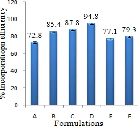
The mean particle size of the various formulations of alginate microspheres was between 0.715 ± 0.007 to 0.890 ± 0.008 mm, shown in Fig.1. The results indicated the proportional increase in the mean particle size of the microspheres with increasing amount of sodium alginate in the formulations A, B, C and D. This could be attributed to an increase in the relative viscosity at higher concentration of sodium alginate and formation of large droplets during the addition of the polymer solution to the cross-linking agents. The incorporation efficiency, increased progressively with increasing sodium alginate concentration Fig. 2. This may be attributed to the greater availability of active binding sites in the polymer chains and, consequently, the greater degree of cross-linking as the quantity of sodium alginate increased [16]. The incorporation efficiencies were generally higher for the formulations cross-linked with Al2(SO4)3 and
BaCl2 as compared to the beads cross-linked with
CaCl2. This may be attributed to the formation of
nonporous alginate beads due to increase in the apparent cross-linking density in presence of Al3+ and Ba2+ which prevent the diffusion of the drug out of the beads at the time of curing. The low incorporation efficiency of alginate beads cross-linked with Ca2+ could be attributed to the formation of porous beads, ensuring the diffusion of the drug out of the beads at the time of curing.
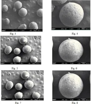
Morphology of the various formulations of alginate microspheres prepared was found to be
discrete and spherical in shape. The SEM photomicrographs of the dried alginate microspheres are shown in Fig.3-8.The surface of the alginate microspheres was rough due to higher concentration of drug uniformly dispersed at the molecular level in the alginate matrices.
The sodium alginate was used at four different concentrations: 2.5 (A), 3.0 (B), 3.5 (C) and 4.0 (D) % w/v. It is observed from the Fig.9 that the steady state release was achieved after an initial lag time and it was directly proportional to the concentration of sodium alginate. Which can elucidate as sustained effect by increasing the concentration of sodium alginate. This type of release behavior agreed with the pulsatile release pattern. The pulsatile [17] drug release is defined as the rapid release of a certain amount of drug within a short time period immediately after a lag time. The pulsed release pattern could be controlled by the disintegration of alginate gel in phosphate buffer. The disintegration of alginate was monitored by the exchange of Ca2+ with Na+ in the dissolution medium. The dissociation of alginate beads in phosphate buffer and the drug release mainly depend upon drug diffusion through the small pores and cracks, so it reveals sodium alginate concentration in the formulation greatly influenced the steady state release of losartan from the alginate beads. The mechanism of gelation or cross-linking of sodium alginate with calcium chloride is solely based on the formation of tight junction between the glucuronic acid residues [18-19]. The number of the apparent cross-linking points formed within the calcium alginate gel beads increased with increasing alginate concentration in the formulation. This increase in the apparent cross-linking density delayed the alginate gel disintegration in phosphate buffer due to the retardation of Ca2+ exchange with Na+ and eventually increasing lag time. By increasing alginate gel density per unit volume was also a cause to affect the decreased pore size within the gels, and thus release Aceclofenac becomes slow.
Then release profile of alginate microspheres with different cross-linking agents depend upon the valance and the size of the cations of the respective cross-linking agent[20]. To investigate this aspect, the sodium alginate (3.5% w/v) beads were prepared via cross-linking in 5% w/v solution of CaCl2 (C), Al2(SO4)3 (E) and BaCl2 (F),
buffer of pH 7.4 have been well depicted in the (Fig.9). The pulsatile release pattern was observed in all cases. The steady state release was achieved after 2.45 h for Ca2+-alginate microspheres, 3.5 h for Ba2+-alginate microspheres and 5.0 h for Al3+-alginate microspheres. It can also be explained, inside the alginate matrices cross-linking agent Ca2+ and Ba2+, being divalent forms two-dimensional bonding structure with sodium alginate. But, Ba2+ has largest size (1.74 A°) as compared to the other two cations (1.14 A° for Ca2+ and 0.68 A° for Al3+), it is expected to form strong alginate microspheres with smaller voids and low water uptake. In case of Ca2+-alginate microspheres, the smaller size of Ca2+ as compared to Ba2+ ensure rapid removal of Ca2+ as calcium phosphate from the microspheres due to ion-exchange process with Na+ of phosphate buffer medium and thus leading to higher water uptake and quick release. Similarly the exchange of larger Ba2+ in the microspheres with Na+ of dissolution medium (phosphate buffer, pH 7.4) and also their removal in the form of insoluble barium phosphate was hindered, thus lead to delayed swelling of the microspheres and slow release. In case of three-dimensional bonding structure Al3+-alginate microspheres, results in extended cross-linking through the whole microsphere producing hard alginate microspheres with low water uptake and thus leading to slow removal of Al3+ due to
ion-exchange with Na+ in the phosphate buffer, which delayed swelling of the beads leading to slow disintegration.
The stability of Aceclofenac in the alginate microspheres was investigated by infrared spectroscopy study (IR). I.R spectrum of Aceclofenac shows prominent peaks of secondary amine at 3316.3cm-1, ester stretching at 1770.5cm-1, Carboxylic vibration at 1717.4cm-1 and the presence of two C-Cl stretching at 749.2cm-1 and 662.5cm-1. Comparison of I.R spectrum of drug loaded alginate microparticles (ALG) shows the presence of all the peaks of drug with a little shifting of secondary amine peaks in 3318.3cm-1. It indicates that drug and excipient (polymer) interaction was not seen in the formulation. Similarly, different ionic crosslinking agents used for preparation of alginate microspheres also indicate that the drug was stable. It has been shown in Fig. 10-13.
The DSC thermo-grams [21] of pure drug and drug-loaded alginate microspheres prepared with different cross-linking agents are shown in Fig.14-17. Losartan exhibited a sharp endothermic peak at 170°C corresponding to its melting point. The peak of the drug did not appear in the thermo- gram of any type of the prepared microspheres containing the drug. It may indicate that the drug was uniformly dispersed at the molecular level in the microspheres as observed by SEM analysis Fig.3-8.
Fig. 3-8: SEM Photograph showing surface morphology of Aceclofenac loaded with Sodium alginate loaded microspheres with different Crosslinking Agents [Fig 3 & 4: With CaCl2 , Fig 5 & 6: With BaCl2 , Fig 7 &
8: with Al2(SO4)3 ]
Fig 9: Effect of sodium alginate conc. with various cross linking agent on release pattern of Aceclofenac microspheres.
-40 -20 0 20 40 60 80 100 120
0 1 2 3 4 5 6 7
%
Re
lease
Time in hour
A
B
C
D
E
Fig 10: FTIR spectra of pure Aceclofenac Fig 11: FTIR spectra of Drug+SA+CaCl2
Fig 12: FTIR spectra of Drug+SA+BaCl2 Fig 13: FTIR spectra of Drug+SA+ Al2(SO4)3
Fig 16: DSC spectra of drug+SA+BaCl2 Fig 17: DSC spectra of drug+SA+ Al2(SO4)3
CONCLUSION
It can be concluded from the above investigation that it is important to achieve high encapsulation efficiency and to control the release of losartan from alginate microspheres. All formulations showed pulsatile release pattern. The alginate microspheres swelled and eventually disintegrated in phosphate buffer of pH 7.4. At the end 100% of losartan was released in the dissolution medium. Therefore, more formulation studies are to be designed for getting best controlled release formulation.
Acknowledgement
Authors wish to give thanks to Giet School of Pharmacy authority for providing suitable research laboratory to carry out this project work and it also my deep greatness to Aceclofenac was procured from Yarrow chem., Mumbai, India, Sodium alginate was purchased from Finolex Ltd. Ahmadabad, Calcium Chloride, Barium Chloride, Aluminium Sulphide purchased from Molychem Ltd Mumbai India.
REFERENCES
[1]. Grau M1, Guasch J, Montero JL, Felipe A, Carrasco E, Juliá S. Pharmacology of the potent new non-steroidal anti-inflammatory agent aceclofenac, Arzneimittelforschung, 41(12), 1991, 1265-76.
[2]. González-Alvaro I1, Carmona L, Díaz-González F, González-Amaro R, Mollinedo F, Sánchez-Madrid F, Laffón A, García-Vicuña R. Aceclofenac, a new nonsteroidal antiinflammatory drug, decreases the expression and function of some adhesion molecules on human neutrophils. J Rheumatol, 23(4), 1996, 723-9.
[3]. Tønnesen HH, Karlsen J. Alginate in drug delivery systems. Drug Dev Ind Pharm, 28(6), 2002, 621-30.
[4]. Hakan Kaygusuz, F. Bedia Erim, Onder Pekcan, Gulşen Akın Evingur. Cation Effect on Slow Release from Alginate Beads: A Fluorescence Study. Journal of Fluorescence; 24(1), 2014, 161-167.
[5]. Neha S Raut, Prasad R Deshmukh, Milind J Umekar, and Nandkishor R Kotagale. Zinc cross-linked hydroxamated alginates for pulsed drug release. Int J Pharm Investig, 3(4), 2013, 194–202.
[6]. Kim MS, Park GD, Jun SW, Lee S, Park JS, Hwang SJ. Controlled release tamsulosin hydrochloride from alginate beads with waxy materials. J Pharm Pharmacol, 57(12), 2005, 1521-8.
[7]. Liakos I, Rizzello L, Bayer IS, Pompa PP, Cingolani R, Athanassiou A. Controlled antiseptic release by alginate polymer films and beads. Carbohydr Polym, 92(1), 2013, 176-83.
[9]. Angadi SC, Manjeshwar LS, Aminabhavi TM. Novel composite blend microbeads of sodium alginate coated with chitosan for controlled release of amoxicillin. Int J Biol Macromol, 51(1-2), 2012, 45-55.
[10].Munish Ahuja, Meenakshi Bhatia, Komal Saini. Sodium alginate–arabinoxylan composite microbeads: preparation and characterization. Journal of Pharmaceutical Investigation, (8), 2016, 1-9.
[11].Xiao CD1, Shen XC, Tao L. Modified emulsion solvent evaporation method for fabricating core-shell microspheres. Int J Pharm. 452(1-2), 2013, 227-32.
[12].Yaswanth Allamneni, BVVK Reddy, P Dayananda Chary, Venkata Balakrishna Rao N, S Chaitanya Kumar, Arun Kumar Kalekar. Performance Evaluation of Mucoadhesive Potential of Sodium Alginateon Microspheres Containing an Anti-Diabetic Drug: Glipizide. International Journal of Pharmaceutical Sciences and Drug Research, 4(2), 2012, 115-122.
[13].Keyur S Patel and Mandev B Patel. Preparation and evaluation of chitosan microspheres containing nicorandil. Int J Pharm Investig, 4(1), 2014, 32–37.
[14].Fatemeh Khonsari, Parvin Zakeri-Milani, and Mitra Jelvehgari. Formulation and Evaluation of In-vitro Characterization of Gastic-Mucoadhesive Microparticles/Discs Containing Metformin Hydrochloride. Iran J Pharm Res. Winter; 13(1), 2014, 67–80.
[15]. Hayton WL, Chen T. Correction of perfusate concentration for sample removal. J Pharm Sci. 71, 1982, 820–1. [16].Mohammad Ali Khosravi Zanjani, Babak Ghiassi Tarzi, Anousheh Sharifan, and Nima Mohammadi.
(Microencapsulation of Probiotics by Calcium Alginate-gelatinized Starch with Chitosan Coating and Evaluation of Survival in Simulated Human Gastro-intestinal Condition. Iran J Pharm Res. summer; 13(3), 2014, 843–852.
[17].Kikuchi A, Okano T. Pulsatile drug release control using hydrogels. Adv Drug Deliv Rev. (54), 2002, 53–77. [18].Rajinikanth PS, Sankar C, Mishra B. Sodium alginate microspheres of metoprolol tartrate for intranasal
systemic delivery: Development and evaluation. Drug Deliv. (10), 2003, 21–8.
[19].Donthidi AR, Tester RF, Aidoo KE. Effect of lecithin and starch on alginate-encapsulated probiotic bacteria. J. Microencapsul. (27), 2010, 67–77.
[20].Ko JA, Park HJ, Hwang SJ, Park JB, Lee JS. Preparation and characterization of chitosan microparticles intended for controlled drug delivery. Int. J. Pharm. (249), 2002, 165–174.