Open Access
Research
Cloning, expression in
Pichia pastoris
, and characterization of a
thermostable GH5 mannan endo-1,4-
β
-mannosidase from
Aspergillus niger
BK01
Do Bien-Cuong
1, Dang Thi-Thu
1, Jean-Guy Berrin
2, Dietmar Haltrich
3,
To Kim-Anh
1, Jean-Claude Sigoillot
2,4and Montarop Yamabhai*
5Address: 1Institute of Biological and Food Technology, Hanoi University of Technology, Hanoi, Vietnam, 2INRA, UMR1163 de Biotechnologie des Champignons Filamenteux, ESIL, 163 avenue de Luminy, CP 925, 13288 Marseille Cedex 09, France, 3BOKU - University of Natural Resources and Applied Life Sciences, Vienna, Austria, 4Université de Provence, UMR1163 de Biotechnologie des Champignons Filamenteux, ESIL, 163 Avenue de Luminy, CP 925, 13288 Marseille Cedex 09, France and 5School of Biotechnology, Institute of Agricultural Technology, Suranaree University of Technology, 111 University Avenue, Nakhon Ratchasima, 30000, Thailand
Email: Do Bien-Cuong - [email protected]; Dang Thi-Thu - [email protected]; Jean-Guy Berrin - [email protected]; Dietmar Haltrich - [email protected]; To Kim-Anh - [email protected]; Jean-Claude Sigoillot - [email protected]; Montarop Yamabhai* - [email protected]
* Corresponding author
Abstract
Background: Mannans are key components of lignocellulose present in the hemicellulosic fraction of plant primary cell walls. Mannan endo-1,4-β-mannosidases (1,4-β-D-mannanases) catalyze the random hydrolysis of β-1,4-mannosidic linkages in the main chain of β-mannans. Biodegradation of
β-mannans by the action of thermostable mannan endo-1,4-β-mannosidase offers significant technical advantages in biotechnological industrial applications, i.e. delignification of kraft pulps or the pretreatment of lignocellulosic biomass rich in mannan for the production of second generation biofuels, as well as for applications in oil and gas well stimulation, extraction of vegetable oils and coffee beans, and the production of value-added products such as prebiotic manno-oligosaccharides (MOS).
Results: A gene encoding mannan endo-1,4-β-mannosidase or 1,4-β-D-mannan mannanohydrolase (E.C. 3.2.1.78), commonly termed β-mannanase, from Aspergillus niger BK01, which belongs to glycosyl hydrolase family 5 (GH5), was cloned and successfully expressed heterologously (up to 243
μg of active recombinant protein per mL) in Pichia pastoris. The enzyme was secreted by P. pastoris and could be collected from the culture supernatant. The purified enzyme appeared glycosylated as a single band on SDS-PAGE with a molecular mass of approximately 53 kDa. The recombinant
β-mannanase is highly thermostable with a half-life time of approximately 56 h at 70°C and pH 4.0. The optimal temperature (10-min assay) and pH value for activity are 80°C and pH 4.5, respectively. The enzyme is not only active towards structurally different mannans but also exhibits low activity towards birchwood xylan. Apparent Km values of the enzyme for konjac glucomannan (low viscosity), locust bean gum galactomannan, carob galactomannan (low viscosity), and 1,4-β-D -mannan (from carob) are 0.6 mg mL-1, 2.0 mg mL-1, 2.2 mg mL-1 and 1.5 mg mL-1, respectively, while
the kcat values for these substrates are 215 s-1, 330 s-1, 292 s-1 and 148 s-1, respectively. Judged from
the specificity constants kcat/Km, glucomannan is the preferred substrate of the A. niger β
-Published: 13 November 2009
Microbial Cell Factories 2009, 8:59 doi:10.1186/1475-2859-8-59
Received: 18 July 2009 Accepted: 13 November 2009
This article is available from: http://www.microbialcellfactories.com/content/8/1/59
© 2009 Bien-Cuong et al; licensee BioMed Central Ltd.
mannanase. Analysis by thin layer chromatography showed that the main product from enzymatic hydrolysis of locust bean gum is mannobiose, with only low amounts of mannotriose and higher manno-oligosaccharides formed.
Conclusion: This study is the first report on the cloning and expression of a thermostable mannan endo-1,4-β-mannosidase from A. niger in Pichia pastoris. The efficient expression and ease of purification will significantly decrease the production costs of this enzyme. Taking advantage of its acidic pH optimum and high thermostability, this recombinant β-mannanase will be valuable in various biotechnological applications.
Background
β-1,4-Mannans are among the main hemicellulose
com-ponents that are widespread in wood, tubers, plant seeds and beans [1,2]. The structural diversity of mannans allows for a wide range of physico-chemical properties. When some of the mannose residues are replaced by glu-cose residues, as in the glucomannans, or substituted with galactose residues, as in galactomannans, the water-solu-bility of the polymers increases, whereas pure mannans are insoluble [3]. In lignocellulosic biomass these polysaccharides associate with lignin and cellulose, pro-tecting the fibers against degradation by cellulases and microbial attack [1]. Lignocellulose is seen as a promising feedstock for the production of second-generation biofu-els. For this application, the carbohydrate components in lignocellulose should be completely converted into etha-nol, and enzymatic degradation of these polysaccharides is an attractive approach.
Thermostability of the employed enzymes is essential dur-ing the pre-treatment step in the conversion of lignocellu-losics biomass to fermentable sugars, since steam is used to make the biomass more accessible to enzymatic attack. Thus, enzymatic hydrolysis can take place directly after the heating step, without the need to significantly pre-cool the system; hence shortening processing time, saving energy, lowering of risk of contamination, and improving saccharification and fermentation yields. Thus, overall economy of the process can be increased [4,5]. Further-more, the saccharification step can be combined with the fermentation (simultaneous saccharification and fermen-tation, SSF) in order to reduce the inhibition of hydrolysis by glucose or other monosaccharides. Since fermentations for bioethanol will be run under aseptic conditions to reduce costs, increased fermentation temperatures will reduce the risk of microbial contamination. Therefore, increased thermostability for the saccharification enzymes is preferable.
Biodegradation of β-mannans by the action of
thermosta-ble mannan endo-1,4-β-mannosidase or 1,4-β-D-mannan
mannanohydrolase (E.C. 3.2.1.78), commonly known as
β-mannanase, offers significant technical advantages for
delignification of kraft pulps [6] and for various industrial applications, such as oil and gas well stimulation [2,7], extraction of vegetable oils or coffee beans [6,8], biocon-versions of non-utilized lignocellulosic substrates rich in mannan into added-value products (e.g., chemicals, feed, prebiotic manno-oligosaccharides) [2,9], or for the pro-duction of second generation biofuels [8-10].
Mannan endo-1,4-β-mannosidases, which are stable and
efficiently functional at high temperatures, are found in several thermophilic bacteria, eubacteria [7,10-12], actin-omycetes [13] and fungi [4,8,14,15]. Some genes
encod-ing thermostable mannan endo-1,4-β-mannosidase of
bacterial origin have been cloned, sequenced and
expressed in E. coli [11,16]. Most of these enzymes belong
to glycosyl hydrolase family 26 (GH26) according to the Carbohydrate Active Enzymes database, http://
www.cazy.org[17]. Mannan endo-1,4-β-mannosidases
that belong to glycosyl hydrolase family 5 (GH5) are found in bacteria [18], fungi [19] as well as higher plants [20]. Up to now, only one thermostable GH5 mannan
endo-1,4-β-mannosidase gene from the fungus Bispora sp.
MEY-1 was cloned and expressed in Pichia pastoris [15].
Our preliminary studies showed that the recently isolated
strain Aspergillus niger BK01 (formerly termed Aspergillus
sp. BK) produced thermostable mannan endo-1,4-β
-man-nosidase activity [21]. However, the high viscosity of the induction media containing guar gum and coffee pulp waste caused difficulties for fermentation and thus limited the scale-up for the production of this enzyme on an industrial scale. Molecular biology technology provides
an opportunity to express and subsequently purify A. niger
mannan endo-1,4-β-mannosidase in host strains capable
of producing large amounts of secreted protein by indus-trial-scale fermentations based on well-established proto-cols [22].
This report describes cDNA cloning and successful heter-ologous expression of a novel thermostable GH5 mannan
endo-1,4-β-mannosidase gene from A. niger BK01 in P.
Results
Cloning and sequence analysis of the mannan endo-1,4-β
-mannosidase gene from A. niger BK01
A 1035-nucleotide sequence encoding the mature
man-nan endo-1,4-β-mannosidase from Aspergillus niger BK01
(GenBank accession number FJ268574) was amplified by RT-PCR as described in the Methods section. The deduced amino acid sequence showed 70 to 93% similarity to
dif-ferent Aspergillus β-mannanases (Fig. 1). The enzyme
belongs to glycosyl hydrolase family 5 [17]. It shows sev-eral characteristics common to all of the mannan
endo-1,4-β-mannosidases in this family, for example a catalytic
center containing catalytically/structurally important Asn, Glu, and His residues. Two catalytic glutamates (Glu196, Glu303) and five out of the six active site residues (Arg81, His129 and His268, Asn195, Tyr270, Trp333) are
con-served, corresponding to the known structures of other
GH5 mannan endo-1,4-β-mannosidases [13,23].
Heterologous expression of the A. niger mannan
endo-1,4-β-mannosidase in P. pastoris
The nucleotide sequence encoding the mature form of A.
niger BK01 mannan endo-1,4-β-mannosidase was cloned
into the pPICZαA vector. The recombinant enzyme was
constructed such that the native signal peptide of the A.
niger β-mannanase was replaced by that of the Saccharomy-ces cerevisiae α-factor signal peptide. The resulting plasmid
was then transformed into P. pastoris by electroporation.
After transformation and primary screening on plates con-taining Zeocin in varying concentrations, antibiotic-resist-ant transformantibiotic-resist-ants were evaluated for their ability to secrete active mannanase on plates containing Azo-carob galactomannan (BMGY-Azo). Among 40 transformants
Amino acid sequence alignment of mannan endo- β-1,4-mannosidase from Aspergillus niger BK01 and related fungi
Figure 1
Amino acid sequence alignment of mannan endo- β-1,4-mannosidase from Aspergillus niger BK01 and related
displaying mannan endo-1,4-β-mannosidase activity on the agar plates, the clone yielding the largest clearing halo was selected for cultivation in liquid culture.
Under appropriate conditions (2% methanol, initial cell
density 6 × 10⁷ cells mL-1, pH 6.0) the highest extracellular
mannan endo-1,4-β-mannosidase activity (669 U mL-1)
could be obtained after a 96-hour incubation at 28°C. No
β-mannanase activity was detected in the culture medium
of the control strain under identical culture conditions. SDS-PAGE analysis of the crude supernatants at the induc-tion period from 24 to 96 h showed a single band of 53
kDa (Fig. 2A), corresponding to mannan endo-1,4-β
-mannosidase activity in the zymogram analysis (Fig. 2B).
The ratio of secreted to intracellular β-mannanase activity
was estimated to be approximately 3:1 (data not shown).
Purification of recombinant mannan endo-1,4-β -mannosidase
The recombinant enzyme could be purified to apparent homogeneity both by immobilized metal affinity (IMAC) chromatography and size exclusion chromatography on Superdex. However, we found that the yield of the puri-fied enzyme after chromatography on the Superdex col-umn was higher (data not shown). This might be due to
instability of the recombinant β-mannanase in the buffer
at pH 7.5, which was used for equilibration and elution during IMAC chromatography. After purification on the Superdex column as described in the Methods section, the
specific activity of the purified enzyme (2570 U mg-1)
showed a 5.5-fold increase compared with the crude
cul-ture supernatant of P. pastoris (Table 1).
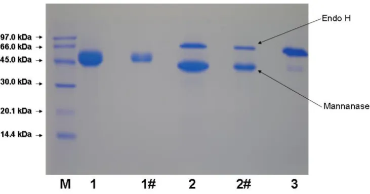
Purified recombinant mannan endo-1,4-β-mannosidase
displayed one single band with an apparent molecular mass of 53 kDa on SDS-PAGE. When the enzyme was treated with Endoglycosidase H, the apparent molecular mass of the deglycosylated enzyme was approximately 47
kDa (Fig. 3), indicating that mannan endo-1,4-β
-man-nosidase is a glycoprotein, which is glycosylated to a degree of approximately 12%. IEF revealed one single iso-form of pI 4.7 (not shown), in good agreement with the theoretical pI deduced from the amino-acid sequence.
Characterization of recombinant mannan endo-1,4-β
-mannosidase
Effect of temperature and pH on enzyme activity and stability The effect of temperature and pH were investigated on
recombinant A. niger BK01 mannan endo-1,4-β
-mannosi-dase using locust bean gum mannan as substrate. The enzyme displayed over 50% activity in the temperature range of 50 to 90°C with an optimal temperature at 80°C for the 10-min assay at pH 6.0 (Fig. 4). The enzyme was stable up to 70°C. Practically all of its activity (>98%) was retained after a 4-h incubation at 70°C, and the half-life time at this temperature was 56 h. At 80°C the half-life
time of activity of A. niger β-mannanase was only 15 min,
and at 90°C about 90% of its activity was lost within 2.5 min of incubation (Fig. 5). The optimal pH for mannan
endo-1,4-β-mannosidase (at 80°C) was pH 4.5, and more
than 80% of its maximal activity was retained within a
SDS-PAGE and zymogram analysis of recombinant A. niger mannan endo- β-1,4-mannosidase
Figure 2
SDS-PAGE and zymogram analysis of recombinant A. niger mannan endo- β-1,4-mannosidase. SDS-PAGE (A) and
zymogram (B) of secreted proteins from recombinant Pichia pastoris expressing the A. niger β -mannanase are shown. Lane M, molecular mass standard protein; Lane 1-6, culture supernatant after induction for 6, 24, 36, 48, 72 and 96 h, respectively; Lane C, control of P. pastoris X33 harbouring the empty pPICZαA vector after induction for 96 h. Twelve μL of culture supernatant was loaded onto each lanes.
rather broad pH range (from pH 4.0-5.5) (Fig. 6). The enzyme was stable in a narrow range around its optimal pH value (pH 4.5-5.0) (Fig. 7)
Effect of metal ions and chemical reagents on mannan endo-1,4-β -mannosidase activity
The relative activities (in parenthesis) of the enzyme after incubation with various reagents revealed that mannan
endo-1,4-β-mannosidase from A. niger BK01 was strongly
inhibited by sodium dodecyl sulfate, SDS (20.5%), and
Mn2+ (34.9%), and slightly inhibited by
phenylmethyl-sulphonylfluoride, PMSF (75.2%), Na+ (72.6%),
Zn2+(86.6%), Mg2+ (87.4%), K+ (89.7%), Ca2+(90.5%)
and Fe2+(95.8%). On the contrary, EDTA (235.6%) and
Cu2+ (110%) could activate this enzyme.
Substrate specificity and kinetics parameters
The relative activity of the purified enzyme on various substrates was determined as described in Methods.
Man-nan endo-1,4-β-mannosidase from A. niger exhibited high
activity on locust bean gum (100%), but showed consid-erably less activity on guar gum (17.9% relative activity)
and birchwood xylan (9.1%). Its activity is negligible on
starch, carboxymethylcellulose and α-cellulose (<0.1%).
The Michaelis-Menten constants were determined for mannan-containing polysaccharides from various sources (Table 2). These substrates have different structures because of the different ratios of monomers found in the backbone and side chain substituents. For example, gluco-mannan from konjac has a glucose to mannose ratio of 0.66 to 1; galactomannan from locust bean gum (Sigma) has a mannose to galactose ratio of 4:1, while those from carob (Megazyme) and guar gum have a mannose to
galactose ratio of 3.76:1 and 1.6:1, respectively. The Km
and kcat values were 0.6 mg mL-1 and 215 s-1 for low
viscos-ity glucomannan from konjac, 2.0 mg mL-1 and 330 s-1 for
locust bean gum galactomannan, 2.2 mg mL-1 and 292 s-1
for low viscosity galactomannan from carob, 1.5 mg mL-1
and 148 s-1 for β-mannan from carob, and 7.7 mg mL-1
and 352 s-1 for guar galactomannan.
Product analysis
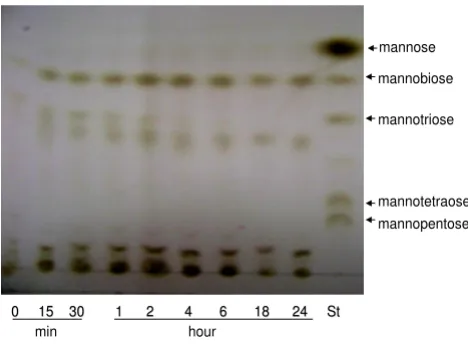
Analysis of oligosaccharide products obtained during enzymatic hydrolysis of locust bean gum using thin layer
Table 1: One-step purification of recombinant β-mannanase from A. niger
Step Total protein (mg) Total activity (U) Specific activity (U mg-1) Degree of purification (fold) Yield
(%)
Culture supernatant 42.6 20870 471 1.0 100
Superdex 75 3.9 10200 2570 5.5 50.7
Deglycosylation of recombinant A. niger mannan endo- β-1,4-mannosidase
Figure 3
Deglycosylation of recombinant A. niger mannan endo- β-1,4-mannosidase. SDS-PAGE analysis of the recombinant
chromatography revealed that the recombinant mannan
endo-1,4-β-mannosidase from A. niger yields
mannobi-ose as its main product, and a small amount of different oligosaccharides (Fig. 8). No trace of mannose could be detected in these hydrolysis experiments.
Discussion
The mannan endo-1,4-β-mannosidase of A.niger BK01
and the hypothetical protein (An05 g01320) of A.niger
CBS513.18 are highly similar with 99% amino acid sequence identity, and 98% DNA sequence similarity. Their amino acid sequences also match the N-terminal
sequence of native A. niger mannan endo-1,4-β
-mannosi-dase that was reported in 1998 [24]. We have thoroughly
searched the databases and could not find any report on
cloning, expression, and characterization of an A. niger
mannan endo-1,4-β-mannosidase. Thus, our work is the
first report on cloning, expression and characterization of this enzyme. In addition, this work also helps verify the function of the hypothetical protein (An05 g01320) of
A.niger CBS513.18 that has been reported to have strong
similarity to mannanase (man1) from A. aculeatus. β
-Mannanases from A. niger BK01 [21], A. niger CBS513.18
and A. niger published by Ademark et al [24] appear to be
Effect of temperature on mannan endo- β-1,4-mannosidase activity
Figure 4
Effect of temperature on mannan endo- β
-1,4-man-nosidase activity. The optimal temperature was deter-mined using 0.5% locust bean gum in 0.1 M citrate-phosphate buffer, pH 4.0, in the 10-min assay.
Effect of temperature on stability of A. niger BK01 β -mannan-ase
Figure 5
Effect of temperature on stability of A. niger BK01 β -mannanase. The temperature stability is shown as relative residual activity after incubation without substrate at differ-ent temperatures and at pH 4.0.
Effect of pH on mannan endo- β-1,4-mannosidase activity
Figure 6
Effect of pH on mannan endo- β-1,4-mannosidase
activity. The optimal pH was determined at 80°C using 0.5% LBG in 0.1 M citrate-phosphate buffer over a pH range of 2.5-7.0.
Effect of pH on stability of A. niger BK01 β-mannanase
Figure 7
closely related and have similar properties, which are
superior than those from A. niger NRRL 337 [25]. Mannan
endo-1,4-β-mannosidases from other Aspergillus species
e.g., those from A. aculeatus, A. fumigatus or A. sulphureus,
have previously been cloned and expressed in various
hosts [26-31]. A. niger β-mannanase has unique
proper-ties that are different from other Aspergillus mannanases.
Amino acid sequence of A. niger BK01 mannan
endo-1,4-β-mannosidases including putative signal peptide can be
found in additional file 1.
Mannan endo-1,4-β-mannosidases or β-mannanases have
been classified into two family of glycoside hydrolases,
i.e., family 5 and 26, based on their sequence similarity
http://www.cazy.org. The A. niger mannan endo-1,4-β
-mannosidase belongs to glycoside hydrolase family 5 (GH5). Analysis of the primary sequence of fungal
man-nan endo-1,4-β-mannosidases belonging to this family
revealed amino acid sequence similarities ranging from
43 to 93%. Some fungal β-mannanases have been shown
to contain a cellulose-binding domain (CBD) at either the C-terminus or N-terminus of the enzyme. For example,
CBD of T. reesei β-mannanase is located at its C-terminus
(amino acid position 373-410) preceded by a serine-, threonine-, and proline-rich region [29], whereas CBD of
A. fumigatus β-mannanase is located at its N-terminus (amino acid position 9-44) as shown in Fig. 1. A role of this domain in the hydrolysis of mannan/cellulose
com-plex substrates has been suggested [32]. However, β
-man-nanases from different fungi including A. niger do not
contain a CBD. The protruding N-terminus of Bispora sp.
as shown in the sequence alignment in Fig. 1 is not similar to a CBD, and its role in catalysis still remains to be explored.
The mature A. niger mannan endo-1,4-β-mannosidase
gene without its signal peptide was expressed in P. pastoris
X33 with the C-terminus of the recombinant enzyme fused to the 6xHis tag for affinity purification by immobi-lized metal affinity chromatography (IMAC). The native
signal peptide was replaced with that of the Saccharomyces
cerevisiae α-factor, allowing secretion of the active enzyme into the extracellular medium with an expression yield of
approximately 243 mg L-1. Since cultivation experiments
were done in shake flasks, one can expect that growth of
P. pastoris in a fermentor under controlled and optimised conditions will result in considerably higher protein
yields [33]. In comparison, the expression level of
Tri-choderma reesei mannan endo-1,4-β-mannosidase in S. cerevisiae was roughly 150 mg L-1 [29] and that of Bispora
sp. MEY-1 [15] and B. subtilis mannan endo-1,4-β
-man-nosidase in P. pastoris GS115 [34] was 1800 mg L-1 and
150 mg L-1, respectively. Moreover, the enzyme activity
during fermentation reached 670 U mL-1, which is
signif-icantly higher than those reported for most mannan
endo-1,4-β-mannosidases [26,34].
Amino acid sequence analysis by the NetNGlyc 1.0 sever program indicated two putative N-glycosylation sites in
the amino acid sequence of mannan endo-1,4-β
-man-nosidase i.e., 195NSS197, and 252NFT254 (according to
num-Table 2: Kinetic parameters for the purified mannan endo-1,4-β-mannosidasea at 70°C
Substrate Vmax(U mg-1) Km(mg mL-1) kcat(s-1) kcat/Km(mg-1 s-1 mL)
Glucomannan 243 ± 15 0.6 ± 0.3 215 358 Locust bean gum 373 ± 19 2.0 ± 0.4 330 165 Galactomannan 330 ± 9 2.2 ± 0.2 292 133
Mannan 167 ± 5 1.5 ± 0.2 148 98.4
Guar gum 398 ± 48 7.7 ± 1.8 352 45.7
a Enzymatic reactions were carried out for 5 min at 70°C in 0.07 M citrate phosphate buffer (pH 4.5).
Thin layer chromatography analysis
Figure 8
Thin layer chromatography analysis. Products from
locust bean gum hydrolysis by recombinant A. niger mannan-ase at various time points were separated on silica plates. Incubation times (hour or minute) are indicated. Lane St indi-cates authentic standards, i.e. a mixture of mannose to man-nopentaose. Ten μL of hydrolysate was spotted onto each lane.
0 15 30 1 2 4 6 18
0 15 30 1 2 4 6 18 24 St 24 St
min hour
mannose
mannobiose
mannotriose
mannotetraose
bering in Fig. 1). The deglycosylation analysis, showed in Fig. 3, confirms the prediction that the enzyme is
glyco-sylated. Purified mannan endo-1,4-β-mannosidase
treated with Endoglycosidase H was calculated to be 43
kDa by SDS-PAGE. This result indicated that the A. niger
mannan endo-1,4-β-mannosidase expressed in P. pastoris
was properly glycosylated, as the enzyme was as active as the native enzyme.
Recombinant A. niger BK01 mannan endo-1,4-β
-man-nosidase efficiently hydrolysed galactomannans,
gluco-mannans and β-1,4-mannans from different sources.
Although determination of kinetic parameters enabled us
to demonstrate that A. niger GH5 mannan endo-1,4-β
-mannosidase was highly active towards structurally differ-ent mannans, this enzyme displayed highest specificity
towards unsubstituted carob glucomannan with a Km and
kcat of 0.6 mg.mL-1 and 215 s-1, respectively. Based on
these kinetic parameters, mannanase activity of this enzyme appears not to be hampered by the presence of
side chains, i.e. galactosyl residues. The enzyme can
hydrolyse galactomannans as well as glucomannans and
unsubstituted β-1,4-mannan. The action of the enzyme
on these substrates as well as on Azo-carob galactoman-nan, a standard substrate for the assessment of mannan
endo-1,4-β-mannosidase activity, indicates its function as
a true endo-β-1,4-mannanase. This activity was confirmed
by TLC analysis of reaction products obtained through hydrolysis of locust bean gum, which gave mannobiose as major product and no detectable mannose, indicating
that the enzyme has no β-mannosidase activity. In
addi-tion to mannan, A. niger mannanase was capable of
degrading birchwood xylan (9.1% relative activity). Its lacks of activity for cellulose is valuable for applications in bleaching pulp and paper [1].
In general, the properties of the recombinant mannan
endo-1,4-β-mannosidase from A. niger are very similar to
those of the enzyme from its natural source with only
some minor differences. The recombinant A. niger
man-nan endo-1,4-β-mannosidase furthermore shows
proper-ties typical of thermostable enzymes, which is in accordance with the wild-type enzyme. The optimal tem-perature of its activity is 80°C, similar to that of the native
enzyme [21]. Optimal temperatures of different fungal β
-mannanases have previously been reported; that of the β
-mannanase from Trichoderma reesei C-30 was found at
75°C [35], whereas β-mannanase produced from A. niger
and A. flavus showed their optimum at 65 and 60°C,
respectively [14]. Two forms of mannan endo-1,4-β
-man-nosidase from the thermotolerant fungus A. fumigatus IMI
385708 showed highest activity at 60°C (pH 4.5) [6]. These rather high optima appear to be a common but
val-uable characteristic of fungal β-mannanases. At such high
temperatures (above 60-65°C), enzymatic digestion may
not only increase the rate of hydrolysis but also reduce microbial contamination of the material being processed [36].
Both native and recombinant A. niger mannan
endo-1,4-β-mannosidase appear to be among the most
thermosta-ble fungal β-mannanases reported to date. The
recom-binant A. niger mannan endo-1,4-β-mannosidase retained
>98% activity after 4 h at 70°C, while mannan
endo-1,4-β-mannosidase from Talaromyces emersonii and Aspergillus
niger NRRL 337 retained 53% and 20% activity after 1 h at 65°C, respectively [36]. The half-life of thermostable
acidic mannan endo-1,4-β-mannosidase from Sclerotium
(Athelia) rolfsii at 70°C and pH 4.5 was 1.5 h [37], while
that of recombinant A. niger mannan endo-1,4-β
-man-nosidase at 70°C was 56 h. Bispora sp. MEY-1 mannan
endo-1,4-β-mannosidase, which was expressed in P.
pas-toris, only retained more than 50% of its initial activity after an incubation at 70°C for 20 min [15].
The strong increase of the enzyme activity in the present of EDTA (1 mM) suggests that metal ions are not present in the active site and are not required for activity. At the
same concentration, the effect of EDTA on A. sulphureus β
-mannanase activity was only moderate [26]. EDTA might protect the enzyme against the detrimental effect of metal ions present in the enzyme preparation, which inhibit the
A. niger β-mannanase to a certain extent while not
affect-ing the A. sulphureus enzyme significantly. For example,
Mn2+ strongly inhibited the A. niger β-mannanase but did
not effect the latter enzyme. Other ions including Na+,
Zn2+, Mg2+, Ca2+, Fe2+ exerted an adverse effect on the
activity of A. niger β-mannanase but did not affect or
slightly increased the A. sulphureus β-mannanase activity
[26]. The β-mannanase activity inhibition in the presence
of PMSF (1 mM) was 24.8%, suggesting the role of a
ser-ine in the catalytic action of A. niger mannan endo-1,4-β
-mannosidase.
Conclusion
To date, this study is the first report on production of a
thermostable GH5 mannan endo-1,4-β-mannosidase
from A. niger using a P. pastoris expression system. This
kind of thermostable enzyme can gain a great deal of interest for industrial applications in large scale due to an increasing demand towards renewable resource utiliza-tion in the near future.
Methods
Strains, plasmids, enzymes, reagents, and growth media
Aspergillus niger strain BK01, isolated from an orange in Vietnam, was identified by the Center of Biotechnology, Vietnam National University, Hanoi, and is preserved in
our laboratory. Escherichia coli strain TOP 10 (Invitrogen)
pPIC-ZαA (Invitrogen) and propagation of recombinant
expres-sion vectors. Pichia pastoris X33 (Invitrogen) was used for
heterologous protein expression. All media and protocols for Pichia are described in the Pichia expression manual (Invitrogen). Manno-oligosaccharides were from Meg-azyme (Bray, Ireland).
Total RNA isolation, cDNA synthesis and cloning of the
mannan endo-1,4-β-mannosidase gene
A. niger BK01 was grown at 37°C for 48 h in a plate con-taining 10 mL liquid modified Czapek medium including
locust bean gum as a sole carbon source (5 g L-1). The
mycelium was then transferred to a plate containing 10
mL β-mannanase induction medium based on modified
coffee pulp waste (21.6 g L-1) and guar gum (10 g L-1) [21].
After a 72-h growth period, the mycelium was harvested. Total RNA was isolated by extracting 50 mg of mycelium using the RNeasy Plant Mini Kit (Qiagen, Valencia, CA). cDNA was synthesized from RNA using One Step RT-PCR Kit (Qiagen) according to the manufacturer's instructions. The primers used were CF (5'-ATG AAG CTT TCC AAC GCC CTC CTC-3') and CR (5'-TTA AGC ACT ACC AAT AGC AGC AAC ATG ATC-3'), which were designed according to the amino acid sequence of the putative
mannan endo-1,4-β-mannosidase from A. niger CBS
513.88 (Genbank accession no. XP_001390707) The thermocycling parameters were: 50°C for 35 min, 95°C for 15 min and 30 cycles of 94°C - 30 s, 54°C - 45 s and 72°C - 90 s, followed by 10 min extension at 72°C.
To subclone the mannan endo-1,4-β-mannosidase cDNA
into the expression vector, the β-mannanase-encoding
sequence was amplified from the previously isolated cDNA with the primers PNf (5'-CTG TGC GAA TTC TCC
TTC GCC AGC ACC TCC G-3'), including an EcoRI site,
and PRv (5'-CTG TGC TCT AGA GCA CTA CCA ATA GCA
GCA ACA TGA TCC -3'), including a XbaI site, using a
mix-ture of Pfu DNA polymerase (3 U/reaction) and Taq DNA
polymerase (3 U/reaction). The thermocycling parameters were 95°C for 2 min, and 30 cycles of 95°C for 45 s, 59°C - 45 s and 72°C - 3 min. The DNA insert was cloned into
EcoRI and XbaI sites of the pPICZαA vector, downstream
of the α-factor signal peptide sequence. Proper
construc-tion was confirmed by restricconstruc-tion digesconstruc-tion and auto-mated DNA sequencing (Macrogen Inc., Korea). The DNA
sequence encoding mature mannan endo-1,4-β
-mannosi-dase was deposited in the GenBank database under acces-sion no. FJ268574.
Expression of the mannan endo-1,4-β-mannosidase gene
in Pichia pastoris
The selected expression plasmid was linearized with SacI
(New England BioLabs, USA), and then transformed into
P. pastoris strain X33 by electroporation [22]. Transform-ants were first screened from YPDS (1% yeast extract, 2%
peptone, 2% dextrose, 1 M sorbitol, 2% agar) plates
con-taining Zeocin™ at a final concentration of 100 μg mL-1,
then on YPDZ plates (1% yeast extract, 2% peptone, 2% dextrose and 2% agar containing Zeocin at final
concen-trations of 150, 300, and 500 μg mL-1) in order to screen
for higher copy numbers of the targeted gene.
Recom-binant strains producing mannan endo-1,4-β
-mannosi-dase were further confirmed by BMGY-Azo plates (1% yeast extract, 2% peptone, 100 mM potassium phosphate
pH 6.0, 1.34% YNB, 4 × 10⁻⁵ % biotin, 0.5% methanol,
2% agar and 0.3% Azo-carob galactomannan). Mut+
phe-notype of P. pastoris recombinants was analysed by
genomic PCR with 5'AOX and 3'AOX primers. The culture
medium from the P. pastoris transformant that had the
highest β-mannanase activity was used for the subsequent
analysis of the recombinant protein. A control was
trans-formed with an empty pPICZαA plasmid.
Purification of recombinant mannan endo-1,4-β -mannosidase
All purification steps were performed at 4°C unless stated otherwise. The crude culture supernatant was obtained by centrifugation of the culture broth at 4000 rpm for 5 min at 4°C. The crude supernatant was concentrated 30-fold by ultrafiltration through a 10-kDa cut-off membrane (Sartorius Stedim Biotech, Germany). The final purifica-tion was done by size exclusion chromatography on a Superdex 75 HR 10/30 column (Amersham Pharmacia Biotech, Sweden) equilibrated with 50 mM potassium phosphate buffer, pH 6.0, and the protein was eluted at a
flow rate of 1 mL min-1. Active fractions were combined
and used in further experiments as the purified β
-man-nanase.
Polyacrylamide gel electrophoresis and zymograms
Sodium dodecyl sulfate polyacrylamide gel
electrophore-sis (SDS-PAGE) was performed in a 12.5% (w v-1)
polyacr-ylamide gel by the method of Laemmli (1970). Proteins were stained with Coomassie brilliant blue G-250.
Zymograms were obtained by co-polymerizing 0.2% (w v
-1) locust bean gum with 12.5% (w v-1) polyacrylamide gel
as previously reported [38]. After electrophoresis, the gel was soaked in 2.5% cold Triton X100 with gentle shaking to remove SDS and re-fold the proteins in the gel. The gel was then washed four times at 4°C in 50 mM potassium phosphate buffer pH 6.0 for 30 min. After incubation for 45 min at 50°C, the gels were stained with Congo red
solution (0.1%, w v-1), and destained with 1 M NaCl. The
activity bands were observed as clear yellow halos.
Deglycosylation of recombinant β-mannanase Purified
mannan endo-1,4-β-mannosidase was deglycosylated by
denaturing the protein at 100°C for 10 min prior to the
Biolabs) at 37°C for 1 h according to the manufacturer's instructions.
β-Mannanase assay and protein quantification
Mannan endo-1,4-β-mannosidase activity was
deter-mined using the 3,5-dinitrosalicylic acid (DNS) method [39]. The reaction was started by mixing 0.1 mL of
appro-priately diluted enzyme sample with 0.9 mL of 5 mg mL-1
locust bean gum in 0.1 M citrate-phosphate buffer, pH 4.0. After 10-min incubation at 50°C, the reaction was stopped by the addition of 5 mL DNS reagent. One unit of
mannan endo-1,4-β-mannosidase activity is defined as
the amount of enzyme releasing 1 μmole of mannose
equivalents per minute. All experiments were done in trip-licate, and average values are reported. Protein concentra-tion was determined with bovine serum albumin as standard, using the Micro-BCA Protein Assay Reagent (Biorad, USA).
Effect of temperature, pH and various reagents on enzyme activity
The optimal temperature for mannan endo-1,4-β
-man-nosidase activity (10-min assay) was determined by the standard activity assay as described above at various tem-peratures ranging from 40 to 100°C. To estimate thermal stability, the enzyme was pre-incubated in 0.1 M citrate-phosphate buffer (pH 4.0) at various temperatures (60-90°C) for different periods of time without substrate and then assayed for residual activity at 50°C using the
stand-ard activity assay. The effect of pH on β-mannanase
activ-ity was determined at 80°C over a pH range of 2.5-7.0 by using 0.1 M citrate-phosphate buffer. To estimate pH sta-bility, the enzyme was diluted/pre-incubated in the same buffer at different pH-values for different periods of time at 50°C without substrate, and then assayed for residual activity at pH 4.0. The effect of ions on mannan
endo-1,4-β-mannosidase activity was examined by incubating the
enzyme with 1 mM of various metal ions in 0.1 M sodium citrate buffer (pH 4.0) at 50°C for 30 min.
Substrate specificity and kinetic parameters
The substrate specificity of purified recombinant mannan
endo-1,4-β-mannosidase was evaluated with the
follow-ing substrates at 5 mg mL-1 in 0.1 M citrate-phosphate
buffer, pH 4.0: locust bean gum (Sigma), guar gum (Sigma), birchwood xylan (Roth), soluble starch
(Sigma-Aldrich), α-cellulose and carboxyl methyl cellulose
(Fluka). Activity was measured by the dinitrosalicylic acid (DNS) method as described above. For the kinetic experi-ments, 0.1 M citrate-phosphate buffer (pH 4.5)
contain-ing 0.1 to 10 mg mL-1 of various mannan substrates (i.e.,
locust bean gum (Sigma), guar gum (Sigma), carob galac-tomannan (low viscosity) (Megazyme), konjac
glucoman-nan (low viscosity) (Megazyme), and carob 1,4-β
-D-mannan (Megazyme), was incubated with the purified
mannan endo-1,4-β-mannosidase at 70°C for 5 min.
Kinetic parameters (Km and vmax) were calculated from the
experimentally obtained data and non-linear regression analysis using the "GraFit" software (Erithacus Software Ltd.).
Thin layer chromatography
Locust bean gum was prepared at a concentration of 1%
(w v-1) in 50 mM sodium citrate buffer, pH 4.5. After
addi-tion of the purified enzyme (7.5 U for 2-mL reacaddi-tions), the solution was incubated at 40°C. Aliquots were removed at various time, and heated to 100°C for 10 min. Hydrolysis products were separated on silica plates 60 F 254 (Merck, Darmstadt, Germany) using a solvent system
consisting of 1-propanol-nitromethane-water (7:1:2, v v
-1). The products were detected by spraying with 5%
sul-phuric acid in ethanol followed by heating at 110°C for about 5 min. Manno-oligosaccharides (mannose, manno-biose, mannotriose, mannotetraose and mannopentose, Megazyme) were used as standards.
Competing interests
The authors declare that they have no competing interests.
Authors' contributions
DBC performed most parts of the experiments and pre-pared the first draft of the manuscript. DTT and TKA directed the study. JBG checked all data, designed the frame of this publication and revised the draft. JCS co-designed the frame of this publication. DTT, TKA, JGB and JCS also co-supervised enzyme characterization. DH
initi-ated expression in Pichia pastoris and edited the
manu-script. MY conceived of the study, supervised molecular biology work, participated in sequence alignment and edited the manuscript. All authors read and approved the final manuscript.
Additional material
Acknowledgements
This work was supported by the National Research Council of Thailand (NRCT), Faculty and Student Exchange Program between Thailand and Neighbour Countries 2007, T2008-219 project (Hanoi University of Tech-nology), Regional Center of Excellence Project (PER) (Agency for French Speaking Universities) and the ASEAN University Network Program
Additional file 1
Complete amino acid sequence of A. niger BK01 mannan endo-1,4-β mannosidase including native signal peptide. Amino acid sequence in FASTA format of the entire mannan endo-1,4-β-mannosidase from
Aspergillus niger BK01, including putative signal peptide.
Click here for file
(AUNP). The authors would like to thank Prof. Truong Nam Hai and Dr. Do Thi Huyen (Vietnam Academy of Science and Technology) for valuable advices on the expression studies; MSc. V.T.K.Thoa for technical assistance; and MY lab members for their friendship and technical advices.
References
1. Dhawan S, Kaur J: Microbial mannanases: an overview of pro-duction and applications. Crit Rev Biotechnol 2007, 27(4):197-216. 2. Gübitz G, Sachslehner A, Haltrich D: Microbial mannanases: sub-strates, production and application. In Glycosyl Hydrolases for Bio-mass Conversion Volume 769. Washington, DC: ACS Symposium Series, American Chemical Society; 2001:239-262.
3. Schroder R, Atkinson RG, Redgwell RJ: Re-interpreting the role of endo-β-mannanases as mannan endotransglycosylase/hydro-lases in the plant cell wall. Ann Bot (Lond) 2009, 104(2):197-204. 4. Turner P, Mamo G, Karlsson EN: Potential and utilization of thermophiles and thermostable enzymes in biorefining.
Microb Cell Fact 2007, 6:9.
5. Viikari L, Alapuranen M, Puranen T, Vehmaanpera J, Siika-Aho M:
Thermostable enzymes in lignocellulose hydrolysis. Adv Bio-chem Eng Biotechnol 2007, 108:121-145.
6. Puchart V, Vrsanská M, Svoboda P, Pohl J, Ogel ZB, Biely P: Purifica-tion and characterizaPurifica-tion of two forms of endo-β -1,4-man-nanase from a thermotolerant fungus, Aspergillus fumigatus
IMI 385708 (formerly Thermomyces lanuginosus IMI 158749).
Biochimica et Biophysica Acta (BBA) - General Subjects 2004,
1674(3):239-250.
7. Duffaud GD, McCutchen CM, Leduc P, Parker KN, Kelly RM: Purifi-cation and characterization of extremely thermostable beta-mannanase, beta-mannosidase, and alpha-galactosi-dase from the hyperthermophilic eubacterium Thermotoga neapolitana 5068. Appl Environ Microbiol 1997, 63(1):169-177. 8. Sachslehner A, Foidl G, Foidl N, Gubitz G, Haltrich D: Hydrolysis of
isolated coffee mannan and coffee extract by mannanases of
Sclerotium rolfsii. J Biotechnol 2000, 80(2):127-134.
9. Sanchez C: Lignocellulosic residues: biodegradation and bio-conversion by fungi. Biotechnol Adv 2009, 27(2):185-194. 10. Jiang Z, Wei Y, Li D, Li L, Chai P, Kusakabe I: High-level
produc-tion, purification and characterization of a thermostable β -mannanase from the newly isolated Bacillus subtilis WY34.
Carbohydrate Polymers 2006, 66(1):88-96.
11. Luthi E, Jasmat NB, Grayling RA, Love DR, Bergquist PL: Cloning, sequence analysis, and expression in Escherichia coli of a gene coding for a beta-mannanase from the extremely ther-mophilic bacterium "Caldocellum saccharolyticum". Appl Envi-ron Microbiol 1991, 57(3):694-700.
12. Talbot G, Sygusch J: Purification and characterization of ther-mostable beta-mannanase and alpha-galactosidase from
Bacillus stearothermophilus. Appl Environ Microbiol 1990,
56(11):3505-3510.
13. Hilge M, Gloor SM, Rypniewski W, Sauer O, Heightman TD, Zimmer-mann W, Winterhalter K, Piontek K: High-resolution native and complex structures of thermostable beta-mannanase from
Thermomonospora fusca - substrate specificity in glycosyl hydrolase family 5. Structure 1998, 6(11):1433-1444.
14. Kote NV, Patil AG, Mulimani VH: Optimization of the production of thermostable endo-beta-1,4 mannanases from a newly isolated Aspergillus niger gr and Aspergillus flavus gr. Appl Bio-chem Biotechnol 2009, 152(2):213-223.
15. Luo H, Wang Y, Wang H, Yang J, Yang Y, Huang H, Yang P, Bai Y, Shi P, Fan Y, et al.: A novel highly acidic beta-mannanase from the acidophilic fungus Bispora sp. MEY-1: gene cloning and over-expression in Pichia pastoris. Appl Microbiol Biotechnol 2009,
82(3):453-461.
16. Politz O, Krah M, Thomsen KK, Borriss R: A highly thermostable endo-(1,4)-beta-mannanase from the marine bacterium
Rhodothermus marinus. Appl Microbiol Biotechnol 2000,
53(6):715-721.
17. Cantarel BL, Coutinho PM, Rancurel C, Bernard T, Lombard V, Hen-rissat B: The Carbohydrate-Active EnZymes database (CAZy): an expert resource for Glycogenomics. Nucleic Acids Res 2009:D233-238.
18. Ethier N, Talbot G, Sygusch J: Gene cloning, DNA sequencing, and expression of thermostable beta-mannanase from
Bacil-lus stearothermophiBacil-lus. Appl Environ Microbiol 1998,
64(11):4428-4432.
19. Aparicio R, Fischer H, Scott DJ, Verschueren KH, Kulminskaya AA, Eneiskaya EV, Neustroev KN, Craievich AF, Golubev AM, Polikarpov I: Structural insights into the beta-mannosidase from T. ree-sei obtained by synchrotron small-angle X-ray solution scat-tering enhanced by X-ray crystallography. Biochemistry 2002,
41(30):9370-9375.
20. Bourgault R, Oakley AJ, Bewley JD, Wilce MC: Three-dimensional structure of (1,4)-beta-D-mannan mannanohydrolase from tomato fruit. Protein Sci 2005, 14(5):1233-1241.
21. Thu DT, Cuong DB: Purification and characterization of an endo-β-1,4-mannanase from Aspergillus sp. BK. Journal of Sci-ence and Technology (Vietnamese) 2004, 42(5):38-43.
22. Cregg JM, ed: Pichia Protocols. 2nd edition. Totowa, New Jersey: Humana Press; 2007.
23. Sabini E, Schubert H, Murshudov G, Wilson KS, Siika-Aho M, Penttila M: The three-dimensional structure of a Trichoderma reesei
beta-mannanase from glycoside hydrolase family 5. Acta Crys-tallogr D Biol CrysCrys-tallogr 2000, 56(Pt 1):3-13.
24. Ademark P, Varga A, Medve J, Harjunpaa V, Torbjorn D, Tjerneld F, Stalbrand H: Softwood hemicellulose-degrading enzymes from Aspergillus niger: Purification and properties of a β -man-nanase. Journal of Biotechnology 1998, 63(3):199-210.
25. Araujo A, Ward P: Hemicellulases of Bacillus species: prelimi-nary comparative studies on production and properties of mannanase and galactanases. Journal of Applied Bacteriology 1990,
68:253-261.
26. Chen X, Cao Y, Ding Y, Lu W, Li D: Cloning, functional expres-sion and characterization of Aspergillus sulphureus beta-man-nanase in Pichia pastoris. J Biotechnol 2007, 128(3):452-461. 27. Duruksu G, Ozturk B, Biely P, Bakir U, Ogel ZB: Cloning,
expres-sion and characterization of endo-beta-1,4-mannanase from
Aspergillus fumigatus in Aspergillus sojae and Pichia pastoris.
Biotechnol Prog 2009, 25(1):271-276.
28. Setati ME, Ademark P, van Zyl WH, Hahn-Hagerdal B, Stalbrand H:
Expression of the Aspergillus aculeatus endo-beta-1,4-man-nanase encoding gene (man1) in Saccharomyces cerevisiae
and characterization of the recombinant enzyme. Protein Expr Purif 2001, 21(1):105-114.
29. Stalbrand H, Saloheimo A, Vehmaanpera J, Henrissat B, Penttila M:
Cloning and expression in Saccharomyces cerevisiae of a Tri-choderma reesei beta-mannanase gene containing a cellulose binding domain. Appl Environ Microbiol 1995, 61(3):1090-1097. 30. van Zyl PJ, Moodley V, Rose SH, Roth RL, van Zyl WH: Production
of the Aspergillus aculeatus endo-1,4-beta-mannanase in A. niger. J Ind Microbiol Biotechnol 2009, 36(4):611-617.
31. Roth R, Moodley V, van Zyl P: Heterologous Expression and Optimized Production of an Aspergillus aculeatus Endo-1,4-β -mannanase in Yarrowia lipolytica. Molecular Biotechnology 2009,
43(2):112-120.
32. Hagglund P, Eriksson T, Collen A, Nerinckx W, Claeyssens M, Stal-brand H: A cellulose-binding module of the Trichoderma reesei
beta-mannanase Man5A increases the mannan-hydrolysis of complex substrates. J Biotechnol 2003, 101(1):37-48.
33. Gasser B, Saloheimo M, Rinas U, Dragosits M, Rodriguez-Carmona E, Baumann K, Giuliani M, Parrilli E, Branduardi P, Lang C, et al.: Protein folding and conformational stress in microbial cells produc-ing recombinant proteins: a host comparative overview.
Microb Cell Fact 2008, 7:11.
34. He X, Liu N, Li W, Zhang Z, Zhang B, Ma Y: Inducible and consti-tutive expression of a novel thermostable alkaline β -man-nanase from alkaliphilic Bacillus sp. N16-5 in Pichia pastoris
and characterization of the recombinant enzyme. Enzyme and Microbial Technology 2008, 43(1):13-18.
35. Arisan-Atac I, Hodits R, Kristufek D, Kubicek CP: Purification, and characterization of a β-mannanase of Trichoderma reesei C-30. Applied Microbiology and Biotechnology 1993, 39(1):58-62. 36. Araujo A, Ward OP: Extracellular mannanases and
galactan-ases from selected fungi. Journal of Industrial Microbiology and Bio-technology 1990, 6(3):171-178.
37. Sachslehner A, Haltrich D: Purification and some properties of a thermostable acidic endo-beta-1,4-d-mannanase from
Sclerotium (Athelia) rolfsii. FEMS Microbiology Letters 1999,
Publish with BioMed Central and every scientist can read your work free of charge "BioMed Central will be the most significant development for disseminating the results of biomedical researc h in our lifetime."
Sir Paul Nurse, Cancer Research UK
Your research papers will be:
available free of charge to the entire biomedical community
peer reviewed and published immediately upon acceptance
cited in PubMed and archived on PubMed Central
yours — you keep the copyright
Submit your manuscript here:
http://www.biomedcentral.com/info/publishing_adv.asp
BioMedcentral 38. Yamabhai M, Emrat S, Sukasem S, Pesatcha P, Jaruseranee N,
Burana-banyat B: Secretion of recombinant Bacillus hydrolytic enzymes using Escherichia coli expression systems. Journal of Biotechnology 2008, 133(1):50-57.
39. Miller GL: Use of Dinitrosalicylic Acid Reagent for Determina-tion of Reducing Sugar. Analytical Chemistry 1959, 31(3):426-428. 40. Larkin MA, Blackshields G, Brown NP, Chenna R, McGettigan PA, McWilliam H, Valentin F, Wallace IM, Wilm A, Lopez R, et al.: Clustal W and Clustal X version 2.0. Bioinformatics 2007,
23(21):2947-2948.
41. Gouet P, Courcelle E, Stuart DI, Metoz F: ESPript: analysis of mul-tiple sequence alignments in PostScript. Bioinformatics 1999,