R E S E A R C H A R T I C L E
Open Access
Postoperative anatomical and functional
outcomes of different stages of high
myopia macular hole
Qing Shao
1,2, Huijuan Xia
1, Florian M. A. Heussen
3, Yanling Ouyang
2, Xiaodong Sun
1and Ying Fan
1*Abstract
Background:Recently it was suggested that high myopia macular holes (HMMH) and macular holes accompanied by retinal detachment occur in the advanced stages of myopia traction maculopathy (MTM), while macular retinoschisis, shallow retinal detachment without holes, and lamellar macular holes occur in the early stages of MTM. Complete vitreous cortex removal associated with internal limiting membrane peeling is now widely used to treat HMMH. However, it remains uncertain at what HMMH stage patients would benefit most from surgical intervention. Our study was aimed to evaluate the postoperative anatomical changes and functional outcomes of high myopia macular holes (HMMH).
Methods:Patients were retrospectively collected between March 2009 and August 2011. Before and 1st, 3rd, and 9th month after 23G pars plana vitrectomy, all patients underwent a complete ophthalmologic examination, spectral domain optical coherence tomography (SD-OCT) and MP-1. At each follow-up, best-corrected visual acuity (BCVA), photoreceptor inner and outer segments (IS/OS) defects, and retinal sensitivity (RS) were investigated. According to different preoperative macular hole morphologies, patients were divided into three groups: Group 1, macular hole with epiretinal membrane (ERM) traction and macular retinoschisis; Group 2, full-thickness macular hole (FTMH); Group 3, FTMH with subretinal fluid.
Results:43 eyes from 43 patients met the inclusion criteria. The mean age was 60 years. BCVA and RS were significantly improved after vitrectomy; the mean IS/OS defect was significantly reduced. At 9 postoperative months, 11 of 43 (25.6 %) eyes achieved IS/OS junction integrity; 9 of these 11 (81.8 %) eyes belonged to Group 1, 2 (18.2 %) belonged to Group 2.
Conclusions:Pars plana vitrectomy combined with ILM peeling and gas tamponade results in limited functional outcomes in patients with HMMH. The appearance of subretinal fluid indicates a worse prognosis for surgical intervention.
Background
Retinopathy in pathological myopia is a major cause of visual impairment, and long axial lengths and posterior staphyloma eventually lead to myopia traction maculo-pathy (MTM) [1]. The term MTM was first used in 2004 to describe a series of posterior pole pathologies in cases of high myopia, such as macular retinoschisis, shallow retinal detachment without retinal holes, lamel-lar maculamel-lar holes, and maculamel-lar holes, with or without
retinal detachment. In 1999, Takano and Kishi specu-lated that the formation of high myopia macular holes (HMMH) resulted from macular retinoschisis or shallow retinal detachment without holes [2]. Panozzo and Mercanti [3] supported this notion and suggested that HMMH and macular holes accompanied by retinal detachment occur in the advanced stages of MTM, while macular retinoschisis, shallow retinal detach-ment without holes, and lamellar macular holes occur in the early stages of MTM. This hypothesis has been confirmed by several other groups [4–8].
Complete vitreous cortex removal associated with in-ternal limiting membrane peeling is now widely used to * Correspondence:[email protected]
1
Department of Ophthalmology, Shanghai First People’s Hospital, Haining Road 100, 200080 Shanghai, China
Full list of author information is available at the end of the article
treat HMMH. However, it remains uncertain at what HMMH stage patients would benefit most from surgical intervention.
In this study, we used spectral domain optical coherence tomography (SD-OCT) and microperimetry pre- and postoperatively in patients diagnosed with HMMH to observe and analyze the retinal microstructure and functional recovery.
Materials and methods Data collection
Patients with a diagnosis of HMMH were retrospectively collected from the Department of Ophthalmology at Shanghai First People’s Hospital, Shanghai Jiaotong University between March 2009 and August 2011. All patients underwent a standard ophthalmologic examination preoperatively and at 1 month, 3 months, and 9 months after pars plana vitrectomy, including volume OCT scan-ning with an SD-OCT (Spectralis, Heidelberg Engineering, Heidelberg, Germany) and the MP-1 microperimeter (Nidek Technologies, Padua, Italy). The imaging protocols used are described below. Patient information, such as age, sex, lens status, axial length, history of ophthalmic diseases or surgeries, and ophthalmic diagnoses, was also collected. The following inclusion criteria were applied: subjective visual symptoms; high myopia (>−6.00 diopters), with an axial length >26.00 mm; and a full-thickness macular hole (FTMH) as confirmed and documented by OCT examin-ation. Patients with diabetes, hypertension, eye surgery within 1 year, one or several peripheral retinal holes or ret-inal detachment were excluded. Best-corrected visual acuity (BCVA) was measured using E-VA charts, and the results were converted to logMAR units. Approval for data collec-tion and analysis was obtained from the institucollec-tional review board of the Shanghai First People’s Hospital. Written con-sent was obtained from all patients. The research adhered to the tenets set forth in the Declaration of Helsinki.
SD-OCT
All OCT scans were performed with the Spectralis OCT (Heidelberg Engineering, Germany). The scan protocol consisted of 19-line scans centered on the fovea covering 30° radial, with a line spacing of 350 μm between the scans. Active eye tracking and line averaging of 10 scans per line (ART 10) were used. The IS/OS defect was measured by OCT. Only gradable records were used in the study. Two graders (Q.S., Y.F.) independently evaluated each OCT image set (Fig. 1).
Microperimetry
The retinal sensitivity was measured using the MP1 (Nidek technology, Padua, Italy).
The eye-tracking system corrected eye movements automatically and obtained images with an infrared
camera. The test area comprised the macular 10° and 40 stimulus locations within this field. A white background, with a red single cross as a fixation target and Gold-mann III white spot stimuli, were used. The luminance range was 0–20 dB. The maximum brightness was 127 cd/m [2], the minimum brightness was 1.27 cd/m [2], and the stimulus duration was 200 ms. A staircase strategy of the 4-2-1 pattern was used. The retinal sensitivity threshold map was automatically registered to the color fundus photo, and the overall average retinal sensitivity was calculated (Fig. 2).
Groupings
Based on the OCT images, the patients were divided into three groups: [9, 10] Group 1, macular hole with epiretinal membrane (ERM) traction, macular retinoschisis; Group 2, FTMH without ERM and subretinal fluid; and Group 3, FTMH with subretinal fluid, with or without ERM (Fig. 3).
Surgical approach
All patients underwent transconjunctival 23G pars plana vitrectomy. The procedures for 7 eyes were combined with phacoemulsification and intraocular lens implant-ation in the same surgery. All surgeries were performed by a single surgeon (Y.F.). To stain the internal limiting membrane (ILM), 0.05 % indocyanine green (ICG) was applied. A 15 % perfluoropropane (C3F8) gas tamponade was used at the completion of vitrectomy. The patients were required to maintain a prone position for 7 to 15 days. Eyes with silicone oil tamponade were not included.
Statistical methods
Data were analyzed using frequency and descriptive sta-tistics. Shapiro-Wilk test was used to assess the normal distribution of the quantitative parameters in this study. To compare differences in pre- and postoperative IS/OS defect, BCVA and RS in each group, Student pairedt-test was used. Multiple linear regression analysis was used to evaluate correlation between IS/OS defect before surgery and BCVA, RS before and after surgery. Statistical analysis was performed using commercially available SPSS soft-ware (Version 19.0, SPSS Inc., Chicago, IL, USA). A two-sided p value of <0.05 was considered to be statistically significant.
Results
Demographic characteristics
Fig. 1aandb: Measurements of foveal microstructure on SD-OCT images. The distance between two one-way arrows was measured as IS/OS defect (see two-way arrow).ais a preoperative image from Group 1, which showed a macular hole with ERM traction, macular retinoschisis;
bis FTMH image before surgery in Group 2
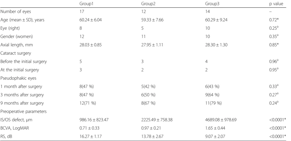
study, and 7 eyes underwent combined procedures with phacoemulsification and intraocular lens implantation at the initial surgery. The between-group differences regarding age, eye, gender, axial length and cataract surgery were not significant (Table 1). The preoperative parameters regarding age, eye, gender, axial length and cataract surgery had no significant effect in the surgical outcomes (Table 3).
Anatomical results
The mean IS/OS defect of all patients measured by OCT was 2537.62 ± 279.75 μm before surgery and 1120.11 ± 167.60μm at 9 months after surgery (p < 0.0001). More-over, the mean IS/OS defect after surgery decreased sig-nificantly from follow-up to follow-up in each group. The mean IS/OS defect of Group 1 before surgery was 986.16 ± 823.47μm, and this value decreased to 318.34 ± 481.72 μm at 9 months after surgery (p < 0.0001). In Group 2, the IS/OS defects before surgery and 9 months after surgery were 2225.49 ± 758.38 μm and 811.76 ± 465.70μm (p < 0.0001), respectively. The means for Group
3 were 4689.08 ± 978.69μm before surgery and 2357.98 ± 924.54μm 9 months after surgery (p < 0.0001) (Table 2).
Functional results
The mean logMAR BCVA of all patients was 1.09 ± 0.53 before surgery and 0.67 ± 0.29 at 9 months after surgery (p < 0.0001). Moreover, the mean BCVA improved significantly after surgery at all follow-ups in each group. The mean BVCA in Group 1 was 0.71 ± 0.33 before surgery and 0.43 ± 0.24 at 9 months after surgery (p < 0.0001). Before surgery, the mean BCVA of Group 2 was 0.97 ± 0.21, whereas this value 9 months after surgery was 0.71 ± 0.19 (p = 0.0002). In Group 3, the mean BCVA values before surgery and 9 months after surgery were 1.65 ± 0.44 and 0.91 ± 0.20 (p < 0.0001), respectively.
In Group 3, before surgery and 9 months after sur-gery, the mean RS values were 9.07 ± 2.07 and 12.69 ± 2.46 (p < 0.0001), respectively, and this difference was statistically significant (Table 2).
Closure Rates
At 9 months after surgery, 11 of 43 (25.6 %) eyes achieved IS/OS junction integrity; 9 (81.8 %) of these 11 eyes belonged to Group 1, 2 (18.2 %) belonged to Group
2. The closure rates were 52.9 % (9 of 17 eyes) in Group 1, 16.7 % (2 of 12 eyes) in Group 2 and 0 in Group 3.
Correlation between preoperative anatomy and postoperative function
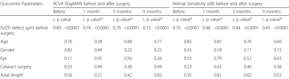
The preoperative IS/OS defect was significantly correlated with the postoperative BCVA and retinal sensitivity at 1 month after surgery (r = 0.71, p < 0.0001). The same result was also observed at 3 and 9 months after surgery (Table 3). Table 1Comparison of Demographic Characteristics of Each Group
Group1 Group2 Group3 p value
Number of eyes 17 12 14 –
Age (mean ± SD), years 60.24 ± 6.04 59.33 ± 7.66 60.29 ± 9.24 0.72*
Eye (right) 8 5 10 0.25a
Gender (women) 12 11 10 0.35a
Axial length, mm 28.03 ± 0.85 27.95 ± 1.11 28.30 ± 1.30 0.85*
Cataract surgery
Before the initial surgery 5 3 4 0.96a
At the initial surgery 3 2 2 0.95a
Pseudophakic eyes
1 month after surgery 8(47 %) 5(42 %) 6(43 %) 0.33a
3 months after surgery 8(47 %) 6(50 %) 9(64 %) 0.27a
9 months after surgery 12(71 %) 8(67 %) 11(79 %) 0.24a
Preoperative parameters
IS/OS defect,μm 986.16 ± 823.47 2225.49 ± 758.38 4689.08 ± 978.69 <0.0001*
BCVA, LogMAR 0.71 ± 0.33 0.97 ± 0.21 1.65 ± 0.44 <0.0001*
RS, dB 16.27 ± 1.17 13.78 ± 2.67 9.07 ± 2.07 <0.0001*
IS/OSphotoreceptor inner segment and outer segment,BCVAbest-corrected visual acuity,logMARlogarithm of minimal angle of resolution,RSretinal sensitivity. P value represents the difference between groups. P < 0.05 was considered statistically significant. *Kruskal-Wallis test.a
Fisher’s exact probability test
Table 2IS/OS defect, BCVA and RS Outcomes of 3 Groups Before and After Surgery
Outcomes Parameter
Before surgery 1 month after surgery 3 months after surgery 9 months after surgery
Mean ± SD Mean ± SD t, p Value* Mean ± SD t, p Value* Mean ± SD t, p Value*
Group1 (17 eyes)
IS/OS defect (μm) 986.16 ± 823.47 593.08 ± 697.79 5.02, < 0.0001 415.23 ± 561.58 5.25, < 0.0001 318.34 ± 481.72 5.08, < 0.0001
BCVA (logMAR) 0.71 ± 0.33 0.49 ± 0.25 4.41, 0.0004 0.45 ± 0.25 5.47, < 0.0001 0.43 ± 0.24 5.76, < 0.0001
RS (dB) 16.27 ± 1.17 17.23 ± 1.09 8.35, < 0.0001 17.47 ± 1.12 8.68, < 0.0001 17.62 ± 1.17 8.95, < 0.0001
Group2 (12 eyes)
IS/OS defect (μm) 2225.49 ± 758.38 1484.31 ± 423.94 5.99, < 0.0001 1097.06 ± 450.65 9.56, < 0.0001 811.76 ± 465.70 10.83, < 0.0001
BCVA (LogMAR) 0.97 ± 0.21 0.85 ± 0.16 2.36, 0.38 0.77 ± 0.23 5.87, 0.0001 0.71 ± 0.19 5.33, 0.0002
RS (dB) 13.78 ± 2.67 15.08 ± 2.32 5.95, < 0.0001 15.42 ± 2.19 6.17, < 0.0001 15.51 ± 2.27 6.34, < 0.0001
Group3 (14 eyes)
IS/OS defect (μm) 4689.08 ± 978.69 3257.98 ± 892.75 4.64, 0.0005 2671.43 ± 985.54 6.15, < 0.0001 2357.98 ± 924.54 7.07, < 0.0001
BCVA (LogMAR) 1.65 ± 0.44 1.29 ± 0.39 2.75, 0. 17 1.05 ± 0.23 5.10, 0.0002 0.91 ± 0.20 6.57, < 0.0001
Discussion
OCT has recently made it possible to explore changes in ocular layers as axial progresses and the globe is stretched. These findings consist of dehiscence of retinal layers known as retinoschisis, tractional ILM detachment, macular holes (lamellar and full thickness), posterior ret-inal detachment and choroidal neovascular membranes [10]. According to these OCT findings, patients in this study were separated into 3 groups.
This study showed that HMMH eyes without subret-inal fluid (Groups 1 and 2) obtained better postoperative anatomical and functional outcomes than eyes with sub-retinal fluid (Group 3). In addition, the HMMH patients with ERM traction, retinoschisis (Group 1) regained more visual function than patients with full-thickness macular holes (Group 2). This result suggests that subretinal fluid represents an advanced damage stage. Other research groups have also reported this observation [4–8]. In our study, Group 1 through 3 approximated HMMH at vary-ing severities stage in its natural course. Indeed, preopera-tive IS/OS defects, BCVA and RS all differed significantly among the three groups, providing clinical significance for the grouping of patients performed in our study.
The integrity of inner and outer segments of photore-ceptors (IS/OS) is closely related to visual recovery after successful MH closure surgery [11–15]. Visual acuity is generally used as a gold standard to indicate visual func-tion, although it represents only one aspect. In this study, microperimetry was applied to quantify foveal and perifoveal retinal sensitivity and correlate these results to the microstructural findings in HMMH [16, 17]. Specif-ically, we used the IS/OS defect as an anatomical param-eter and BCVA and retinal sensitivity (RS) as functional parameters to evaluate the relationship between micro-structure and function in the HMMH eyes that under-went surgery.
We found that in all 3 groups, the IS/OS defect decreased significantly as time progressed, indicating a
potential regeneration of photoreceptors. Similarly, BCVA and RS measurements gradually reached signifi-cant improvement after surgery. Interestingly, the pre-operative IS/OS defect was significantly correlated with both the preoperative and postoperative BCVA and RS values (Table 3). Although great care must be taken with small patient cohorts, our study revealed the potential prognostic value of the size of the IS/OS defect in conjunction with other known factors (e.g., retinal or RPE atrophy) and the preoperative diameters of macular holes.
Furthermore, 52.9 % (9 of 17 eyes) eyes in Group 1 approached postoperative closure, only 16.7 % (2 of 12 eyes) in Group 2 and 0 in Group 3 at 9 months after surgery. The minimum preoperative diameter of IS/OS defect in each group was 305.88 μm in Group 1, 1258.82μm in Group 2, and 2317.64μm in Group 3. As this parameter has showed a potential prognostic value, this result explained the significant differences in post-operative closure rates between groups.
Subretinal fluid is a generally regarded as a negatively prognostic complication of HMMH, because, in theory, chronic fluid may aggravate damage to photoreceptors. Assuming that subretinal fluid is a severity-dependent complication in the evolution of HMMH, it is even more important to perform effective surgery at a less damage stage for optimal postoperative functional recovery and restoration. Our study suggests that the appearance of ERM traction and consequent retinoschisis without sub-retinal fluid in HMMH, indicates a better prognosis for surgical intervention. Although some patients may show no subjective symptoms for years at this stage [18–20], visual acuity impairment should indicate surgery in the affected eye.
Based on its low complication rate, transconjunctival 23-gauge (23G) pars plana vitrectomy, as performed in our cases, was an effective treatment for HMMH. None-theless, our study lacks long-term results exceeding 9 months. Indeed, vitrectomy in myopic cases is Table 3Regression Analysis Between IS/OS Defect Before Surgery and BCVA, RS Before and After Surgery
Outcomes Parameters BCVA (logMAR) before and after surgery Retinal Sensitivity (dB) before and after surgery
Before 1 month 3 months 9 months Before 1 month 3 months 9 months
r, p value r, p value* r, p value* r, p value* r, p value* r, p value* r, p value* r, p value*
IS/OS defect (μm) before surgery
0.89 <0.0001 0.74 <0.0001 0.79 <0.0001 0.73 <0.0001 0.70 <0.0001 0.48 <0.0001 0.44 <0.0001 0.43 <0.0001
Age 0.76 0.28 0.88 0.77 0.85 0.85 0.76 0.69
Gender 0.82 0.49 0.23 0.25 0.35 0.18 0.11 0.13
Eye 0.11 0.95 0.56 0.26 0.53 0.79 0.52 0.63
Cataract surgery 0.33 0.94 0.38 0.49 0.23 0.43 0.46 0.38
Axial length 0.56 0.21 0.42 0.82 0.35 0.81 0.62 0.52
associated with a postoperative risk for retinal detach-ment and subsequent visual loss [21], which we fortu-nately did not observe within these 9 months. Generally, MH closure is less frequently observed following vitrec-tomy in HMMH compared to emmetropic cases [22]. These less favorable results may be explained by the presence of persistent macular traction because of con-tinued elongation and staphyloma formation in patho-logic myopia. Based on this hypothesis, other groups have suggested that an additional procedure, such as an episcleral macular buckle, may be useful to counteract the posterior traction [21, 23]. Although a variety of pro-cedures are available for HMMH, there is still no uni-fied standard treatment method. Therefore, additional longer-term studies are required.
ICG toxicity and damage to the retina has been reported in in vitro and in vivo studies, and following macular surgery. Toxic effects can occur to retinal glial cells, to the nerve fiber layer, to retinal ganglion cells, and to the optic nerve. ICG at concentrations higher than 1.25 % or application of the dye in air are very likely causing retinal damage [24]. In this study, a concentration of was applied in every case with a less than 30 seconds dyeing time to minimize the ICG toxicity as far as possible.
Our study has a number of limitations. First, none of the eyes in Group 3 and very few in Group 2 closed, whereas the postoperative BCVA were all significantly improved. This may result from the underestimation of the preoperative visual acuity due to cataract. The im-proved paracentral vision thanks to the regeneration of IS/OS structure as time progressed could also be a part of the explanation. Second, ICG toxicity and damage to the retina has been reported in in vitro and in vivo stud-ies, and following macular surgery. Toxic effects can occur to retinal glial cells, to the nerve fiber layer, to ret-inal ganglion cells, and to the optic nerve. ICG at con-centrations higher than 1.25 % or application of the dye in air are very likely causing retinal damage [24]. In this study, a concentration of was applied in every case with a less than 30 seconds dyeing time to minimize the ICG toxicity as far as possible. Third, this study lacks long-term results exceeding 9 months. Further prospective, long-term study would be needed for detection of further anatomical and functional changes.
Conclusions
In conclusion, pars plana vitrectomy combined with ILM peeling and gas tamponade results in limited functional outcomes in patients with HMMH. In HMMH, the appearance of subretinal fluid indicates a worse prognosis for surgical intervention.
Abbreviations
BCVA:best-corrected visual acuity; HMMH: high myopia macular hole; ICG: Indocyanine green; ILM: internal limiting membrane;
IS/OS: photoreceptor inner and outer segments; MTM: myopic traction maculopathy; MP1: Microperimetry 1; OCT: optical coherence tomography; RS: retinal sensitivity; FTMH: full-thickness macular hole; SD-OCT: spectral domain OCT; ERM: epiretinal membrane.
Competing interests
Dr. Ying Fan receives research support from the National Basic Research Program of China (973 Program) 2011CB707500. All authors declare that they have no competing interests.
Authors’contributions
QS carried out the collection, analysis and interpretation of data; participated in drafting the manuscript. HX was involved in data collection, analysis and revision of the draft. FH and XS participated in its design and coordination and helped to revise the draft. YF was responsible for the preliminary thoughts and analyses, original study design, coordination and revision of the manuscript. All authors read and approved the final manuscript.
Author details
1
Department of Ophthalmology, Shanghai First People’s Hospital, Haining Road 100, 200080 Shanghai, China.2Department of Ophthalmology, Charité-Universitätsmedizin, Berlin, Germany.3Department of Ophthalmology, The Royal Liverpool University Hospital, Liverpool, UK.
Received: 14 January 2015 Accepted: 31 July 2015
References
1. Panozzo G, Mercanti A. Optical coherence tomography findings in myopic traction maculopathy. Arch Ophthalmol. 2004;122:1455–60.
2. Takano M, Kishi S. Foveal Retinoschisis and Retinal Detachment in Severely Myopic Eyes With Posterior Staphyloma. Am J Ophthalmol. 1999;128:472–6. 3. Panozzo G, Mercanti A. Vitrectomy for myopic traction maculopathy. Arch
Ophthalmol. 2007;125:767–72.
4. Smiddy WE, Kim SS, Lujan BJ, Gregori G. Myopic traction maculopathy: spectral domain optical coherence tomographic imaging and a
hypothesized mechanism. Ophthalmic Surg Lasers Imaging. 2009;40:169–73. 5. Gaucher D, Haouchine B, Tadayoni R, Massin P, Erginay A. Long-term follow-up
of high myopic foveoschisis: natural course and surgical outcome. Am J Ophthalmol. 2007;143:455–62.
6. Ikuno Y, Tano Y. Early macular holes with retinoschisis in highly myopic eyes. Am J Ophthalmol. 2003;136:741–800.
7. Shimada N, Ohno-Matsui K, Baba T, Futagami S, Tokoro T. Natural course of macular retinoschisis in highly myopic eyes without macular hole or retinal detachment. Am J Ophthalmol. 2006;142:497–500.
8. Matsumura N, Ikuno Y, Tano Y. Posterior vitreous detachment and macular hole formation in myopic foveoschisis. Am J Ophthalmol. 2004;138:1071–3. 9. Hayashi K, Ohno-Matsui K, Shimada N, Moriyama M, Kojima A. Long-term
pattern of progression of myopic maculopathy: a natural history study. Ophthalmology. 2010;117:1595–611.
10. Hooshang F, Fedra H, Mohammad RE. Optical coherence tomographic findings in highly myopic eyes. J Ophthalmic Vis Res. 2010;5:110–21. 11. Inoue M, Watanabe Y, Arakawa A, Sato S, Kobayashi S. Spectral-domain
optical coherence tomography images of inner/outer segment junctions and macular hole surgery outcomes. Graefes Arch Clin Exp Ophthalmol. 2009;247:325–30.
12. Sano M, Shimoda Y, Hashimoto H, Kishi S. Restored photoreceptor outer segment and visual recovery after macular hole closure. Am J Ophthalmol. 2009;147:313–8.
13. Wakabayashi T, Fujiwara M, Sakaguchi H, Kusaka S, Oshima Y. Foveal microstructure and visual acuity in surgically closed macular holes: spectral-domain optical coherence tomographic analysis. Ophthalmology. 2010;117:1815–24.
14. Oh J, Smiddy WE, Flynn Jr HW, Gregori G, Lujan B. Photoreceptor inner/outer segment defect imaging by spectral domain OCT and visual prognosis after macular hole surgery. Invest Ophthalmol Vis Sci. 2010;51:1651–8. 15. Fujimoto S, Ikuno Y, Nishida K. Postoperative optical coherence
tomographic appearance and relation to visual acuity after vitrectomy for myopic foveoschisis. Am J Ophthalmol. 2013;156:968–73.
17. Chang LK, Koizumi H, Spaide RF. Disruption of the photoreceptor inner segment-outer segment junction in eyes with macular holes. Retina. 2008;28:969–75.
18. Baba T, Ohno-Matsui K, Futagami S, Yoshida T, Yasuzumi K. Prevalence and characteristics of foveal retinal detachment without macular hole in high myopia. Am J Ophthalmol. 2003;135:338–42.
19. Coppé AM, Ripandelli G, Parisi V, Varano M, Stirpe M. Prevalence of asymptomatic macular holesin highly myopic eyes. Ophthalmology. 2005;112:2103–9.
20. Ratiglia R, Osnaghi S, Bindella A, Pirondini C. Posterior traction retinal detachment in highly myopic eyes: clinical features and surgical outcome as evaluated by optical coherence tomography. Retina. 2005;25:473–8. 21. Ando F, Ohba N, Touura K, Hirose H. Anatomical and visual outcomes after
episcleral macular buckling compared with those after pars plana vitrectomy for retinal detachment caused by macular hole in highly myopic eyes. Retina. 2007;27:37–44.
22. Wu TT, Kung YH. Comparison of anatomical and visual outcomes of macular hole surgery in patients with high myopia vs. non-high myopia: a case–control study using optical coherence tomography. Graefes Arch Clin Exp Ophthalmol. 2012;250:327–31.
23. Chen YP, Chen TL, Yang KR, Lee WH, Kuo YH. Treatment of retinal detachment resulting from posterior staphyloma-associated macular hole in highly myopic eyes. Retina. 2006;26:25–31.
24. Gandorfer A, Haritoglou C, Kampik A. Toxicity of indocyanine green in vitreoretinal surgery. Dev Ophthalmol. 2008;42:69–81.
Submit your next manuscript to BioMed Central and take full advantage of:
• Convenient online submission
• Thorough peer review
• No space constraints or color figure charges
• Immediate publication on acceptance
• Inclusion in PubMed, CAS, Scopus and Google Scholar
• Research which is freely available for redistribution