An integrative taxonomic approach reveals a new
species of
Eranthis
(Ranunculaceae) in North Asia
Andrey S. Erst1,2, Alexander P. Sukhorukov3, Elizaveta Yu. Mitrenina2, Mikhail V. Skaptsov4, Vera A. Kostikova1, Olga A. Chernisheva5, Victoria Troshkina1, Maria Kushunina3, Denis A. Krivenko2,5, Hiroshi Ikeda6,
Kunli Xiang7,8, Wei Wang7,8
1 Central Siberian Botanical Garden, Siberian Branch of Russian Academy of Sciences, 101 Zolotodolinskaya Str., 630090, Novosibirsk, Russia 2 Tomsk State University, 36 Lenin Ave., 634050, Tomsk, Russia 3 Lomo-nosov Moscow State University, Leninskie Gory 1/12, 119234, Moscow, Russia 4 South-Siberian Botanical Garden, Altai State University, 61 Lenin Ave., Barnaul, 656049, Russia 5 Siberian Institute of Plant Physio-logy and Biochemistry, Siberian Branch of Russian Academy of Sciences, 132 Lermontov Str., 664033, Irkutsk, Russia 6 The University Museum, The University of Tokyo, Hongo 7-3-1, Bunkyo-ku, Tokyo 113-0033, Japan 7 State Key Laboratory of Systematic and Evolutionary Botany, Institute of Botany, Chinese Academy of Sciences, 100093, Beijing, China 8 University of Chinese Academy of Sciences, 19 Yuquan Road, Beijing, 100049, China
Corresponding author:Andrey S. Erst ([email protected])
Academic editor:M. Pellegrini | Received 3 December 2019 | Accepted 6 February 2020 | Published 4 March 2020
Citation: Erst AS, Sukhorukov AP, Mitrenina EYu, Skaptsov MV, Kostikova VA, Chernisheva OA, Troshkina V, Kushunina M, Krivenko DA, Ikeda H, Xiang K, Wang W (2020) An integrative taxonomic approach reveals a new species of Eranthis (Ranunculaceae) in North Asia. PhytoKeys 140: 75–100. https://doi.org/10.3897/phytokeys.140.49048
Abstract
A new endemic species, Eranthis tanhoensissp. nov., is described from the Republic of Buryatia and Irkutsk Province, Russia. It belongs to Eranthis section Shibateranthis and is morphologically similar to E. sibirica and E. stellata. An integrative taxonomic approach, based on cytogenetical, molecular and bio-chemical analyses, along with morphological data, was used to delimit this new species.
Keywords
Biochemistry, cytology, integrative taxonomic approach, morphology, phylogeny, Ranunculales, Russia http://phytokeys.pensoft.net
Copyright Andrey S. Erst et al. This is an open access article distributed under the terms of the Creative Commons Attribution License (CC BY 4.0), which permits unrestricted use, distribution, and reproduction in any medium, provided the original author and source are credited.
Introduction
The genus Eranthis L. (Ranunculaceae) consists of eight to ten species distributed in southern Europe and temperate Asia (Lee et al. 2012; Park et al. 2019). Most species have narrow distributions and only one European species, E. hyemalis (L.) Salisb., has been widely cultivated in gardens and become naturalised in Britain (Boens 2014) and North America (Parfitt 1997). Eranthis are perennial herbs with tuberous rhizomes, ba-sal long-petiolate leaves with the blades divided into several or many palmate segments (leaflets) that are entire or lobate; unbranched scapes carrying a solitary, bisexual and actinomorphic flower supported by three verticillate leaf-like bracts forming an invo-lucre; (4–)5–8 yellow, white or pink, caducous sepals; 5–10(–15) yellow or white, bifid petals shorter than sepals; nectaries located at the middle or upper part of the petals; > 10 stamens; and 3–10 follicles with several smooth seeds in each fruitlet (Parfitt 1997). All species are early-blooming plants, with anthesis from March to May (depending on the altitude), but E. hyemalis has been found at full anthesis in mid-January in gardens (Sukhorukov, pers. obs. in Mainz, Germany, 2019 and Leiden, Netherlands, 2020).
On the basis of morphology, the genus has been divided into two sections: E. sect.
Eranthis and E. sect. Shibateranthis (Nakai) Tamura (Tamura 1987). The type section
is characterised by annual tubers, yellow sepals and emarginate or slightly bilobate upper petal margins without swellings (nectaries), whereas the members of section
Shibateranthis have long-lived tubers, white sepals and bilobate or forked petal margins
with swellings (Tamura 1995). Molecular phylogenetic analysis. based on nrITS and chloroplast trnL-trnF interspacer region, supports the subdivision of the genus into these sections (Park et al. 2019). Furthermore, they are geographically separated, with section Eranthis occurring in Europe (E. hyemalis) and SW & W Asia (E. cilicica Schott & Kotschy, E. longistipitata Regel) and section Shibateranthis distributed in temperate N & E Asia (E. albiflora Franch., E. byunsanensis B.Y.Sun, E. lobulata W.T.Wang, E.
pinnatifida Maxim., E. pungdoensis B.U.Oh, E. sibirica DC. and E. stellata Maxim.:
Park et al. 2019). Two additional species with yellow sepals, E. bulgarica (Stef.) Stef. (Stefanoff 1963) and E. iranica Rukšāns & Zetterl. (Rukšāns and Zetterlund 2018), have been described from Bulgaria and Iran, respectively, but have not yet been in-cluded in molecular analysis.
Recent studies have revealed the genetic diversity, phylogeny and presumed origin of some narrowly distributed Korean and Japanese species with further conclusions about their taxonomic status (Lee et al. 2012; Oh and Oh 2019). The taxonomic and genetic diversity of Eranthis in the Asiatic part of Russia is insufficiently studied. To date, only two species have been found in Russia: E. sibirica and E. stellata (both belonging to sect. Shibateranthis) from South Siberia and Far East Russia (Malyshev 2005). High genetic polymorphism of E. sibirica across populations near Baikal Lake was discovered only recently (Protopopova et al. 2015) and this fact has inspired us to conduct a new study of Eranthis in the Asiatic part of Russia.
wheth-er any undescribed species wwheth-ere present thwheth-ere. The relationship between E. sibirica, E.
stellata and a new species, described and named below as Eranthis tanhoensis Erst, sp.
nov. is explored here.
Materials and methods
Plant material
More than 300 herbarium specimens were collected during field investigations in the Republics of Khakassia and Buryatia and the Irkutsk Province during 2018 and 2019. Fieldwork was conducted during different seasons to observe the species in both their flowering and fruiting stages. The specimens were deposited in the E and NS her-baria (herbarium abbreviations according to Thiers 2019+). Revision of herbarium materials was undertaken in the herbaria at IRK, LE, MW, NS, NSK, PE, VBGI and VLA. Drawings of the new species, Eranthis tanhoensis, are based on images of the type specimen (NS-0000948!) and paratype (NS-0000949!). The flowering and fruiting times and habitats are provided as cited on the collectors’ labels. Maps of records were made with SimpleMappr (http://www.simplemappr.net). Conservation analysis was performed using criteria from the International Union for the Conservation of Nature (IUCN 2019). The Extent of Occurrence (EOO) and Area of Occupancy (AOO) of each species were estimated using GeoCat (Bachman et al. 2011).
Molecular analysis
We sampled 15 individuals of E. tanhoensis and six of E. sibirica. Two individuals of E.
stellata and one each of E. pinnatifida and E. longistipitata were also included. The details
option using 1000 replicates. BI analysis was conducted in MrBayes v3.2.1 (Ronquist et al. 2012). Data partitioning and nucleotide substitution models were determined using PartitionFinder 2.1.1 (Lanfear et al. 2016). Two independent analyses, consist-ing of four Markov Chain Monte Carlo chains were run, samplconsist-ing one tree every 1000 generations for 10 million generations. Runs were completed when the average standard deviation of split frequencies reached 0.01. The stationarity of the runs was assessed us-ing Tracer v1.6 (Rambaut et al. 2014). After removus-ing the burn-in period samples (the first 25% of sampled trees), a majority rule (> 50%) consensus tree was constructed.
Morphological analysis
The morphology of vegetative and reproductive structures was examined on well-de-veloped specimens. For numerical analysis, 25 specimens at flowering and 25 speci-mens at fruiting stages were examined for each species (more than 150 specispeci-mens altogether). For each species, we studied different populations from across the range, including populations from the type localities of E. stellata and E. sibirica. As E. stellata
often does not produce basal leaves at flowering, we studied this character in a limited number of samples. The morphological characters were measured using AxioVision 4.8 software (Carl Zeiss, Munich, Germany).
The missing values in the original data table were restored using multidimensional linear regression, in accordance with recommendations of Myers (2000) and Lee and Carlin (2010). A one-way analysis of variance (ANOVA), according to Chambers et al. (1992), was used to identify the distinguishing morphometric features of each species. The differences were considered significant at P-value < 0.05. As multiple statistical test-ing was performed, the calculated P-value was adjusted ustest-ing the procedure proposed by Benjamini and Hochberg (1995). The principal component analysis was used to visu-alise the distribution of the analysed individuals over the space of morphometric char-acters. This method was employed only for those characters that displayed significant intergroup differences, according to the results of the ANOVA. For scale adjustment, the logarithmic transformation of data was used. The results of the principal component analysis were visualised using the Factoextra package (Kassambara and Mundt 2017).
Cytogenetic analysis
Somatic chromosomes were studied in root tip cells. Tubers were germinated in wet moss at ~15 °C for 2–4 weeks. Newly formed 1–2 cm long roots were excised and pretreated in a 0.5% colchicine solution for 2–3 h at 15 °C. Roots were fixed in a mixture of 96% ethanol and glacial acetic acid (3:1). Root tips were stained with 1% aceto-haematoxylin and the squash method was employed for investigation of the karyotype (Smirnov 1968).
Zeiss, Munich, Germany) and photographed using an Axio Imager A.1 microscope (Carl Zeiss, Germany) with AxioVision 4.7 software (Carl Zeiss, Germany) and Ax-ioCam MRc5 CCD–camera (Carl Zeiss, Germany) at 1000× magnification in the Laboratory for Ecology, Genetics and Environmental Protection (Ecogene) of the Na-tional Research Tomsk State University. KaryoType software (Altinordu et al. 2016) was used for karyotyping, whereas Adobe Photoshop CS5 (Adobe Systems, USA) and Inkscape 0.92 (USA) were used for image editing. Karyotype formulae were based on measurements of mitotic metaphase chromosomes taken from photographs. The meas-urements were performed on 5–10 metaphase plates. The symbols used to describe the karyotypes followed those of Levan et al. (1964): m = median centromeric chromo-some with arm ratio of 1.0–1.7 (metacentric chromochromo-some); sm = submedian cen-tromeric chromosome with arm ratio of 1.7–3.0 (submetacentric chromosome); st = subterminal centromeric chromosome with arm ratio of 3.0–7.0 (subtelocentric chro-mosome); t = terminal centromeric chromosome with arm ratio of 7.0–∞ (acrocentric chromosome); T = chromosome without obvious short arm, i.e. with arm ratio of ∞.
Flow cytometry
Flow cytometry with propidium iodide (PI) staining was used to determine the abso-lute DNA content. The relative DNA content in the nucleus (C-value) in representa-tives of three Eranthis species – E. stellata, E. sibirica and E. tanhoensis from different populations, was determined in this study. In total, more than 70 samples from 15 populations were studied (see Suppl. material 1: Table S1). Silica gel-dried leaf material (0.5–1.0 cm2) was chopped with a sharp razor blade in a 1 ml cold nuclei extraction buffer composed of 50 mM Hepes, 10 mM sodium metabisulphite, 10 mM MgCl2, 0.5% polyvinylpyrrolidone, 0.1% bovine serum albumin, 0.3% Tween20, 0.2% Triton X-100, 50 μg/ml RNase, 1 μg/ml β-mercaptoethanol and 50 μg/ml propidium iodide (PI). The samples were filtered through 50 μm nylon membranes into sample tubes and incubated in the dark at 4 °C for 15 min. Samples were measured using a Partec CyFlow PA flow cytometer equipped with a green laser, at 532 nm wavelength. The ab-solute nuclear DNA content, the 2C-value according to Greilhuber et al. (2005), was calculated as the ratio of the mean fluorescence intensity of the nuclei of the sample to that of an external standard multiplied by the total nuclear DNA content of the stand-ard. The possible effect of secondary metabolites on the binding of the intercalating dye was evaluated by measuring the fluorescence of Allium fistulosum L. leaf samples prepared as described above, but with the addition of the supernatant from Eranthis
samples, centrifuged without PI. The samples were measured three times at 10 min intervals. If no variation in the average values of the detection channels was observed for the A. fistulosum peak, the effect of secondary metabolites was considered negligible.
L. ‘Inovec’ 2С = 26.90 pg (Doležel et al. 1992; Skaptsov et al. 2016). We used the Statistica 8.0 software (StatSoft, Inc.), Flowing Software 2.5.1 (Turku Centre for Bio-technology) and CyView software (Partec, GmbH) for data analyses. Flow cytometry was performed at the Laboratory for Bioengineering of the South-Siberian Botanical Garden, Altai State University (Barnaul, Russia).
High-performance liquid chromatography (HPLC) analysis of individual phenolic compounds in ethanol leaf extracts
In order to determine the composition of phenolic compounds, air-dried plant material was mechanically ground to obtain a homogenous powder and then samples of ~0.2 g were extracted three times using 70% aqueous ethanol solution for 30 min in a water bath at 72 °C. Next, the combined extract was concentrated in porcelain dishes to 5 ml. The solutions were filtered and stored at 4 °C until analysis. Analysis of phenolic com-ponents was performed using an Agilent 1200 HPLC system equipped with a diode array detector and a ChemStation system for the collection and processing of chromato-graphic data (Agilent Technology, Palo Alto, CA, USA). The separation was performed on a Zorbax SB-C18 column (5 μm, 4.6 × 150 mm) at 25 °C. The methanol content of the mobile phase in an aqueous solution of phosphoric acid (0.1%) varied from 50–52% over 56 min (van Beek 2002). The eluent flow rate was 1 ml/min. Detection wavelengths were 255, 270, 340 and 360 nm. Groups of phenolic substances were identified by their spectral characteristics (Bate-Smith 1962; Mabry et al. 1970). For identification of the phenolic components in plant extracts, standard samples of salicylic and chlorogenic acids, quercetin, kaempferol, orientin (Sigma-Aldrich Chemie GmbH, Munich, Ger-many), gentisic and caffeic acids (Serva Heidelberg, GerGer-many), hyperoside and vitexin (Fluka Chemie AG, Buchs, Switzerland) were used. The samples were analysed twice.
Results and discussion
Molecular phylogenetic analysis
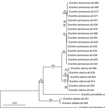
Bayesian and ML analyses of the combined dataset produced highly consistent topolo-gies. Eranthis sibirica and the new species E. tanhoensis formed a sister clade of that of
E. pinnatifida. The monophyly of each species, E. tanhoensis sp. nova, E. sibirica and E.
stellata, was strongly supported (Fig. 1).
Morphological analysis
Figure 1. ML tree inferred from the combined cpDNA and ITS data. The numbers above branches are bootstrap values (BS > 50%) and numbers under branches are Bayesian posterior probabilities (PP > 0.50).
E. stellata (Suppl. material 1: Table S2). The basal and involucral leaves in Eranthis spp.
undergo changes at fruiting and, for this reason, the lengths of all leaves, their segments and segment lobes were measured both at the flowering and fruiting stages. In Suppl. material 1: Table S2, an asterisk (*) indicates the characters used in the numerical analy-sis. An ANOVA was conducted only for quantitative characteristics. As basal leaves are often absent at the time of flowering and there were no samples with basal leaves in herbarium collections, there were limited data on these characteristics of E. stellata.
and in 9 out of 13 parameters at fruiting. The principal component analysis revealed that the first two main components accounted for 83.1% and 81.8% of the variance in the entire data array of the parameters measured at flowering and fruiting, respectively and showed the best species discrimination. The highest variability of morphometric characters was found at flowering in E. sibirica (Fig. 2A) and at fruiting in E. tanhoensis
(Fig. 2B). As signified by the directions of the vectors indicating the gradients in the character values, at flowering, E. sibirica differed from E. tanhoensis by having lower values for characters (18), (22), (24) and (31) and a higher value for character (9). At fruiting, E. sibirica was characterised by having lower values for parameters (19), (17), (23) and (25) and higher values for parameters (10), (40) and (29), in comparison with those of E. tanhoensis. E. sibirica differed from E. stellata by having higher values for characters (1), (16), (29), (30) and (32) at flowering and (10) and (14) at fruiting. The pattern of overlap between the species differed between flowering and fruiting plants. For instance, E. tanhoensis was reliably distinguished from E. sibirica only at fruiting (the ellipses enclosing the samples did not overlap; Fig. 2B). In addition to numerical parameters, the new species was also distinguished by qualitative characters.
Cytogenetic analysis
Table 1. Chromosome numbers (2n), ploidy level (nx), karyotype formulas, and C-values (C ± SD) of the three studied Eranthis species.
Voucher
number Species Voucher information 2n nx Karyotype formulae 2C±SD, pg 1Cx±SD, pg
1 E. sibirica Republic of Khakassia, Bolshoi
On river 28 4x 2n2m/sm + 6 sm = 20m (2 sat) + 38.83±1.03 9.71±0.26 2 E. sibirica Irkutsk Province, Kuitun river 28 4x 2n = 20m (2 sat) +
2m/sm + 6 sm 38.19 0.28± 9.55 ± 0.14 3 E. sibirica Irkutsk Province, Slyudyanka
river 42 6x 2n = 30m + 12sm (2 sat) 55.75±0.28 9.23±0.14 4 E. sibirica Irkutsk Province, Burovschina
river 42 6x 2n = 30m + 12sm (2 sat) 55.76±0.47 9.27±0.23 5 E. sibirica Irkutsk Province, Utulik river 42 6x 2n = 30m + 12sm
(2 sat) 55.31±0.45 9.22±0.25 6 E. tanhoensis Irkutsk Province, Mamai river 14 2x 2n = 10m (2sat) +
4sm 24.88±0.54 12.44±0.27 7 E. tanhoensis Republic of Buryatia, Duliha
river 14 2x 2n = 10m (2sat) + 4sm 24.97±0.43 12.49±0.22 8 E. tanhoensis Republic of Buryatia,
Tolbazikha river 14 2x 2n = 10m (2sat) + 4sm 24.77±0.52 12.38±0.26 9 E. tanhoensis Irkutsk Province, Malye
Mangaly river 14 2x 2n = 10m (2sat) + 4sm + 0–8B 24.15±0.11 12.07±0.06 10 E. tanhoensis Irkutsk Province, Semirechka
river 14 2x 2n = 10m (2sat) + 4sm 25.31±0.15 12.41±0,29 11 E. tanhoensis Republic of Buryatia,
Osinovka river (Tanhoi village) 14 2x 2n = 10m (2sat) + 4sm 25.11±0.32 12.56±0.16 12 E. tanhoensis Republic of Buryatia, Mishiha
river 14 2x 2n = 10m (2sat) + 4sm + 0–4B 25.25±0.15 12.07±0.07 13 E. tanhoensis Republic of Buryatia,
Shestipalikha river 14 2x 2n = 10m (2sat) + 4sm 25.53±0.18 12.77±0.09 14 E. stellata Primorsky Krai, Vladivostok,
Studencheskaya railway station 16 2x 24sm (2sat) + 2tn = 16 = 10m + 31.76±0.61 15.88±0.31
15 E. stellata Primorsky Krai, Malaya
Sedanka river 16 2x 24sm (2sat) + 2tn = 16 = 10m + 31.88±0.67 15.94±0.34
16 E. stellata Primorsky Krai, “13th
km” railway station 16 2x 24sm (2sat) + 2tn = 16 = 10m + – – 17 E. stellata Primorsky Krai, Russkiy Island 16 2x 2n = 16 = 10m +
4sm (2sat) + 2t 28.47±0.46 14.23±0.23
Eranthis sibirica. Two cytotypes, with basic chromosome number x = 7, were
re-vealed. Eranthis sibirica from the Republic of Khakassia (1) and Irkutsk Province (2) were tetraploid with 2n = 4x = 28 (Fig. 3A, B). Three populations from the Irkutsk Province (3, 4 and 5) were hexaploid with 2n = 6x = 42 (Fig. 3C). Metacentric and submetacen-tric chromosome types were present in all examined E. sibirica specimens. The karyo-type formula of tetraploid plants was 2n = 20m(2sat) + 2m/sm + 6sm and 2n = 30m + 12sm(2sat) in hexaploid plants. No B chromosomes were identified in this species.
Eranthis tanhoensis. We determined the chromosome numbers in specimens of eight
of B chromosomes. The maximum number of B chromosomes appeared to be eight (9). B chromosomes in this species were represented by two types: small (2.3–2.5 μm) metacentrics and dot-shaped 1.3–1.5 μm long chromosomes, which were obviously acrocentric. The karyotype formula of E. tanhoensis was 2n = 10m (2sat) + 4sm + 0–8B.
Eranthis stellata. In all four studied populations of E. stellata, the basic chromosome
number was x = 8. This species was diploid with 2n = 2x = 16, which is typical of the genus (Table 1; Fig. 3F). Five pairs of chromosomes were metacentric, two pairs were submetacentric and one pair was acrocentric (Fig. 3F). The karyotype formula of E. stel-lata was 2n = 10m + 4sm (2sat) + 2t. No B chromosomes were observed in this species. The basic chromosome number x = 8 has been reported for the entire genus Eranthis
(Langlet 1932; Kurita 1955; Tak and Wafai 1996; Gömürgen 1997; Yuan and Yang 2006; Kim et al. 2011; Marhold et al. 2019). Our results are consistent with previously pub-lished data (Yuan and Yang 2006), with insignificant differences in the karyotype formula. However, we showed, for the first time, that E. sibirica and E. tanhoensis are distinguished from other species of the genus by the basic chromosome number x = 7. Such differences in basic chromosome numbers (x = 7 and x = 8) have been found in some other genera of Ranunculaceae, for example, Anemone L. and Ranunculus L. (Rice et al. 2015). Our results regarding the chromosome numbers in E. sibirica (2n = 28 and 2n = 42) differed from the data reported by other researchers for this species (2n = 32: Krogulevich (1976) or 2n = 16: Gnutikov et al. (2016, 2017)). Eranthis tanhoensis was found to have 2n = 14. Based on the incongruence of the chromosome data with previous and recent analyses, we assume that some populations of E. sibirica and E. tanhoensis may have diverse cytotypes. Both species clearly differed from E. stellata by the absence of acrocentrics. All three species were char-acterised by five metacentrics and two submetacentrics per monoploid chromosome set.
Flow cytometry
The average absolute DNA content of hexaploid samples of E. sibirica was 2C = 55.33 ± 0.52 pg and that of tetraploid samples was 2C = 38.19 ± 0.28 pg. In diploid E.
tan-hoensis, the average absolute DNA content was 2C = 25.02 ± 0.28 pg. The average
absolute DNA content of diploid E. stellata was 2C = 31.47 ± 0.46 pg. The monoploid DNA content of the E. sibirica cytotypes was similar: 1Cx = 9.55 ± 0.14 pg in tetra-ploids and 1Cx = 9.25 ± 0.20 pg in hexaploids. The monoploid DNA content of E.
tanhoensis was 1Cx = 12.49 ± 0.16 pg and that of E. stellata was 1Cx = 15.77 ± 0.20 pg.
HPLC analysis of individual phenolic compounds
Phenolic compounds are often used in chemotaxonomic studies owing to their wide dis-tribution in plants, structural diversity and chemical stability (Braunberger et al. 2015; Radušienė et al. 2018). They have also been reported as promising chemotaxonomic markers for Ranunculaceae (Hao 2018). However, data on the significance of these substances for the taxonomy of Eranthis is still insufficient. Only a few studies of the phytochemical characteristics of certain Eranthis species, considered as medicinal plants, have been published (Djafari et al. 2018; Hao 2018; Watanabe et al. 2003, 2019).
Twenty four phenolic compounds were detected in 70% ethanol extracts of plant leaves of the three Eranthis species (E. sibirica, E. stellata and E. tanhoensis) using HPLC (Fig. 4). Phenolic acids (chlorogenic, gentisic, caffeic and salicylic acids), flavonols (quercetin, kaempferol and hyperoside) and flavones (orientin and vitexin) were identi-fied amongst these compounds. All three species were very similar in the composition of the phenolic compounds extracted from their leaves; however, there were specific com-pounds for each taxon. The common comcom-pounds present in all studied plants were chlo-rogenic acid, phenolic acids (Fig. 4, peak 3: tR, min = 10.0, λmax, nm = 250, 290 sh, 335; peak 12: tR, min = 20.9, λmax, nm = 240, 290 sh, 335 and peak 23: tR, min = 44.3, λmax, nm = 255, 300, 330) and flavonols (Fig. 4, peak 9: tR, min = 15.1, λmax, nm = 255, 360 and peak 15: tR, min = 32.7, λmax, nm = 270, 310, 365). Almost all plants contained
ferol, phenolic acids (Fig. 4, peak 14: tR, min = 25.4, λmax, nm = 255, 300 and peak 16: tR, min = 34.5; λmax, nm = 250, 290, 330) and flavonols (Fig. 4, peak 13: tR, min = 22.3, λmax, nm = 255, 360 and peak 18: tR, min = 38.7, λmax, nm = 255, 305, 360). Eranthis sibirica
leaves contained about 15 to 20 phenolic compounds, whereas, in E. stellata leaves, their number varied from 16 to 18. Phenolic compounds were less diverse in E. tanhoensis
leaves than in leaves of other species, whose numbers varied from 13 to 16 substances. The chromatographic profile of E. sibirica differed from that of E. tanhoensis in the presence of caffeic acid, orientin, vitexin and flavone (peak 6: tR, min = 9.4, λmax, nm = 270, 310) in 70% ethanol leaf extracts (Fig. 4). Caffeic acid, orientin and flavone (peak 6) were generally absent from leaves of E. tanhoensis, whereas vitexin was found in some sam-ples in trace amounts. The leaves of E. tanhoensis from almost all the studied populations contained quercetin, which was not detected in E. sibirica. Distinguishing compounds in leaf extracts of E. stellata were gentisic acid, phenolic acid (Fig. 4, peak 4: tR, min = 7.1, λmax, nm = 250, 300) and flavone (Fig. 4, peak 21: tR, min = 42,2; λmax, nm = 210, 310), which were absent from the two other species. Vitexin, hyperoside and salicylic acids were not found in E. stellata leaves. All samples of E. stellata contained orientin and caffeic acid, which were characteristic of E. sibirica and quercetin, which was typical of E. tanhoensis.
Taxonomy
The analysis of the data presented above allowed us to distinguish a new species from specimens previously identified as E. sibirica.
Eranthis tanhoensis Erst, sp. nov.
urn:lsid:ipni.org:names:77206949-1
Figs 5, 6A–D, 7B
Type. Russia, Republic of Buryatia, Kabansky district, Osinovka River near Tanhoi village, 51°33'06.2"N, 105°05'34.7"E, 458 m a.s.l., 01 May 2019, A.S. Erst, D.A.
Krivenko, & O.A. Chernysheva s.n. (holotype, NS-0000948!, isotypes TK, IRK, E).
Figure 6. Morphological differences amongst A–DEranthis tanhoensisE–HEranthis sibirica; and I–L Er-anthis stellataA, E, I flower position B, F, J flowers C, G, K involucral bracts and follicles D, H, L basal leaves.
at flowering and fruiting stages; each segment with 5–21 teeth (at both flowering and fruiting stages), acute at the apex. Pedicels 0.5–1.5 cm long, elongated in fruiting (3.5– 5.5 cm long), densely covered with papillate and large hemispherical trichomes. Flowers
bisexual, actinomorphic, solitary, erect, 2–4 cm diam. Sepals 4–7, deciduous in fruit, white or light pink at margin, flat, narrowly obovate or elliptic, 1.1–2.6 × 0.5–1.3 cm.
Petals 5–15 × 0.6–0.8 cm long, bicoloured, white, tubular, two-lipped with bilobate or
forked lips, each lobe of abaxial lip acute at the apex and with globular yellow swellings (nectaries: Fig. 7B). Stamens 36–45, 0.7–1.1 cm long; filaments filiform, white; an-thers white. Follicles 3–10, 0.8–1.4 cm long, on short (0.3–0.5 mm) stalks, divergent towards the end of fruiting; stylodium 0.1–0.3 mm long, straight or slightly curved.
Notes. Turczaninow (1842) described the species E. uncinata Turcz., growing at higher altitudes and distinguished from E. sibirica by the number of petals (5–6, not strictly 5), by the shape of the stylodium (recurved rather than straight), smaller flow-ers and more dissected leaf blades. However, our studies have shown that these mor-phological characters are variable and all variations can be found both in the foothill and alpine plants. Shipchinskiy (1937) merged E. uncinata with E. sibirica. However, he described two varieties: E. sibirica DC. var. nuda Schipcz. with glabrous pedicels
(= E. sibirica var. sibirica) and E. sibirica DC. var. glandulosa Schipcz. with
glandular-pubescent pedicels. These varieties were not validly published under ICN Article 39.1 (Turland et al 2018). Nakai (1937) attributed E. sibirica and E. uncinata to the genus
Schibateranthis Nakai (≡Eranthis sect. Schibateranthis (Nakai) Tamura).
Affinity. The new species belongs to E. sect. Shibateranthis (Nakai) Tamura and it is sister to E. sibirica, according to the results of molecular phylogenetic analysis (Fig.
1). E. tanhoensis is morphologically similar to E. sibirica and E. stellata (Figs 5–9) in
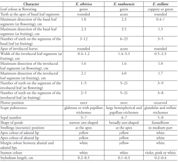
having white sepals, tubular two-lipped petals with bilobate or forked lips, apically acute lobes with abaxial lip and globular yellow swellings (nectaries) at the top or in the central part. The differences amongst the three species are presented in Table 2.
The new species differs from other related species by dense glandular pubescence of the flower stems, rounded or widely rhombic (not obovate or lanceolate) leaf blade seg-ments, acute, rather than rounded teeth apices of the basal and stem leaves, a large num-ber of teeth and width of the segments of the basal and stem leaves (see also 2). Addition-ally, all three species growing in Russia have different distribution patterns (Figs 10, 11).
Phenology. Flowering time: April–early May; fruiting time: late May–June.
Distribution (Fig. 10): Eranthis tanhoensis is endemic to southern Baikal (Khamar-Daban range of the Republic of Buryatia and Irkutsk Province).
Habitat and ecology.Eranthis tanhoensis can be found at 350–2400 m a.s.l., where it grows in fir, Siberian pine, spruce and birch forests, on riverbanks, beside streams (up to 1500 m a.s.l.) and in subalpine meadows (at higher altitudes).
Etymology. The specific epithet of the new species is derived from the type local-ity, Tanhoi village, Republic of Buryatia, Russia.
(NS-0000949!); Kabansky district, Mishikha river, 51°37'46.7"N, 105°32'05.2"E, 480 m a.s.l., 01 May 2019, A.S. Erst, D.A. Krivenko & O.A. Chernysheva 31 (NS-0000950!); Kabansky district, Mishikha river, 51°37'46.7"N, 105°32'05.2"E, 480 m a.s.l., 01 May 2019, A.S. Erst, D.A. Krivenko & O.A. Chernysheva 31a (NS-0000951!); Kabansky district, Mishikha river, 51°37'32.6"N, 105°32'03.4"E, 478 m a.s.l., 20 Jun 2019, A.S.
Erst, D.A. Krivenko, E.Yu. Mitrenina & O.A. Chernysheva s.n. (NS-0000952!); Kabansky
district, Dulikha river, 51°32'04.9"N, 105°01'43.2"E, 461 m a.s.l., 01 May 2019, A.S.
Erst, D.A. Krivenko & O.A. Chernysheva 14 (NS-0000953!); Kabansky district, Dulikha
river, 51°32'04.9"N, 105°01'43.2"E, 461 m a.s.l., 20 Jun 2019, A.S. Erst, D.A. Krivenko,
E.Yu. Mitrenina & O.A. Chernysheva (NS-0000954!); Kabansky district, Shestipalikha
river, 51°32'46.4"N, 105°04'28.9"E, 465 m a.s.l., 01 May 2019, A.S. Erst, D.A.
Krivenko & O.A. Chernysheva s.n. (NS-0000955!); Kabansky district, Shestipalikha river,
51°32'46.4"N, 105°04'28.9"E, 465 m a.s.l, 21 Jun 2019, A.S. Erst, D.A. Krivenko, E.Yu.
Mitrenina & O.A. Chernysheva (NS-0000956!); Kabansky district, Tolbazikha river,
Table 2. Morphological differences among E. sibirica, E. tanhoensis, and E. stellata.
Character E. sibirica E. tanhoensis E. stellata
Leaf colour at flowering green green coppery or green
Teeth at the apex of basal leaf segments rounded acute rounded Maximum dissection of the basal leaf
segments (at flowering), cm 1.0 2.3 0.4–?
Maximum dissection of the basal leaf
segments (at fruiting), cm 2.3 3.5 1.3
Number of teeth on the segments of the
basal leaf (at fruiting) 3–12 6–25 3–5
Apex of involucral leaves rounded acute rounded
Width of the involucral leaf segments (at
fruiting), cm 0.4–1.2 1.4–5.3 0.5–2.3
Maximum dissection of the involucral
leaf segments (at flowering), cm 1.6 1.6 1.0
Maximum dissection of the involucral
leaf segments (at fruiting), cm 2.1 4.0 1.7
Number of teeth on the segments of the
involucral leaf (at flowering) 1–5 5–21 3–9
Number of teeth on the segments of the
involucral leaf (at fruiting) 2–5 5–21 3–8
Flower position erect erect recurved
Scape pubescence glabrous or with papillate
trichomes large hemispherical and papillate trichomes glandular and stellate trichomes
Sepal number 5–7 4–7 5–8
Shape of petals narrow urn-shaped broadly urn-shaped funnelform Swellings (nectaries) position at the apex at the apex in medium part
Apex colour of adaxial lip yellow yellow white
Apex colour of abaxial lip yellow yellow white
Margin colour between abaxial and
adaxial lips white yellow white
Stamen colour white white violet, pink or white
Figure 10. General distribution of Eranthis sibirica (dots) and E. tanhoensis (stars), based on herbarium materials.
51°26'21.06"N, 104°41'09.82"E, 471 m a.s.l., 02 May 2019, A.S. Erst, D.A.
Krivenko & O.A. Chernysheva s.n. (NS-0000957!); Kabansky district, Tolbazikha river,
51°26'21.06"N, 104°41'09.82"E, 471 m a.s.l., 20 Jun 2019, A.S. Erst, D.A. Krivenko,
E.Yu. Mitrenina & O.A. Chernysheva s.n. (NS-0000958!); Irkutsk Province: Slyudyansky
district, Semirechka river, 51°28'56.92"N, 104°19'43.47"E, 470 m a.s.l., 02 May 2019,
A.S. Erst, D.A. Krivenko & O.A. Chernysheva 048 (NS-0000959!); Slyudyansky district,
Semirechka river, 51°28'56.92"N, 104°19'43.47"E, 470 m a.s.l., 21 Jun 2019, A.S. Erst,
D.A. Krivenko, E.Yu. Mitrenina & O.A. Chernysheva s.n. (NS-0000960!).
Preliminary conservation status. Although the species seems to have a small dis-tribution area in southern Baikal Lake, the populations observed in 2018 and 2019 consisted of numerous individuals producing viable fruits and no threats to the habi-tats were observed in the field studies. The EOO of E. tanhoensis was estimated for an area of more than 1372 km2, while the AOO was 72 km2. Preliminary conservation status, according to IUCN’s Extent of Occurrence criteria indicates the species as En-dangered (EN) (IUCN 2019).
Key to the Eranthis species growing in Asiatic Russia
1 Maximum dissection of basal leaf segments ~0.4 cm long at flowering stage, 1.3 cm long at fruiting stage; scape with stellate hairs; involucral leaves green or coppery at flowering; maximum dissection of the involucral leaves 1.7 cm long at fruiting; flowers recurved; petals narrowly funnelform, swellings (nec-taries) located in medium part of adaxial lip lobes, apex of abaxial and adaxial lips white; anthers violet, pink or white ...E. stellata – Maximum dissection of basal leaf segments at least 1.0 cm long at flowering,
2.3 cm long at fruiting stage; scape without stellate hairs; involucral leaves green at flowering; maximum dissection of the involucral leaves 2.1 cm long or more at fruiting; flowers erect, petals urn-shaped, swellings (nectaries) lo-cated at the apex of adaxial lip lobes, apex of abaxial and adaxial lips yellow; anthers white ...2
2 Apex of basal and involucral leaves rounded; maximum dissection of basal leaf segments 1.0 cm long at flowering and 2.3 cm long at fruiting; segments of involucral leaves at fruiting 0.4–1.2 cm wide; maximum dissection of the involucral leaves at fruiting 2.1 cm long; each segment of involucral leaves with 1–5 teeth; scape glabrous or papillate; petals narrowly urn-shaped, mar-gins between abaxial and adaxial lips white ...E. sibirica – Apex of basal and involucral leaves acute; maximum dissection of basal leaf
Acknowledgements
We thank Mark Newman, Marco Pellegrini, Andriy Novikoff, Colin Pendry, Christoph Dobeš and Johannes Walter for discussion of some parts of the manu-script and valuable comments, the staff of the herbaria visited, as well as Valentin Yakubov for the images of Eranthis stellata and Roman Annenkov for preparing Fig. 3. We are indebted to Natalya Pridak for all the black and white drawings. The samples of E. longistipitata were kindly provided by Evgeny Boltenkov. The research was supported by the Russian Foundation for Basic Research, grant 18-34-20056 mol_a_ved. The work of Alexander Sukhorukov and Maria Kushunina was also supported by a Moscow State University (MSU) Grant for Leading Sci-entific Schools “Depository of the Living Systems” in the framework of the MSU Development Programme.
References
Altinordu F, Peruzzi L, Yu Y, He X (2016) A tool for the analysis of chromosomes: KaryoType. Taxon 65(3): 586–592. https://doi.org/10.12705/653.9
Bachman S, Moat J, Hill A, de la Torre J, Scott B (2011) Supporting Red List threat assess-ments with GeoCAT: Geospatial conservation assessment tool. ZooKeys 150: 117–126.
https://doi.org/10.3897/zookeys.150.2109
Bate-Smith EC (1962) The phenolic constituents of plants and their taxonomic significance I. Dicotyledons. Journal of the Linnean Society of London. Botany 58(371): 95–173.
https://doi.org/10.1111/j.1095-8339.1962.tb00890.x
Benjamini Y, Hochberg Y (1995) Controlling the false discovery rate: A practical and powerful approach to multiple testing. Journal of the Royal Statistical Society. Series B. Methodo-logical 57(1): 289–300. https://doi.org/10.1111/j.2517-6161.1995.tb02031.x
Boens W (2014) The genus Eranthis, heralds of the end of winter! International Rock Gardener 49: 1–24.
Braunberger C, Zehl M, Conrad J, Wawrosch C, Strohbach J, Beifuss U, Krenn L (2015) Fla-vonoids as chemotaxonomic markers in the genus Drosera. Phytochemistry 118: 74–82.
https://doi.org/10.1016/j.phytochem.2015.08.017
Chambers JM, Freeny A, Heiberger RM (1992) Analysis of variance; designed experiments. In: Chambers JM, Hastie TJ (Eds) Statistical Models in S. Wadsworth & Brooks/Cole, Pacific Grove, California, 145–194. https://doi.org/10.1201/9780203738535
Djafari J, McConnell MT, Santos HM, Capelo JL, Bertolo E, Harvey SC, Lodeiro C, Fernán-dez-Lodeiro J (2018) Synthesis of gold functionalised nanoparticles with the Eranthis
hyemalis lectin and preliminary toxicological studies on Caenorhabditis elegans. Materials
(Basel) 11(8): 1363. https://doi.org/10.3390/ma11081363
Gnutikov AA, Protopopova MV, Pavlichenko VV, Chepinoga VV (2016) Eranthis sibirica DC. In: Marhold K (Ed.) IAPT/IOPB chromosome data 22. Taxon 65(5): 1201. https://doi. org/10.12705/655.40
Gnutikov AA, Protopopova MV, Chepinoga VV, Konovalov AD, Zolotovskaya ED, Pavlichen-ko VV (2017) Eranthis sibirica DC. In: Marhold K (Ed.) IAPT/IOPB chromosome data 26. Taxon 66(6): 1489. https://doi.org/10.12705/666.30
Gömürgen AN (1997) Chromosome numbers and karyotype analysis of Eranthis hyemalis (L.) Salisb. In: Tsekos I, Moustakas M (Eds) Progress in botanical research. Proceedings of the 1st Balkan Botanical Congress. Kluwer Academic Publishers, Dordrecht, 489–492. https://
doi.org/10.1007/978-94-011-5274-7_111
Greilhuber J, Doležel J, Lysák MA, Bennett MD (2005) The origin, evolution and proposed stabilization of the terms ‘Genome Size’ and ‘C-Value’ to describe nuclear DNA contents. Annals of Botany 95(1): 255–260. https://doi.org/10.1093/aob/mci019
Hao DC (2018) Ranunculales medicinal plants: biodiversity, chemodiversity and pharma-cotherapy. Academic Press, Cambridge. https://doi.org/10.1016/B978-0-12-814232-5.00007-1
IUCN (2019) The IUCN Red List of Threatened Species. Version 2019-2. http://www.iucn-redlist.org. [downloaded on 18.07.2019]
Kassambara A, Mundt F (2017) Factoextra: Extract and visualize the results of multivariate data analyses. R package version 1.0.5. https://CRAN.R-project.org/package=factoextra
Kearse M, Moir R, Wilson A, Stones-Havas S, Cheung M, Sturrock S, Buxton S, Cooper A, Markowitz S, Duran C, Thierer T, Ashton B, Meintjes P, Drummond A (2012) Geneious basic: An integrated and extendable desktop software platform for the organization and analysis of sequence data. Bioinformatics (Oxford, England) 28(12): 1647–1649. https:// doi.org/10.1093/bioinformatics/bts199
Kim SY, Lee KJ, Kim MH (2011) Chromosome information of endangered species and im-portant biological resources (I). The Bulletin of National Institute of Biological Resources 2(2): 10–26.
Krogulevich RE (1976) Chromosome numbers of some plant species from Tunkinsky Alps (Eastern Sayan). Proceedings of the Siberian Branch of the USSR Academy of Sciences, Biological Sciences 15(3): 46–52. [in Russian]
Kurita M (1955) Cytological studies in Ranunculaceae IV. The karyotype analysis in Actaea and some other genera. Japanese Journal of Genetics 30(3): 124–127. https://doi.org/10.1266/ jjg.30.124
Lanfear R, Frandsen PB, Wright AM, Senfeld T, Calcott B (2016) PartitionFinder 2: New methods for selecting partitioned models of evolution for molecular and morphological phylogenetic analyses. Molecular Biology and Evolution 34(3): 772–773. https://doi. org/10.1093/molbev/msw260
Langlet O (1932) Über Chromosomenverhältnisse und Systematik der Ranunculaceae. Svensk Botanisk Tidskrift 26: 381–400.
Lee CS, Yeau SH, Lee NS (2012) Taxonomic status and genetic variation of Korean endemic plants, Eranthis byunsanensis and Eranthis pungdoensis (Ranunculaceae) based on nrD-NA ITS and cpDnrD-NA sequences. Journal of Plant Biology 55(2): 165–177. https://doi. org/10.1007/s12374-011-9201-8
Leitch IJ, Bennett MD (2004) Genome downsizing in polyploid plants. Biological Journal of the Linnean Society. Linnean Society of London 82(4): 651–663. https://doi.org/10.1111/ j.1095-8312.2004.00349.x
Levan A, Fredga K, Sandberg AA (1964) Nomenclature for centromeric position of chromo-somes. Hereditas 52(2): 201–220. https://doi.org/10.1111/j.1601-5223.1964.tb01953.x
Mabry TJ, Markham KR, Thomas MB (1970) The systematic identification of flavonoids. Springer, Berlin–Heidelberg. https://doi.org/10.1007/978-3-642-88458-0
Malyshev LI (2005) Ranunculaceae. In: Baykov KS (Ed.) Conspectus florae Sibiriae: plantes vasculares. Nauka, Novosibirsk, 20–35. [in Russian]
Marhold K, Kučera J, de Almeida EM, Alves LIF, Araneda-Beltrán C, Baeza CM, Banaev EV, Batista FRC, et al. (2019) IAPT chromosome data 30. Taxon 68(5): 1124–1130. https:// doi.org/10.1002/tax.12156
Myers WR (2000) Handling missing data in clinical trials: An overview. Drug Information Journal 34(2): 525–533. https://doi.org/10.1177/009286150003400221
Nakai T (1937) Plants dedicated to Prof. Shibata. Botanical Magazine Tokyo 51(605): 362– 366. https://doi.org/10.15281/jplantres1887.51.362
Oh A, Oh BU (2019) The speciation history of northern- and southern-sourced Eranthis (Ra-nunculaceae) species on the Korean peninsula and surrounding areas. Ecology and Evolu-tion 9(5): 2907–2919. https://doi.org/10.1002/ece3.4969
Parfitt BD (1997) Eranthis Salisb. In: Flora of North America Editorial Committee (Eds) Flora of North America North of Mexico, vol. 3. Oxford University Press, New York and Oxford. Park SY, Jeon MJ, Ma SH, Wahlsteen E, Amundsen K, Kim JH, Suh JK, Chang JS, Joung
YH (2019) Phylogeny and genetic variation in the genus Eranthis using nrITS and cpIS single nucleotide polymorphisms. Horticulture, Environment and Biotechnology 60(2): 239–252. https://doi.org/10.1007/s13580-018-0113-0
Protopopova MV, Pavlichenko VV, Gnutikov AA, Adelshin RV, Chepinoga VV (2015) Ap-plication of genetic markers for ecological status assessment of the relict plant species of Baikal Siberia. RUDN Journal of Ecology and Life Safety 4: 28–36. http://journals.rudn. ru/ecology/article/view/12867 [in Russian]
Radušienė J, Marksa M, Karpavičienė B (2018) Assessment of Solidago×niederederi origin based on the accumulation of phenolic compounds in plant raw materials. Weed Science 66(3): 324–330. https://doi.org/10.1017/wsc.2018.8
Rambaut A, Suchard MA, Xie D, Drummond AJ (2014) Tracer v1.6. http://beast.bio.ed.ac. uk/Tracer
Rice A, Glick L, Abadi Sh, Einhorn M, Kopelman NM, Salman-Minkov A, Mayzel J, Chay O, Mayrose I (2015) The Chromosome Counts Database (CCDB) – a community re-source of plant chromosome numbers. The New Phytologist 206(1): 19–26. https://doi. org/10.1111/nph.13191
infer-ence and model choice across a large model space. Systematic Biology 61(3): 539–542.
https://doi.org/10.1093/sysbio/sys029
Rukšāns J, Zetterlund H (2018) Eranthis iranica (Ranunculaceae) Rukšāns & Zetterlund – new species of winter aconite from Iran. International Rock Gardener 108: 2–19.
Shipchinskiy NV (1937) Eranthis Salisb. In: Shishkin BK (Ed.) Flora of the USSR, vol. 7. Iz-datelstvo AN SSSR, Moscow–Leningrad, 60–62. [in Russian]
Skaptsov MV, Smirnov SV, Kutsev MG, Shmakov AI (2016) Problems of a standardization in plant flow cytometry. Turczaninowia 19(3): 120–122. https://doi.org/10.14258/turczani-nowia.19.3.9 [in Russian]
Smirnov YA (1968) Accelerated method for studying somatic chromosomes in fruit trees. Tsi-tologia 10(12): 1601–1602. [In Russian]
Stamatakis A (2006) RAxML-VI-HPC: Maximum likelihood-based phylogenetic analyses with thousands of taxa and mixed models. Bioinformatics (Oxford, England) 22(21): 2688– 2690. https://doi.org/10.1093/bioinformatics/btl446
Stefanoff B (1963) Weitere Materialien zur Flora Bulgariens. Izvestiya na Botanicheskiya Insti-tut 11: 151–157.
Tak MA, Wafai BA (1996) Somatic chromosome structure and nucleolar organization in
Anem-one coronaria L., Ranunculus asiaticus L. and Eranthis hyemalis Salisb. (Ranunculaceae).
Phytomorphology 46: 377–385.
Tamura M (1987) Eranthis and Shibateranthis. Acta Phytotaxonomica et Geobotanica 38: 96–97. Tamura M (1995) Eranthis. In: Hiepko P (Ed.), Die Natürlichen Pflanzenfamilien, vol. 17(4).
Duncker und Humblot, Berlin, 253–255.
Thiers B (2019+) Index Herbariorum: a global directory of public herbaria and associated staff. New York: New York Botanical Garden’s Virtual Herbarium. http://sweetgum.nybg.org/ ih/ [accessed 15.10.2019]
Turczaninow NS (1842) Flora Baicalensi-Dahurica. Bulletin de la Société Imperiale des Natu-ralistes de Moscou 15(1): 64–66.
Turland NJ, Wiersema JH, Barrie FR, Greuter W, Hawksworth DL, Herendeen PS, Knapp S, Kusber WH, Li DZ, Marhold K, May TW, McNeill J, Monro AM, Prado J, Price MJ, Smith GF (Eds) (2018) International Code of Nomenclature for algae, fungi, and plants (Shenzhen Code) adopted by the Nineteenth International Botanical Congress, Shenzhen, China, July 2017. Koeltz Botanical Books, Glashütten. [Regnum Vegetabile 159] https:// doi.org/10.12705/Code.2018
van Beek TA (2002) Chemical analysis of Ginkgo biloba leaves and extracts. Journal of Chroma-tography. A 967(1): 21–35. https://doi.org/10.1016/S0021-9673(02)00172-3
Watanabe K, Mimaki Y, Sakuma C, Sashida Y (2003) Eranthisaponins A and B, two new bisdesmosidic triterpene saponins from the tubers of Eranthis cilicica. Journal of Natural Products 66(6): 879–882. https://doi.org/10.1021/np030071m
Watanabe K, Mimaki Y, Fukaya H, Matsuo Y (2019) Cycloartane and oleanane glycosides from the tubers of Eranthis cilicica. Molecules (Basel, Switzerland) 24(1): 69. https://doi. org/10.3390/molecules24010069
Supplementary material 1
Tables S1–S4
Authors: Andrey S. Erst, Alexander P. Sukhorukov, Elizaveta Yu. Mitrenina, Mikhail V. Skaptsov, Vera A. Kostikova, Olga A. Chernisheva, Victoria Troshkina, Maria Kus-hunina, Denis A. Krivenko, Hiroshi Ikeda, Kunli Xiang, Wei Wang
Data type: measurement.
Explanation note: Table S1. List of samples characters used in molecular (M), cy-togenetical (C) and biochemical (B) analyses. Table S2. Morphological characters of Russian Eranthis species. An asterisk indicates characters used in the numerical analysis. Table S3. The results of the variance analysis for plant characters in the flowering stage. The values in parentheses are adjusted P-values; the characters in bold are those without significant interspecific differences. Table S4. The results of the variance analysis for plant characters in the fruiting stage. The values in pa-rentheses are adjusted P-values; the characters in bold are those without significant interspecific differences.
Copyright notice: This dataset is made available under the Open Database License
(http://opendatacommons.org/licenses/odbl/1.0/). The Open Database License
(ODbL) is a license agreement intended to allow users to freely share, modify, and use this Dataset while maintaining this same freedom for others, provided that the original source and author(s) are credited.