ISSN: 2220-1181
Official Journal of The South African Society of Anaesthesiologists
South Afr J Anaesth Analg • 2019;25 No. 5 • PACSA 2019 Supplement
Fatigue and the paediatric anaesthetist
NT Hlongwane-Gukuta
S1
Neonatal pharmacology
T Chimhundu-Sithole
S4
Difficult airway management in children
J Peyton, R Park
S10
The physiologically difficult airway
P Mogane
S17
Clearing the air: Medical marijuana in adolescents with chronic pain
S Mayet
S23
“There’s a child with a heart problem on my orthopaedic list”:
An approach to anaesthesia for children with congenital heart
disease presenting for non-cardiac surgery
MEA Kemp
S27
Approach to blood conservation strategies
P Motshabi Chakane
S31
Point-of-care ultrasound in neonatal anaesthesia –
current applications and future practice
MW Gibbs
S35
http://creativecommons.org/licenses/by-nc/3.0
PACSA SUPPLEMENT
Introduction
The healthcare environment in the state and private sectors is currently very challenging for clinicians. This may compound the problem of occupational fatigue that we currently face as a community.
The problem of occupational fatigue is not unique to the healthcare industry or anaesthesia specifically. To prevent industrial accidents, many industries such as seafaring, mining, public safety, nuclear power, military and transportation have put measures in place to reduce occupational fatigue and improve occupational health.1 We could learn a lot from them.
Although a search of the literature did not reveal studies relating to paediatric anaesthetists specifically, much research has been done in relation to the impact of fatigue on performance in medicine and anaesthesia in general. What follows is a brief review of some of the literature and how I think it relates to paediatric anaesthesia.
Definition and types of fatigue
Occupational or work-related fatigue is extreme tiredness and reduced functional capacity that is experienced during and at the end of the workday, incorporating dimensions of physical, mental and emotional work fatigue.1 Howard et al.2 define fatigue as inability or unwillingness to continue effective performance of a mental or physical task and is a summary descriptor for the varied effects and labels used to describe the cognitive, behavioural, and physiological outcomes of sleep loss and circadian disruption.
Why is (paediatric) anaesthesia more prone to
occupational fatigue?
Our work as anaethetists demands sustained mental focus at high levels for long periods which contributes to fatigue, both mental and physical.1 Paediatric anaesthesia requires the same if not hyper-vigilance and thus this cohort may be at higher risk for occupational fatigue. Although this is recognised as a fact, fatigue among anaesthesiologists is accepted as norm or mostly ignored.1 However, the risk to patient safety cannot be ignored and is well described.
Anaesthesiology by its nature involves crises.3 We work in a complex and dynamic environment where surgery and the patient constantly challenge even the best anaesthetist.3 This
is more pronounced in paediatric anaesthesia, where the heterogeneity of the patients, among other characteristics, often adds to the complexity.
Patients often have poorly structured underlying problems and their responses to interventions cannot always be accurately predicted. Vigilance is required in terms of the progress of surgery because catastrophic complications cannot always be anticipated.3 Added to this patient and surgical stress is time pressure, where the scarcity of theatre time requires anaesthetists to make hasty decisions at times in an attempt to meet efficiency goals, which may not always be for a particular patient’s benefit.3
Factors predisposing to fatigue in (paediatric)
anaesthesia
In addition to the sustained mental focus anaesthesia demands, the following factors are cited as major contributors to occupational fatigue1,4:
• Long working hours (more than 12.5 hours) and the duration and frequency of night shifts and the sleep deprivation linked to it
• Physical demands (standing for long periods, volume and turnover of patients)
• Undiagnosed hypovolaemia and hypoglycaemia due to lack of adequate breaks to allow for food and fluid intake
• Unrelenting cognitive demands especially linked to patient acuity and the complexity of surgical procedures undertaken • Interpersonal dynamics
• Prevailing environmental factors (noise, temperature, shortages etc.)
• Reduced tolerance to night shift work (seen as prolonged recovery times) with advancing age
• Personal and family challenges
Individuals working in state hospitals are subject to government regulations on working hours and an overburdened health system teeming with patients, which may not always align with recommendations based on scientific research. Individuals working in the private sector are forced by various factors to make poor choices in personal scheduling in order to ‘please’ the surgeon and thus keep their list. These factors contribute to fatigue due to the prolonged periods of work which may result and the reduced time for rest between shifts.1
Fatigue and the paediatric anaesthetist
NT Hlongwane-Gukuta
Southern African Journal of Anaesthesia and Analgesia 2019; 25(5) Supplement
Sanders et al. reported Epworth Sleepiness Scale scores in the mild sleepiness category among trainees in the Wits Anaesthesia department, which were still much higher than the average population in keeping with the long working hours and regular night shifts required of trainees.5
Why should we be concerned about occupational
fatigue?
Fatigue has been shown to reduce several aspects of cognitive performance required for the delivery of safe anaesthesia, by 25% from baseline after more than 24 hours of being awake.4 Reduced cognitive performance can include any of the following: • Reduced attention and vigilance with attention lapses • Impaired memory and decision-making
• Slowed cognitive throughput
• Prolonged reaction time with lowered optimal responding • Lapses in attention to detail
• Errors of omission
• Compromised problem solving
• Reduced motivation and disrupted communications4
Anger and depression culminating in absence of compassion for the patient can also result.4
Patient safety is compromised when fatigue-related errors occur, namely drug errors, errors in medical judgment and delayed reaction to events.1 In United States (US) and Australian studies, anaesthetists reported fatigue as a contributing factor to 2.7% of anaesthesia incidents and 50% of medical judgment errors.1 In the South African context, Sanders et al.5 reported that 48.6% of anaethetists in their study admitted to making clinical errors related to sleepiness.
Of equal importance is the danger a fatigued anaesthetist poses to themselves and the general public when they drive home after a night shift for instance, running the real risk of causing a car accident.1 In June 2016, a similar incident was reported in South Africa, where a fatigued intern caused a fatal car accident.6
Another potential danger to the anaesthetist is percutaneous injury, although this may have other causes such as sudden patient movement, poor lighting or lapses in concentration.4,5
Countermeasures to fatigue
It is impossible to eradicate fatigue and its consequences but measures can be employed to reduce it. Fatigue risk management (FRM) uses our current understanding of sleep physiology to develop a set of countermeasures that can reduce the impact of sleep deprivation or circadian shifts on physician performance.7 The transportation sector has utilised such programmes for years to deal with fatigue among pilots, commercial drivers and train engineers to name a few.7
The average adult requires approximately eight hours of sleep in a 24-hour period, which is difficult to ensure given our long hours and night shift work as anaesthetists in most settings.4
Reduction of overall work hours would be the most effective strategy against the negative effects of fatigue, but for obvious reasons, it is the most difficult to implement.7 The disablers to this strategy include existing nursing and support staff schedules, the demand for service delivery in the state sector and the requirements of surgeons for instant service in the private sector. In general, studies suggest an association between long work shifts of more than 24 hours’ duration and a reduction in alertness and performance compared to shorter shifts.7 Thus, scheduling which allows for adequate breaks during shifts in suitable rest facilities as well as power napping at work may help stave off the resultant fatigue.1
In the South African Society of Anaesthesia (SASA) Practice Guidelines on workload, corrective strategies to mitigate fatigue are outlined in detail and are in keeping with international literature.4 Of particular importance are the fatigue-alleviating strategies suggested in this document which include: day sleeps before a night shift, naps of at least 40 minutes when feeling excessively fatigued and before driving home, and improved structuring of call and shift rosters. Regarding scheduling in particular, the following suggestions are made:
• Work activities should not exceed 80 hours per week averaged over six weeks
• A minimum of 10 hours’ rest between consecutive duties should be allowed for
• Ensuring that continuous shifts on call do not exceed 17 hours of anaesthesia provision at any one time
• Between 10–25% of available working time should be allocated towards non-clinical activities such as continuous professional development (CPD) courses etc.
Mental strain at work could be reduced by creating awareness about the effects of external stressors on work performance and active attempts to reduce them.1 Assistance with work for women, decreasing demands for older colleagues and increasing flexibility of scheduling for those with families have all been suggested as strategies for reducing fatigue.1
Other strategies for mitigating fatigue include: caffeine, strategic naps, controlled exposure to bright or blue-enriched light during extended or overnight shifts and appropriate use of recovery sleep.7 The judicious use of alarms and timers on monitors can also be seen as helpful in offsetting the negative effects of fatigue, although studies to prove this are lacking.
intake, exposure to bright light and washing ones’ face with cold water have been suggested as countermeasures to sleep inertia.7
Microbreaks including a short walk and some brief shoulder, back and neck exercises have been studied among surgeons but could possibly improve alertness in anaesthetists performing anaesthesia for very long surgeries.7
Caffeine at a dose of 200 mg six hourly can enhance performance and alertness in fatigued individuals during night shifts.7 Caution is advised as it may impair the quality of rest during breaks.4
Bright and/or blue-enriched light has been shown to counter fatigue by resetting the circadian phase and rapidly increasing alertness and cognitive performance. These benefits are dependent on intensity, duration and the wavelength of the light. Most benefit is derived from the combination of bright light and either stimulants (i.e. caffeine) or strategic napping.7
The relevance of these strategies varies depending on whether the (paediatric) anaesthetist is still in training or a qualified consultant, and whether said consultant is in state or private practice. This is because generally, like our international counterparts, our registrar training years are characterised by long work hours mainly due to night call shifts and the need to prepare for examinations.7 In addition, a (paediatric) anaesthetist in private practice, depending on how they have structured their practice, may have either very little after hours work or a large amount if they work with a busy surgeon in solo practice. Thus, each anaethetist can utilise whichever measures are relevant to them.
Culture and awareness
As clinicians, we often continue to work when most other professionals would either call in sick or get medical advice, presumably because of the demands of our caring profession. This culture of deifying continuing to work while impaired needs to be discouraged. Instead, we must foster a safe culture of vulnerability by speaking out when impaired, by any factors including fatigue, without fearing stigmatisation.
A workplace culture that promotes help-seeking behaviour needs to be nurtured, where a fatigued individual feels no shame in requesting help when feeling vulnerable, and accepts help when others observe fatigue-related behaviour.1 However, it is essential that fighting fatigue is not kept as a ‘matter between colleagues’ but that organisational structures are involved to effect change at policy level.1
Similar to the aviation industry where pilots use a mnemonic checklist to screen for impairment by analysing the influence of potential performance-shaping factors and remaining grounded if impaired, we could use the I’M SAFE approach. I’M SAFE is a simple personal checklist to determine one’s ability to perform safely concerning these factors: Illness, Medication, Stress,
Alcohol/drugs, Fatigue, Eating and Elimination.3 The reality though is that it is not always possible to be excused from work on account of impairment owing to these factors due to various real and perceived pressures, especially in private practice. A change in culture at individual and organisational levels is needed.
Summary
Occupational fatigue must be addressed as a matter of urgency at individual and institutional level for the sake of both patient and clinician safety. A culture of awareness and support must be encouraged especially in our resource-constrained environment where strategies and guidelines for reducing fatigue suggested in the literature are not able to be implemented.
References
1. Stuetzle K, Palvin B, Smith N. Survey of occupational fatigue in anaesthetists in Australia and New Zealand. Anaesthesia and Intensive Care Medicine. 2018;46(4):414-23.
2. Howard S, Katz J, Berry A. Fatigue in anesthesia: implications and strategies for patient and provider safety. Anesthesiology. 2002;97(5):1281-94.
3. Oberfrank S, Rall M, Dieckemann P, Kolbe M, Gaba D. Avoiding patient harm in anesthesia: human performance and patient safety. In: Miller R, editor. Clinical Anesthesia. 1. Philadelphia: Churchill Livingstone Elsevier; 2010. p. 105-78. 4. Rantloane A, Raff M. Practice Guidelines 2018 Revision. Southern African Journal
of Anaesthesiology and Analgesia. 2018;24(2 (Supplement 2)):S1-119.
5. Sanders M, Perrie H, Scribante J. The perceptions and effects of sleep deprivation in a department of anaesthesiology. Sleep Medicine Research. 2018;9(1):53-7. 6. Phaliso S. Tired doctor’s car crash victim dies. 2017.
Southern African Journal of Anaesthesia and Analgesia 2019; 25(5):S4-9 Open Access article distributed under the terms of the Creative Commons License [CC BY-NC 3.0]
http://creativecommons.org/licenses/by-nc/3.0
South Afr J Anaesth Analg
ISSN 2220-1181 EISSN 2220-1173 © 2019 The Author(s)
PACSA SUPPLEMENT
Introduction
While specifically defined as birth to one month of age, neonates are in practice a heterogenous group including extreme pre-term babies born at 22 weeks up to those 50 weeks post menstrual age (PMA) and weights varying from 0.5–5kg.1 They have well recognised pharmacological differences from older children and adults and their biological systems evolve with maturity. This variability is a core component of their clinical pharmacology. Providers caring for neonates should pay close attention to factors contributing to variability specifically patient size, maturation and, to a lesser extent, organ function.2,3 Pharmacokinetic (PK) and pharmacodynamic (PD) variability is also due to differences in patient age, genetic polymorphisms, inter-individual variation, comorbidities and drug co-administration. Appreciation of these differences is essential to provide safe and effective pharmacotherapy.
Disasters due to poor understanding of neonatal pharmacology (chloramphenicol and gray baby syndrome; benzyl alcohol toxicity and gasping syndrome) historically remind us how critical it is to pay close attention to differences in neonates.1 Unfortunately, evidence-based pharmacotherapy in neonates is still limited. Clinical studies in this population remain restricted by difficulties with ethics, recruitment of adequate numbers, technical challenges, perceived high vulnerability, and concerns of potential adverse effects later in life. Advances in population-based modelling, micro-sampling, pooling of data from multiple institutions, and improved computer programs have helped address some of these challenges.4 This article will attempt to summarise our understanding of PK and PD in neonates undergoing anaesthesia. Appreciation of these differences may help to improve pharmacological safety and further research in neonatal anaesthesia.
Pharmacokinetics (PK) and the neonate:
“What the
body does to the drug”
By definition PK is the study of drug disposition by patients. It considers absorption (a), distribution (d), metabolism (m), and elimination (e) of administered drugs.4
Absorption
Absorption links a drug’s physicochemical properties and patient considerations that influence translocation from its exposure site to either the blood stream or effect compartment.2 Absorption can occur via various routes.
Enteral route: Administration by mouth is a common route for drug administration in neonates. Gastric pH and volume of secretions is variable after birth, and absorption is therefore often delayed. Gastric emptying and motility do not mature until six to eight months. This can directly affect the ability of a drug to dissolve, altering the ionised/unionised components.2 This is worsened by feeds that are calorie dense or containing long-chain fatty acids and in congenital gastrointestinal abnormalities such as pyloric stenosis and duodenal atresia. Slower gastric emptying and reduced clearance may influence dosing of medications commonly administered enterally. For example, paracetamol should be administered in decreased doses and frequency in neonates. Co-administration of opioids can further slow down emptying.
Transdermal route: Compared to older children neonates have increased absorption. Exposure to commonly administered drugs such as corticosteroids, local anaesthetic creams, and antiseptics like betadine can easily reach toxic levels. This is due to a greater relative skin surface area, higher cutaneous perfusion, and thinner stratum corneum. Lidocaine-prilocaine creams can be especially toxic as neonates are predisposed to forming higher amounts of methaemoglobin due to reduced methaemoglobin reductase activity and the presence of foetal haemoglobin which is more readily oxidised. This has resulted Neonatal physiology differs from that of older children and adults and has a direct implication on the use of anaesthetic drugs. Their clinical pharmacology is dynamic and diverse as there is ongoing maturation of enzymes, anatomical and physiological systems which leads to drug response variability. In order to properly dose anaesthetic drugs in this patient population it is important to appreciate their unique physiological characteristics, pharmacokinetics, pharmacodynamics and consider potential drug adverse effects. The use of postmenstrual rather than postnatal age has been shown to be a valid measure for maturation. In this article, the unique neonatal pharmacological features pertaining to anaesthesia will be reviewed.
Keywords: neonate, anaesthesia pharmacology, pharmacokinetics, pharmacodynamics
Neonatal pharmacology
T Chimhundu-Sithole
in a general unwillingness to use lidocaine-prilocaine cream in neonates.5
Rectal route: In neonates, administration via this route results in variable plasma concentrations because of irregular motility of the lower gastrointestinal tract and inconsistent depth of drug insertion. Varying depth of insertion affects plasma concentration because absorption via the upper rectal veins undergoes first pass metabolism unlike the middle and inferior veins which bypass this.2
Inhalational and intramuscular route: Anaesthetic delivery by the inhalational route is determined by functional residual capacity (FRC) and alveolar ventilation. In the neonate there is higher minute ventilation (MV) to FRC (MV:FRC) ratio and alveolar ventilation. Rapid wash-in is further supported by the presence of higher cardiac output with a greater proportion distributed to the vessel-rich organs. In the presence of cardiac right-left shunt (intrapulmonary or cyanotic heart disease), inhalational induction is slower especially with the least soluble agents like sevoflurane and nitrous oxide. However, left-right shunting has a minimal effect on induction unless there is reduced cardiac output or peripheral perfusion.
Drug effect after intramuscular administration is faster because of increased neonatal muscle capillary density and higher cardiac output.5,6
Epidural route: The epidural space in infants compared to adults has increased vascularity and a smaller absorptive surface for local anaesthetics. Epidural levobupivacaine absorption T ½ decreases from birth till six months of age. There is also reduced levobupivacaine clearance (via CYP3A4) leading to delayed time to maximum plasma concentration (Tmax). In combination, this may contribute to increased rostral spread and subsequent longer duration of caudal analgesia seen in this population.7 Chloroprocaine is a potentially safer alternative to bupivacaine in neonates because of its much shorter elimination T ½.
Distribution (V
d)
Distribution relates to transfer of a drug from one location in the body to another.6,8
Vd(l/kg) = total amount of a given drug________________________ concentration
Vd is a theoretical value and does not necessarily represent uniform drug distribution throughout the body. Maturational physiological changes that occur in body composition, regional blood flow, organ size and plasma protein concentration can all affect distribution. Many of the drugs used in anaesthesia do not have one simple volume of distribution.6,8
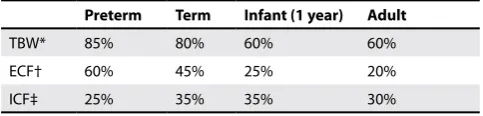
Body composition: For preterm and term neonates the Vd of water-soluble drugs is larger as compared to older children and a larger loading dose is required (e.g. aminoglycosides, cefazolin, paracetamol and neuromuscular blocking drugs [NMBD]).8 (See Table I for the fluid composition of neonates compared to adults).
Despite having a higher initial dose, reduced clearance capacity results in lower maintenance dose to avoid accumulation. Fentanyl (lipophilic) will have a much higher Vd compared to total body volume because fat holds more drug compared to the same volume of blood. If Vd is large then the dose required to achieve a target concentration is also large. However, larger doses may cause more significant adverse effects and are not given because a prolonged effect can result from reduced clearance.2
Table I.Fluid composition in neonates compared to adults2 Preterm Term Infant (1 year) Adult
TBW* 85% 80% 60% 60%
ECF† 60% 45% 25% 20%
ICF‡ 25% 35% 35% 30%
* Total body water † Extracellular fluid ‡ Intracellular fluid All as % of total body weight
Fat contributes 3% of total body weight in a 1.5 kg premature neonate and 12% at term. By the time the infant is four to five months old this would have doubled.5 Drugs relying on redistribution to fat and muscle like thiopentone and propofol can have prolonged and higher concentration in plasma in the preterm.
Cerebrospinal fluid contributes a greater proportion of body composition in neonates as compared to older children explaining the larger doses of spinal anaesthesia drug required in this population (1 mg/kg in infants < 5 kg compared to 0.3 mg/ kg for those > 15 kg).9
Protein binding: Protein binding is decreased in neonates. Concentrations of albumin and α1-acid glycoprotein (AAG) are lower in neonates (0.32–0.92g/l) but by six months values are similar to adults.10,11 Decreased quantity of drug-protein binding results in increased free drug concentrations and subsequent enhanced effect for drugs with typically high protein binding.
Albumin concentrations are lowest in preterm neonates. Drugs such as thiopentone that typically bind to albumin, have lower induction doses in neonates than children due to less protein binding (13% unbound drug in newborns versus 7% in adults).12 Jaundice is also common in premature infants. Elevated bilirubin competes with drugs like phenytoin for protein binding. Phenytoin administration in jaundice may lead to higher free drug concentration and increased risk of kernicterus (immature blood-brain barrier).12
Southern African Journal of Anaesthesia and Analgesia 2019; 25(5) Supplement
(CYP3A4) is the main parameter reduced in infants and there is more inter-individual variability in concentration as compared to reduced protein binding. In neonates, continuous epidural infusions will not generally run beyond 48 hrs (dose of 0.2 mg/kg/hr less than 0.4 mg/kg/hr in older children).13,14
Blood-brain barrier (BBB): The integrity of the BBB improves gradually with age. Foetal and neonatal brains may be more easily accessed by small molecules and even more so with certain disease states such as sepsis, hypoxia, and acidosis. Unbound lipophilic drugs such as bupivacaine can passively diffuse across the BBB.2 Combined with reduced protein binding, this may explain the increased risk of toxic levels leading to seizures in neonates.
Fentanyl is actively transported across the BBB by an ATP-binding cassette protein like P-glycoprotein. Modulation of this glycoprotein can influence onset of action, maximum effect, and duration of analgesic response.
Metabolism and elimination
Significant covariates, when considering neonatal drug metabolism are size, maturation and the effect of disease on organ function.Allometry describes the non-linear relationship between size and function. Use of allometric models enables prediction of paediatric doses from adult ones and target-controlled infusions have the potential to use allometric scaling.1,5 With the exception of remifentanil, allometry alone is insufficient in predicting clearance of most drugs in neonates and infants and there is a need to add a model accounting for maturation. Since maturation of clearance starts before birth, PMA is probably the better predictor than postnatal age (PNA) for drug
elimination.5 For example, CYP2D6, CYP3A4, CYP1A2 display ontogeny in the second and third trimester of pregnancy.15 Organ function changes associated with normal growth and development can be determined from pathological changes as function decreases with disease.1 Organ function may be increased by enzyme inducers like phenobarbitone (CYP1A2, CYP2C9, CYP2C19, CYP3A4, UDP glucuronsyltransferase [UGT]).1
The hepato-biliary (metabolic clearance) and renal (elimination clearance) systems are the main routes of clearance for drugs and metabolites. Immaturity in these two systems has an effect on neonatal drug handling.
Hepatic metabolic clearance: Hepatic elimination is governed by phenotypic variation and relates to inherent, disease-related and genetic factors. During infancy the main determinant is age-dependent phenotypic enzymatic activity. Development of these systems can alter drug clearance significantly.
There are three categories of isoenzymes in neonates with most being in class iii16-18:
i. Mature at birth with decreasing activity with age (CYP3A7, SULT1A3/1A4)
ii. Moderate maturation at birth with increased activity with increasing age (CYP3A5, CYP 2C19, SULT1A1)
iii. Little to modest maturation at birth with increasing activity with age (CYP2D6, CYP3A4, CYP2C9, CYP1A2)
Metabolising enzymes are divided into Phase I (non-synthetic reactions like oxidation, reduction and hydrolysis) and Phase II reactions (synthetic or conjugation reactions making water soluble compounds excreted in urine).
Important for the Phase I reactions are the cytochrome P450 group of enzymes which are often not fully mature in the neonate and also subject to variation due to genetic polymorphisms. Clearance of levobupivacaine depends on CYP3A4 and CYP1A2 for ropivacaine which are both immature in the neonate. This means this population requires reduced epidural infusion rates of these drugs.7 Altered phenotypic expression of CYP2D6 enzymes affects tramadol metabolism and formation of the major (M1) metabolite.19 Plasma cholinesterase activity influencing succinylcholine metabolism is also influenced by genetic polymorphism.5
Phase II reactions show limited activity during foetal life and some reactions like acetylation, glycination and glucuronidation are not mature at birth.2 These systems are complicated. For example, UGT clearance is immature at PMA 24 weeks but reaches maturity by the first year of life. UGT has isoforms maturing at different rates (see Figure 1 showing clearance maturation profiles of drugs mainly metabolised by UGT).5 Lack of understanding of UGT maturity led to gray baby syndrome with chloramphenicol in the 1960s.
Maturation processes can also be affected by illnesses. Morphine clearance is reduced in the very sick neonate and propofol clearance is lower in children after cardiac surgery. Concomitant drug use also affects metabolism. Ketamine’s sedative effects are less in children on long term phenobarbitone (CYP3A4 induction).
Renal clearance: Renal elimination is reflected by diuresis, GFR and renal tubular activity. At PMA of 25, GFR is only 10% of the mature value, 35% at term, and by one year it is 90% of the adult value.4 Renal inefficiency in the neonate is due to low perfusion pressure, incomplete glomerular development and inadequate osmotic load for the counter-current mechanisms. Drugs almost exclusively cleared by GFR (cephalosporins, aminoglycosides, D-tubocurarine) have lower maintenance dose which is predicted by PMA. PMA is used because it more accurately estimates the time course of renal maturation. Immaturity of clearance has some therapeutic use in the management of apnoea. When using theophylline, N7-methylation development to produce caffeine is well developed. However, oxidative demethylation (CPY1A2) is deficient. The produced caffeine is effective in controlling apnoea.20
Pulmonary elimination: In the lungs, anaesthetic absorption is determined by alveolar ventilation, FRC, blood-gas solubility and cardiac output. These also have a bearing on elimination kinetics. Washout will be more rapid due to reduced distribution to fat and muscle.
Some agents undergo hepatic metabolism (halothane much more than isoflurane and sevoflurane). However, hepatic elimination is very small at typical anaesthetic concentrations.5
Table II summarises some PK considerations in neonates.4
Table II.Illustrations of the impact of neonatal physiology on the pharmacokinetics (absorption, distribution, metabolism, elimination) of specific drugs commonly administered to neonates8
Compound Pharmacokinetics Relevance
Iodine disinfectant Skin more permeable, skin surface per kg weight higher (a) Higher absorption may suppress thyroid function
Inhalational gases Higher alveolar ventilation/FRC ratio (a) Faster wash-in
Cefazolin Lower protein-binding capacity results in higher distribution volume (d) and higher free plasma fraction (e)
Lower GFR (e)
Peak concentration is lower
Bactericid effect relates to free concentration
Lower clearance, prolonged duration of bactericid effect
Bupivacaine Lower protein-binding capacity (d)
Lower clearance (e)
Free concentration related to adverse effects Accumulation during continuous administration
Propofol Lipophilic compound, lower distribution volume (d)
Glucuronidation for metabolic clearance (m)
Peak concentration is lower, redistribution more limited Accumulation during continuous or repeated administration
More profound hypotension due to immature (para) sympathetic balance
Paracetamol Water soluble compound, higher distribution volume (d) Glucuronidation for metabolic clearance (m)
Peak concentration is lower, less effective analgesia likely Accumulation during repeated administration possible
Midazolum Clearance to metabolite (1-hydroxy) is low (m)
Elimination clearance of (1-hydroxy) midazolam is low (e)
Metabolite is also sedative
Lower clearance results in prolonged sedation
EMLA cream Skin more permeable, skin surface/kg higher (a) Higher absorption, may induce local anaesthetic related
seizures
Increased risk of methaemoglobinaemia
Codeine Clearance to metabolite (morphine) is low (m)
Elimination of codeine and metabolite is low (e)
Shorter or reduced analgesic effect
Accumulation of codeine or metabolite more likely, prolonged or more pronounced analgesia
NMBD Increased distribution volume (d)
Lower clearance (m)
Lower concentration at end plate, compensated by lower acetylcholine
Southern African Journal of Anaesthesia and Analgesia 2019; 25(5) Supplement
Pharmacodynamics (PD) and the neonate: “What the
drug does to the body”
Pharmacodynamics is the study of the drug effects (therapeutic and adverse) on patients. Despite there being significant differences in this population, drug responses in children have some commonalities with adults once developmental PK characteristics are considered.21
MAC for volatile anaesthetics is generally less in neonates than infants. Peak is at six months and then decreases to adult values by adolescence (see Table III). The variability in drug responses among the different volatile agents is influenced by the change
in number of GABAA receptors and developmental shifts in the regulation of chloride transporters in the brain.2
Response to vasoactive drugs is also age-dependent. PD differences can be attributed to developmental changes in myocardial structure, cardiac function, and receptor function.
Table IV highlights some of the PD differences in neonates.
Components of ideal general anaesthesia include unconsciousness, analgesia and muscle relaxation. Measuring these pharmacodynamic outcomes in neonates is harder compared to children or adults. For example, unconsciousness is assessed by monitoring the anaesthesia depth with Table III.Inhaled anaesthetic agents’ pharmacology1
Halothane Enflurane Isoflurane Sevoflurane Desflurane
A* N† A N A N A N A N
MAC 0.75 0.87 1.7 – 1.2 1.6 2.05 3.2 7.0 9.2
Solubility: Blood-gas 2.4 2.14 1.9 1.78 1.4 1.19 0.66 0.66 0.42 –
Solubility: Brain-blood 1.9 1.5 1.3 0.9 1.6 1.3 1.7 – 1.2 2.7
*adult †neonate
Table IV.Pharmacodynamic differences of common drugs used in anaesthesia5,23-26
Drug PD difference Reason Comments
Propofol Profound hypotension of about 20 minutes in neonates given 3 mg/kg
Unclear Needs further PD and
PK investigation
Morphine Increased sensitivity Functional expression of mu receptors is developmentally regulated
–
Local anaesthetics Amide agents induce shorter block duration Need higher dose for subarachnoid block (see text)
Myelination, spacing of nodes of Ranvier and length of nerve exposed
–
NMBD Increased sensitivity to effects
Succinylcholine induces bradycardia
Immature neuromuscular junction –
Inotropes Dopamine can be used in the presence of pulmonary hypertension
Signs of α-receptor stimulation may occur at lower doses than β-receptor stimulation
Fewer dopamine receptors in pulmonary vs systemic vasculature
β receptor maturation lags behind α maturation
Dopamine popular in the neonatal population compared to adults
Sedatives Bolus midazolam associated with hypotension (especially if given with fentanyl)
– –
Thiopentone Dose 3.4 mg/kg
(compared with 6.3 mg/kg in infants and 4-7 mg/kg in adults)
Uncertain PK and PD Uncertain cause
? immature cerebral cortical function ? rudimentary dendritic abnormalities ? relatively few synapses
–
Paracetamol Poorly defined PD
Early exposure may be related to later development of atopy-related syndromes ? early PDA closure
Unknown link –
Prokinetics Not very useful in very preterm neonates but useful at full term
Age-dependent expression of intestinal motilin
Modulation of antral contractions in the neonate
–
Bronchodilators Ineffective Paucity of bronchial smooth muscle that can cause bronchospasm
–
Calcium channel blockers Can cause life-threatening bradycardia and hypotension
Cardiac calcium stores in the endoplasmic reticulum are lower because of immaturity
electroencephalogram (EEG) or bi-spectral index in adults. However, use of these devices cannot yet be supported in children and EEGs are different in the various categories of paediatric patients.22
Adverse drug effects (ADE): Drug dosage errors are more common in children with the problem being further aggravated by narrow error margins in delivery and dilution.27 Off-label drug administration in neonates is still common with limited evidence-based pharmacotherapy. Therefore, it is requisite to design and participate in trials in the PK and PD of compounds commonly used by paediatric anaesthetists using suitable formulations and assessment methods.
In addition to the potential ADE that can occur in adults, neonates are potentially prone to particular effects because of immaturity of their physiology. Exposure to stimuli at a sensitive point of development may result in permanent effects. There are concerns that exposure of neonates to anaesthesia may cause increased neuronal apoptosis and long-term memory deficits. Implicated drugs are N-methyl D-aspartate antagonists (ketamine and nitrous oxide) and GABAA agonists (benzodiazepines, propofol, all volatile anaesthetic agents and barbiturates).28 Translating these observations to humans has proven difficult. The FDA issued a warning on drugs used for anaesthesia in 2016 raising concerns among parents of children undergoing anaesthesia. The General Anaesthesia Spinal (GAS) study indicated that a sevoflurane-based anaesthetic of less than an hour does not increase the danger of adverse neurological outcome at two years of age.29 The Paediatric Anaesthesia Neuro-Development Assessment (PANDA) study showed no significant differences in full-scale IQ at 10 years of age between exposed (general anaesthesia) and unexposed siblings. Scores assessed memory, language, attention, motor processing speed and behaviour among other things.30 The area of long-term effects of anaesthesia in children is one of ongoing research and debate.
Conclusion
Neonates have significant pharmacological differences compared to adults due to rapidly maturing physiological systems. Paediatric anaesthetists have to be knowledgeable in the PK and PD of neonates through studying available literature and participating in ongoing research in this area.
Acknowledgements
The author has no competing interests. I would like to acknowledge support and guidance from Faye Evans (Boston Children’s Hospital). This review is part of the 2019 Paediatric Anaesthesia Community of South Africa (PACSA) congress in Johannesburg.
References
1. Anderson BJ, Larsson P, Lerman J. Anesthesia and ancillary drugs and the neonate. (chapter 3) In: Lerman J, editor. Neonatal Anesthesia. Springer; 2015:67-113.
2. Anderson BJ. Neonatal pharamacology. Anaesthesia and Intensive Care Medicine, Volume 18, Issue 2, 68-74.
3. Anderson BJ. My child is unique; the pharmacokinetics are universal. Pediatr Anesth. 2012;22:530-8.
4. Martin LD, Jimenez N, Lynn AM. A review of perioperative anesthesia and analgesia for infants: updates and trends to watch [version 1; peer review: 2 approved] F1000Research 2017, 6(F1000 Faculty Rev):120 (https://doi. org/10.12688/f1000research.10272.1).
5. Anderson BJ. Pharmacology in the very young: anaesthetic implications. Eur J Anaesthesiol 2012;29:261-270.
6. Smits A, Kulo A, De Hoon JN, et al. Pharmacokinetics of drugs in neonates: pattern recognition beyond compound specific observations. Curr Pharm Des 2012;18:3119-3146.
7. Chalkiadis GA, Anderson BJ. Age and size are the major covariates for prediction of levobupivacaine clearance in children. Paediatr Anaesth 2006;16:275-282. 8. Allegaert K, Van de Velde M, Van den Anker J. Neonatal clinical pharmacology.
Pediatr Anesth. 2014;24:30-38.
9. Tronci R, Dadure C. Paediatric spinal anaesthesia. In: Homer R, Walker I, Bell G; editors. Update in Anaesth 2015;30:112-115.
10. Luz G, Innerhofer P, Bachmann B, et al. Bupivacaine plasma concentrations during continuous epidural anesthesia in infants and children. Anesth Analg 1996; 82:231-234.
11. Luz G, Wieser C, Innerhofer P, et al. Free and total bupivacaine plasma concentrations after continuous epidural anaesthesia in infants and children. Paediatr Anaesth 1998;8:473-478.
12. Sumpter A, Anderson BJ. Pediatric pharmacology in the first year of life. Curr Opin Anaesthesiol 2009;22:469-475.
13. Bosenberg AT, Thomas J, Cronje L, et al. Pharmacokinetics and efficacy of ropivacaine for continuous epidural infusion in neonates and infants. Paediatr Anaesth 2005;15:739-749.
14. Larsson BA, Lonnqvist PA, Olsson GL. Plasma concentrations of bupivacaine in neonates after continuous epidural infusion. Anesth Analg 1997;84:501-505. 15. Anderson BJ, Holford NH. Mechanism-based concepts of size and maturity in
pharmacokinetics. Annu Rev Pharmacol Toxicol 2008;48:303-332.
16. Van den Anker JN. Developmental pharmacology. Dev Disabil Res Rev 2010;16:233-238.
17. De Wildt SN. Profound changes in drug metabolism enzymes and possible effects ondrug therapy in neonates and children.Expert Opin Drug Metab Toxicol 2011;7:935-948.
18. Hines RN. Developmental expression of drug metabolizing enzymes: impact on disposition in neonates and young children. Int J Pharmdoi doi: 10.1016/j. ijpharm.2012.05.079.
19. Allegaert K, Anderson BJ, Verbesselt R, et al. Tramadol disposition in the very young: an attempt to assess in vivo cytochrome P-450 2D6 activity. Br J Anaesth 2005; 95:231-239.
20. McNamara DG, Nixon GM, Anderson BJ. Methylxanthines for the treatment of apnea associated with bronchiolitis and anesthesia. Paediatr Anaesth 2004;14:541-550.
21. Stephenson T. How children’s responses to drugs differ from adults. Br J Clin Pharmacol 2005;59:670-673.
22. Davidson AJ, Sale SM, Wong C, et al. The electroencephalograph during anesthesia and emergence in infants and children. Paediatr Anaesth 2008;18:60-70.
23. Westrin P, Jonmarker C, Werner O. Thiopental requirements for induction of anesthesia in neonates and in infants one to six months of age. Anesthesiology 1989;71:344-346.
24. Radford D. Side effects of verapamil in infants. Arch Dis Child 1983;58:465-466. 25. Seri I, Tulassay T, Kiszel J, et al. Cardiovascular response to dopamine in
hypotensive preterm neonates with severe hyaline membrane disease. Eur J Pediatr 1984;142:3-9.
26. Cuevas L, Yeh TF, John EG, et al. The effect of low-dose dopamine infusion on cardiopulmonary and renal status in premature newborns with respiratory distress syndrome. Am J Dis Child 1991;145:799-803.
27. Bang SR. Neonatal anesthesia: how we manage our most vulnerable patients. Korean J Anesthesiol 2015 October 68(5): 434-441. http://dx.doi.org/10.4097/ kjae.2015.68.5.434.
28. Ruzzi S, Ori C, Jevtovic-Todorovic V. Timing versus duration: determinants of anesthesia-induced developmental apoptosis in the young mammalian brain. Ann N Y Acad Sci 2010;98:145-58.
29. Davidson AJ, Disma N, De Graaff JC, et al. Neurodevelopmental outcome at 2 years of age after general anaesthesia and awake-regional anaesthesia in infancy (GAS): an international multicenter, randomized controlled trial. Lancet. 2016 Jan 16;387(10015):239-250.
Southern African Journal of Anaesthesia and Analgesia 2019; 25(5):S10-16
Open Access article distributed under the terms of the Creative Commons License [CC BY-NC 3.0]
http://creativecommons.org/licenses/by-nc/3.0
South Afr J Anaesth Analg ISSN 2220-1181 EISSN 2220-1173
© 2019 The Author(s)
PACSA SUPPLEMENT
It is fortunate that the majority of children will have airways that are simple to manage. However, in a small number of cases difficulty may be encountered. The focus of any airway management technique is to provide adequate oxygenation and ventilation. In a cooperative adult, this can be achieved by performing awake intubation techniques, however in children it is often impossible to manage them without performing anaesthesia or deep sedation. In this situation, there are three main ways that airway management is accomplished:
• Face mask ventilation
• Supraglottic airway device ventilation • Endotracheal intubation
There is no formal definition of what constitutes a difficult airway, but from a practical perspective it should be thought of as difficulty with any of the techniques used to provide oxygenation and ventilation. The area of difficulty where all three of the main techniques used to oxygenate a patient converge is the most worrying situation (Figure 1). The incidence of children who are difficult to intubate also experiencing failure of supraglottic device and mask ventilation is unknown, but it can rapidly lead to a ‘can’t intubate, can’t oxygenate’ (CICO) event with immediate life-threatening consequences. Multiple guidelines have been produced to aid anaesthetists in the management of the paediatric difficult airway (e.g. https://www.das.uk.com/ guidelines/paediatric-difficult-airway-guidelines and http:// www.anzca.edu.au/documents/ps56-2012-guidelines-on-equipment-to-manage-a-diff.pdf ).
This review will discuss each aspect of airway management and the existing evidence that should be used when deciding how to approach a child with a difficult airway.
Pre-anaesthetic assessment and preparation
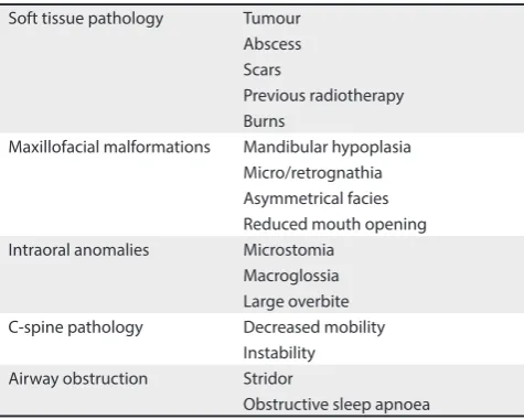
Before initiating anaesthesia a thorough medical history and physical examination should be performed. There are several factors that may point to a child being at risk for having a difficult airway.1 These are summarised in Table I. Additionally, it
is important to ask about previous anaesthetics and if possible, interrogate previous anaesthetic records for a formal description of previous airway management techniques. It should be noted that approximately 20% of difficult intubations in children are unanticipated,2 so every anaesthetic plan should include
back-up contingencies to cope with unexpected difficulty with airway management. Children who weigh less than 10 kg are also more likely to experience complications related to airway management.2 When difficulty is anticipated, planning should
take into account the location where the airway management will occur, the equipment required, and personnel needed to minimise complications. The safest place to manage an anticipated difficult airway is in the operating room.3 The
equipment required will vary depending on the circumstances. Advanced airway equipment from the anaesthetic team (e.g. flexible bronchoscopes, videolaryngoscopes) can be supplemented by the presence of our ENT surgical colleagues
Difficult airway management in children
J Peyton, R ParkDepartment of Anaesthesia, Critical Care and Pain Medicine, Boston Children’s Hospital and Harvard Medical School, Boston, United States of America
Corresponding author, email: [email protected]
Figure 1.What makes an airway difficult? Difficult endotracheal
intubation
Supraglottic device failure Difficult
mask ventilation
Table I. Factors that may predict a difficult airway in children1 Soft tissue pathology Tumour
Abscess Scars
Previous radiotherapy Burns
Maxillofacial malformations Mandibular hypoplasia Micro/retrognathia Asymmetrical facies Reduced mouth opening Intraoral anomalies Microstomia
Macroglossia Large overbite C-spine pathology Decreased mobility
Instability Airway obstruction Stridor
and their equipment (e.g. rigid bronchoscopes, tracheostomy). Another essential feature of the management of the difficult airway is clear communication between all those involved, particularly if the airway needs to be shared with a surgical team. Any plan should be fully discussed with the nursing and surgical staff, and if needed, explicit recognition of everyone’s role, and when roles may be exchanged (e.g. when the surgical team should take over attempts at intubation if the anaesthetic team have been unsuccessful).
Difficult mask ventilation
Difficulties ventilating children with a face mask occur in approximately 6% of cases.4 Physical features that should
be observed during the physical examination that may be associated with difficult mask ventilation include:
• Micro/retrognathia • Craniofacial abnormalities • Cervical spine abnormalities • Obesity
• Obstructive sleep apnoea (OSA)
Positioning the patient ‘head up’ at approximately 30 degrees and the use of airway adjuncts such as oral or nasopharyngeal airways may improve the ability to ventilate via a face mask. If the patient has significant anatomical abnormalities, such as a base of tongue tumour or a neck mass, that make it difficult to bypass the obstruction with an airway adjunct, maintaining spontaneous breathing may be safer than a technique reliant on positive pressure ventilation.
Infants and neonates who experience difficult mask ventilation are at risk of developing significant gastric distension. This can impact on the ability to oxygenate, cause rapid oxygen desaturation through atelectasis and decreased functional residual capacity, resulting in less time available to attempt definitive airway management.
If difficult mask ventilation occurs there are several strategies that can be used to try to improve it:
• Early use of airway adjuncts such as oral/nasopharyngeal airways
• Two-person technique with a two-handed jaw thrust and a second person manually ventilating
• Change of head and/or patient position • Early decompression of the stomach
• Early use of alternative technique, particularly a supraglottic airway
Difficult supraglottic device ventilation
Supraglottic devices were first described for use in adults in 1983.5 Over the intervening decades many different supraglottic
devices have been created for use in children. At the time of writing these include6,7:
• AirQ and AirQ SP
• Ambu AuraGain, Ambu Aura-i and Ambu AuraOnce • Cobra
• I-gel
• Laryngeal tube
• LMA Classic, Flexible, ProSeal, Supreme and Unique • PRO-Breathe
• SLIPA • Softseal
When considering their use in children with difficult airways, the main concern is the risk that a device will fail to provide adequate oxygenation and ventilation. The available evidence examining the failure rates of different devices in children with normal airways is summarised in Table II. The rate of failure depends on the type of device and the individual child. Anatomical features associated with the presence of a difficult airway will tend to increase the risk of a supraglottic device failing.
Table II.Failure rates of supraglottic devices in children7
Device Failures/total cases % (95% CI)
AirQ 0/126 0 (0-3.0)%
AirQ SP 1/69 1.4 (0.26–7.8)%
Ambu AuraGain 0/50 0 (0–7.1)% Ambu Aura-i 0/32 0 (0–10.7)% Ambu AuraOnce 2/132 1.5 (0.42–5.4)%
Cobra 4/301 1.3 (0.52–3.4)%
I-gel 37/1 079 3.4 (2.5–4.7)% Laryngeal tube 2/108 1.9 (0.51–6.5)% LMA Classic 4/1 118 0.36 (0.14–0.92)% LMA Flexible 0/69 0 (0–5.3%)% LMA ProSeal 6/1 211 0.50 (0.23–1.1)% LMA Supreme 9/488 1.8 (0.97–3.5)% LMA Unique 2/410 0.49 (0.1–1.8)% PRO-Breathe 6/100 6.0 (2.8–12.5)%
SLIPA 0/50 0 (0–7.1)%
Softseal 0/36 0 (0-9.6)%
Total 75/5 379 1.4 (1.1–1.7)%
It is not possible to choose a single supraglottic device to recommend over others for use in children. In general, second-generation devices (those with oesophageal and laryngeal outlets) are considered superior to the original supraglottic devices as they demonstrate:
• Higher seal pressures • Increased ease of insertion
• Oesophageal lumens allow access to the stomach to help prevent aspiration and enabling decompression of the stomach whilst continuing to ventilate
Southern African Journal of Anaesthesia and Analgesia 2019; 25(5) Supplement
meta-analysis by Mihara et al. in 2017 compared the current supraglottic devices available for use in children.7 In this study,
the authors concluded that the LMA-ProSeal may overall be the best supraglottic airway device for use in children. However, if the intent is to use the supraglottic device as a conduit to facilitate tracheal intubation, the LMA-ProSeal could be considered a poor choice when compared to the AirQ laryngeal airway, or the i-Gel.
It is also important to emphasise the early use of supraglottic devices when confronted by an unanticipated difficult airway in children. They may be life-saving when used to facilitate oxygenation during airway management and, as mentioned above, have been used as a conduit to facilitate tracheal intubation when used in combination with flexible bronchoscopy.8
Difficult tracheal intubation
Difficult tracheal intubation in the paediatric population is estimated to occur in 0.28–1.35%2,9 of patients. Predictors of
difficult intubation in children include extremes of weight, younger age, increased illness severity as measured by the American Society of Anesthesia (ASA) classifications, and types of surgery such as cardiac or oromaxillofacial surgery that may also serve as a surrogate for associated congenital abnormalities.1,2 Nearly 20% of difficult intubations are not
anticipated.3 Common physical examination findings associated
with difficult intubation include micrognathia, limited mouth opening and cervical spine immobility.3 In 2012 the Pediatric
Difficult Intubation Registry (PeDIR) was formed under the auspices of the Society for Pediatric Anesthesia in the USA. This registry is a multinational database that, at the time of writing, contains over 4 000 cases of difficult paediatric intubation, that has been used to gather data on this vulnerable population. The registry revealed that severe hypoxia occurred in 9% of these children, with cardiac arrest occurring in nearly 2%.2 Every cardiac
arrest was preceded by hypoxia. This demonstrated that during difficult tracheal intubation, maintenance of oxygen saturations should be our first priority.
Tracheal intubation can be accomplished by many different techniques.
Direct laryngoscopy (DL)
DL remains the most commonly chosen technique for tracheal intubation in children. It was used in 98% of cases in the Apricot study examining over 31 000 anaesthetics in 261 institutions in Europe,9 and was the first choice technique in nearly half of
the patients in the PeDIR.2 Unfortunately, DL has a low success
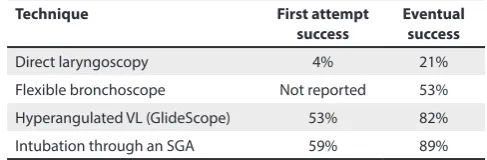
rate in children who are difficult to intubate, with first attempt success rates of 4% and eventual success rates of only 21%.10
Given these poor success rates, DL has a limited role in the management of anticipated difficult intubation. If used as a first choice, it is imperative that back-up plans are in place to ensure a rapid progression to more advanced techniques. However, complications associated with intubation are related to the number of attempts at intubation,10,11 so choosing a technique
with higher first pass success rates is sensible (Table III).
Table III.Success rates of different intubation techniques from the Pediatric Difficult Intubation Registry2,8,10
Technique First attempt
success Eventual success
Direct laryngoscopy 4% 21%
Flexible bronchoscope Not reported 53% Hyperangulated VL (GlideScope) 53% 82% Intubation through an SGA 59% 89%
Videolaryngoscopy (VL)
Videolaryngoscopes use video cameras embedded within the laryngoscope blade to obtain a view of the larynx. They can be thought of as two distinct types:
1. Hyperangulated videolaryngoscopes 2. Standard bladed videolaryngoscopes
Hyperangulated devices cannot be used to directly visualise the larynx because they do not allow alignment of the oral, pharyngeal and laryngeal axes in the same fashion as standard laryngoscopy blades. Hyperangulated VLs look around the curve of the airway and rely solely on the view provided by the video camera. Hyperangulated blades include the GlideScope, Airtraq, Pentax AWS, Truview and the Storz C-Mac D-Blade.
Standard blade videolaryngoscopes are identical to traditional DL blades, but have a camera mounted distally within the blade. This allows DL to be performed, but also provides a second point of view (video-assisted DL–VADL) that may give a better view of the larynx, and allow others to view the endotracheal tube passing through the vocal cords. Standard laryngoscope VL systems include the Storz C-Mac Macintosh and Miller blades, McGrath Mac blades and the UE Scope. VL has been shown to achieve better views of the larynx when compared to DL,12,13
but there has been a suggestion that it may increase the time taken to intubate by approximately five seconds.14 This has not
been shown to increase complications, and in particular there is no evidence that VL use is associated with a greater incidence of hypoxia.
In the PeDIR database, the hyperangulated GlideScope (GVL) was the most frequently used video system, accounting for 76% of all VL use. Park et al. compared GVL with DL use in children in the PeDIR and found that GVL had much higher initial and eventual success rates. The initial success rate with the GVL was 53%, with an eventual success rate of 82%, compared to just 4% and 21% with DL.10 Interestingly, the success rates of
GVL were significantly lower in children weighing less than 10 kg with initial success rates of 39% and eventual success in 73%. In adults, the success rate of GVL following failed DL is greater than 90%,15 so the success rate of GVL in children and
using GVL or DL. This was confirmed in a prospective study comparing videolaryngoscopy and direct laryngoscopy in patients predicted to be difficult intubations, which also showed no difference in rates of airway trauma or desaturation.16
One of the drawbacks of hyperangulated VL is that even if the larynx is clearly visualised, it may not be possible to pass the ETT into the trachea due to the angulation of the larynx with respect to the laryngoscope blade. Different methods to combat this have been described, including using stylets in different configurations to pre-shape the ETT,17-19 or using a flexible
bronchoscope as a manipulatable stylet to enter the trachea.20
Flexible bronchoscopic intubation (FBI)
Awake flexible bronchoscopic intubation has been shown to be a safe and effective method of securing potential difficult airways in adults, with a failure rate of ~1%.21 Most children
will not tolerate awake or even sedated airway management, so FBI is most commonly performed after induction of general anaesthesia. Despite advances in videolaryngoscopy, FBI remains an essential technique for difficult airway management in children. It may be the only option (aside from tracheostomy) for patients with limited or no mouth opening that precludes laryngoscopy or supraglottic airway placement. FBI was the choice for initial airway management in ~1/3 of the patients in the PeDIR.2
There are limited data assessing the safety and effectiveness of FBI in children who are difficult to intubate. Within the PeDIR, FBI had a first pass success rate of 38% in patients weighing less than 10 kg and 54% in those more than 10 kg. In a mannequin study simulating a difficult airway in a child with Robin sequence, Fiadjoe et al. compared first attempt intubation success between Glidescope and FBI amongst attending anaesthesiologists at two major paediatric centres. They found no difference in intubation success rates.22 FBI has also been described as a successful
technique in real infants with Robin sequence,23,24 but it should
also be considered an important part of combined techniques such as intubation via a supraglottic airway, or when used with VL.
Supraglottic airway as a conduit to flexible
bronchoscopic intubation
A supraglottic airway can often bypass the causes of upper airway obstruction and in most cases provides direct access to the larynx. It is possible to perform FBI through a supraglottic device by passing a flexible bronchoscope through the lumen of the device and into the trachea. An endotracheal tube can then be railroaded over the flexible bronchoscope into the trachea. This technique has the advantage of allowing continuous oxygenation to occur via the SGA, and may even allow continuous ventilation depending on the size of the ETT and FBI used.25 In neonates and young infants, awake supraglottic
airway placement is generally well-tolerated and allows for assessment of adequate placement prior to induction.24-26
Among the FBI-SGA patients entered into the PeDIR, rates of hypoxia were significantly lower when continuous ventilation
was used during intubation (7% vs. 25%, p = 0.04).8 The AirQ
laryngeal airway was the most commonly used SGA to facilitate FBI in children. In a study comparing the AirQ assisted technique with a ‘free-hand’ approach in children younger than two years of age, no differences were found in the number of attempts needed to intubate, or the time taken. However, there were less adjustments needed to optimise the view of the larynx if the AirQ was used.27
Other combined techniques
There are numerous case reports describing techniques combining different airway management techniques. As an example, laryngeal visualisation is often possible with hyperangulated videolaryngoscopes, but navigating the endotracheal tube into the trachea can be problematic. Flexible bronchoscopic intubation combined with hyperangulated VL allows for two vantage points to view the airway and the flexible bronchoscope can be used as a movable stylet to guide the ETT into the trachea.20,28 The ability to view the glottis with the GVL
whilst advancing the ETT over the flexible scope can help identify and solve problems that may occur, with the aim of decreasing potential trauma from blind, forceful ETT advancement.
Other combined techniques described include both hyperangulated videolaryngoscopy and video-enhanced direct laryngoscopy in combination with an optical stylet, light wand, or flexible bronchoscope.29-31
Supplemental oxygen administration during airway
management
Children can experience rapid oxygen desaturation during airway management. This occurs because of their high rate of oxygen consumption coupled with a lower functional residual capacity. The use of supplemental oxygen during routine airway management is not currently recommended, however in the setting of a difficult airway, it should be used. When supplemental oxygen is administered during intubation, a significant increase in the time to oxygen desaturation has been demonstrated.32-34
Techniques have included administering oxygen via nasal cannula,35 through the laryngoscope32,34 and through specific
equipment designed to deliver high flow, humidified oxygen (e.g. Transnasal Humidified Rapid Insufflation Ventilatory Exchange [THRIVE]).33,36 The THRIVE system has been shown to
maintain oxygen saturations for at least twice as long as the expected age-dependent apnoea times in healthy children.33
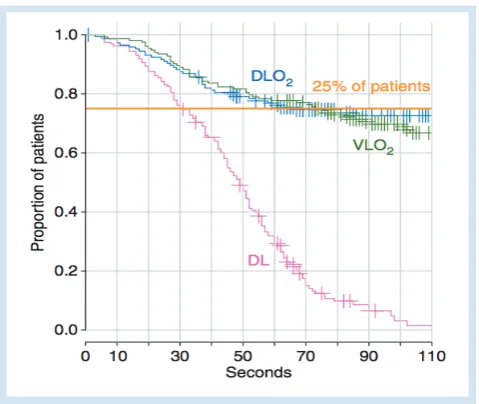
Steiner et al. examined the use of ‘deep’ oxygen insufflation via a PCD Truview videolaryngoscope, through its side-port attached to an oxygen supply, and a modified traditional direct laryngoscope blade. They compared the time to oxygen desaturation using these devices compared to traditional direct laryngoscopy without oxygen supplementation, in apnoeic children.34 The graph below illustrates their results, showing that
Southern African Journal of Anaesthesia and Analgesia 2019; 25(5) Supplement
Riva et al. also studied the effect of oxygen administration via standard nasal cannula versus the use of the THRIVE system in children. They studied apnoeic children receiving 100% inspired oxygen using THRIVE or via low-flow nasal cannula (0.2 litre kg-1) and 30% oxygen using THRIVE.37 Their results
demonstrated an increase in apnoea times in both groups given 100% oxygen, but not in the 30% group. These studies support the use of supplemental oxygen administration during intubation attempts to increase the time to desaturation and increase the time available for practitioners to secure the airway.
There is clear evidence that the use of supplemental oxygen can increase the time to oxygen desaturation during airway management. It should be used whenever a difficult airway is encountered. A recent editorial by Fiadjoe and Litman also addressed this issue,38 concluding that oxygen supplementation
should be used on all expectedly difficult or prolonged intubation attempts in children. The benefit-to-risk ratio is too great to ignore.
Muscle relaxation
Compared with the adult literature, there is less evidence in children to support the use of neuromuscular blockade (NMB) to optimise intubating conditions. A recent Cochrane review
included 34 studies evaluating the influence of neuromuscular blockade on outcomes in tracheal intubation in adolescents and adult patients. Avoidance of NMB was statistically significantly associated with difficult direct laryngoscopy (RR 13.27, 95% CI 8.19–21.49, P = 0.00001).39 There is more limited evidence in
children that neuromuscular blockade improves intubating conditions. In contrast to the adult reviews on this topic, only seven studies met criteria for inclusion for a recent meta-analysis evaluating NMBA use and intubating conditions in children.40 This study concluded that muscle relaxants may be
recommended for intubation over opioids to improve intubation conditions. Of note, all the included studies in this meta-analysis compared intubating conditions between patients receiving muscle relaxant to those receiving a combination of opioids and volatile anaesthetics. The doses of opioids administered in these studies could be expected to render patients apnoeic, therefore the conclusions from these studies may not be applicable in answering questions concerning safety and effectiveness in spontaneously breathing patients. The characteristics of the trials included in the meta-analysis are outlined in Table IV.
There is no evidence that maintaining spontaneous breathing decreases the risk of complications and hypoxia during airway management. Indeed, the opposite may be true, in that complications such as laryngospasm and hypotension from the higher doses of anaesthesia required may occur when neuromuscular blockade is avoided.
The most recent study using data from the PeDIR examined the differences in complications in patients who were breathing spontaneously versus those who underwent controlled ventilation with and without muscle relaxation. The initial hypothesis was that those breathing spontaneously would experience less complications than those who were rendered apnoeic, however, the opposite was found to be true. The spontaneously breathing group was more than twice as likely to experience complications than the apnoeic group. Interestingly there were no differences in complications between the group that was paralysed and those rendered apnoeic without neuromuscular blocking agents, so it is possible that the complications seen in the spontaneously breathing group relate to inadequate depth of anaesthesia, although it is not possible to confirm this with a retrospective review.
Figure 2.Kaplan-Meier curves of time to 1% reduction in saturation from the baseline. Time to 1% reduction in saturation was censored at the end of intubation.34
Table IV. Characteristics of the trials included in the meta-analysis40