BioMedCentral
BMC Biology
Open Access
Research article
Compensation by tumor suppressor genes during retinal
development in mice and humans
Stacy L Donovan
†1, Brett Schweers
†1, Rodrigo Martins
1, Dianna Johnson
2and
Michael A Dyer*
1,2Address: 1Department of Developmental Neurobiology, St. Jude Children's Research Hospital, Memphis, TN 38105, USA and 2Department of Ophthalmology, University of Tennessee Health Science Center, Memphis, TN 38163, USA
Email: Stacy L Donovan - [email protected]; Brett Schweers - [email protected]; Rodrigo Martins - [email protected]; Dianna Johnson - [email protected]; Michael A Dyer* - [email protected]
* Corresponding author †Equal contributors
Abstract
Background: The RB1 gene was the first tumor suppressor gene cloned from humans by studying genetic lesions in families with retinoblastoma. Children who inherit one defective copy of the RB1
gene have an increased susceptibility to retinoblastoma. Several years after the identification of the human RB1 gene, a targeted deletion of Rb was generated in mice. Mice with one defective copy of the Rb gene do not develop retinoblastoma. In this manuscript, we explore the different roles of the Rb family in human and mouse retinal development in order to better understand the species-specific difference in retinoblastoma susceptibility.
Results: We found that the Rb family of proteins (Rb, p107 and p130) are expressed in a dynamic manner during mouse retinal development. The primary Rb family member expressed in proliferating embryonic retinal progenitor cells in mice is p107, which is required for appropriate cell cycle exit during retinogenesis. The primary Rb family member expressed in proliferating postnatal retinal progenitor cells is Rb. p130 protein is expressed redundantly with Rb in postmitotic cells of the inner nuclear layer and the ganglion cell layer of the mouse retina. When Rb is inactivated in an acute or chronic manner during mouse retinal development, p107 is upregulated in a compensatory manner. Similarly, when p107 is inactivated in the mouse retina, Rb is upregulated. No changes in p130 expression were seen when p107, Rb or both were inactivated in the developing mouse retina. In the human retina, RB1 was the primary family member expressed throughout development. There was very little if any p107 expressed in the developing human retina. In contrast to the developing mouse retina, when RB1 was acutely inactivated in the developing human fetal retina, p107 was not upregulated in a compensatory manner.
Conclusion: We propose that intrinsic genetic compensation between Rb and p107 prevents retinoblastoma in Rb- or p107-deficient mice, but this compensation does not occur in humans. Together, these data suggest a model that explains why humans are susceptible to retinoblastoma following RB1 loss, but mice require both Rb and p107 gene inactivation.
Published: 03 May 2006
BMC Biology 2006, 4:14 doi:10.1186/1741-7007-4-14
Received: 02 March 2006 Accepted: 03 May 2006
This article is available from: http://www.biomedcentral.com/1741-7007/4/14
© 2006 Donovan et al; licensee BioMed Central Ltd.
Background
The timing of cell cycle exit is coordinated with cell-fate specification and differentiation in the developing central nervous system (CNS) to ensure that the appropriate number of neurons and glia are generated in the correct proportion. If too many neural progenitor cells exit the cell cycle during the early stages of development, then the overall number of cells is reduced, and the ratio of cell types shifts [1,2].
Genetically engineered mice carrying targeted deletions of tumor suppressor genes that regulate cell cycle progres-sion have been used to study the coordination of prolifer-ation and cell-fate specificprolifer-ation during development. Mice lacking the retinoblastoma susceptibility gene, Rb1
[3-5], were among the first characterized. In embryonic day (E) 13.5 Rb-/- mouse embryos, ectopic mitosis was
observed outside the ventricular zone of the developing hindbrain, and concurrent apoptosis was observed in the region where postmitotic cells normally migrate and dif-ferentiate [4,5]. In contrast, the E13.5 Rb-/- retina was
indistinguishable from E13.5 wild-type or Rb -hetero-zygous retinas [3-6].
Lee and colleagues proposed that specific neuronal sub-lineages have unique requirements for Rb during develop-ment [5]. To explain the specific defects in neurogenesis in Rb-deficient animals, Jacks and colleagues suggested that other, closely related Rb proteins such as p107 play a more prominent role in certain lineages [4]. For example, tissues uniquely dependent on Rb (e.g., developing hind-brain) may exhibit defects in Rb-/- embryos, but lineages
that are not (e.g., embryonic retina) appear normal in its absence.
Although the Rb-deficient embryonic mouse retina devel-ops normally up to E13.5, Rb may play an important role at later stages of development [7]. Using Cre-expressing retroviruses and a retinal progenitor-specific Cre trans-genic mouse line (Chx10-Cre) mated to RbLox mice [8], we
previously showed that the retinal defects first identified by Maandag and colleagues reflect a cell-autonomous requirement for Rb at specific developmental stages in ret-inal progenitor cells and rod photoreceptors [6,9]. Two other groups published similar findings using Cre trans-genic mouse lines with broader expression in the retina [10] and throughout the CNS [11]. Other phenotypes characterized in Rb-/- embryos were recently found to be
secondary non-cell-autonomous effects caused by changes in extraembryonic tissues [12].
Analysis of mice carrying targeted deletions of p107 and
Rb (Rb+/-; p107-/-) indicated that p107 is important for
ret-inal development [13]. Mild retret-inal dysplasia was observed in the Rb+/-; p107-/- mice at 4 to 6 months of age,
and histologic analysis of postnatal day (P) 4 retinas sug-gested that dysplasia originated during development [13]. Subsequent studies using chimeric mice generated from
Rb-/-; p107-/- embryonic stem cells [14] and those
involv-ing conditional inactivation of Rb in the developing retina [10,15] or CNS [11] have not elucidated individual roles of Rb or p107 in retinogenesis.
When progenitor cell proliferation is disrupted during development, secondary effects on cell-fate specification and differentiation are likely to occur [1,2]. Therefore, dis-tinguishing the roles of Rb and p107 in proliferation from those in cell-fate specification is a major challenge. In addition, elucidating the cell-autonomous and non-cell-autonomous [12] roles of the Rb family during develop-ment can be challenging [16]. Moreover, as several inves-tigators have proposed [3-5,13,17], individual Rb proteins are likely to have both unique and overlapping roles during development, thereby making it difficult to identify their individual contributions. Compensation and redundancy can further complicate analyses and obscure the individual roles of Rb proteins during devel-opment [2,18,19]. Interestingly, the response to acute inactivation of Rb in mouse embryonic fibroblasts (MEFs) differs from that seen when Rb is inactivated in a chronic manner [20]. This finding increases the complexity of the genetic analyses of Rb family function in vivo.
Relatively little is known about the expression or the role of the Rb family during human fetal retinal development. Therefore, we do not know if Rb, p107 and p130 are play-ing different roles in the developplay-ing human and mouse retinas or if cell cycle regulation through the Rb family is evolutionarily conserved.
In this study, we used genetic tools (replication-incompe-tent retroviruses, in vivo electroporation, and SiRNA anal-yses) combined with cell cycle analysis to investigate the individual cell-autonomous roles of Rb and p107 in the developing retina. We also sought to compare Rb family expression and intrinsic genetic compensation during ret-inal development in mice and humans to explain why humans are susceptible to retinoblastoma following RB1
inactivation, but mice are not.
Results
Expression of the Rb family during mouse retinal development
BMC Biology 2006, 4:14 http://www.biomedcentral.com/1741-7007/4/14
step toward distinguishing between these two possibili-ties, we analyzed the expression of Rb, p107 and p130 over seven stages of mouse retinal histogenesis. Real-time RT-PCR and immunoblot analyses of E14.5 to P0 mice indicated that p107 is the primary Rb protein expressed in
levels of p107 mRNA and protein decreased at P0, those of Rb and p130 increased (Fig. 1B–D) [6]. However, a low level of Rb was detected in a subset of embryonic retinal progenitor cells and newly postmitotic (transition) cells at the basal and apical surfaces of the developing inner
neu-Dynamic expression of the Rb family during mouse retinal development Figure 1
Dynamic expression of the Rb family during mouse retinal development. (A-C) Real-time PCR analysis of p107, Rb and p130 was done at seven stages of mouse retinal development. Data sets of three retinas per stage were analyzed twice, normalized to Gapdh and averaged. The standard deviations were within 5% of the mean. (D) Immunoblot analysis was done at five stages of mouse retinal development. For negative controls, MEF lysates prepared from knockout embryos were used. Positive controls were taken from 293T cells ectopically expressing the full-length cDNAs. Data were normalized to actin. (E-I) Immunofluorescent detection of Rb (green) in E14.5 and E17.5 retinas was overlaid on the nuclear counterstain (red). (J)
Radioactive in situ hybridization analysis of Rb (blue silver grains) levels at P0 demonstrated broad expression in the onbl and inbl. This finding was consistent with immunofluorescent detection (green) of the protein (K). (L-O) Immunofluorescent detection of p107 (green) in E14.5 and E17.5 retinas was overlaid on the nuclear counterstain (red). (P) Radioactive in situ hybridization for p107 (blue silver grains) at E17.5 demonstrated broad expression in the onbl and little expression at P3 (Q).
The p107 mRNA and protein were restricted to proliferat-ing retinal progenitor cells in the outer neuroblastic layer (ONBL) (Fig. 1L–Q), and p130 mRNA and protein were restricted to a subset of postnatal retinal progenitor cells, postmitotic neurons and glia in the inner nuclear layer
(INL) and ganglion cell layer (GCL) during the final stages of retinal histogenesis (Fig. 1R–U and data not shown) [6]. These data indicated that the embryonic retinal pro-genitor cells primarily express p107 (Fig. 1V), which is consistent with a lack of any retinal phenotype in Rb
-/-Expression of the Rb family during the cell cycle Figure 2
Expression of the Rb family during the cell cycle. (A-D) The E14.5 retinal explants were maintain in culture in the pres-ence of [3H]thy for 1 h, washed and then maintained in culture for different periods. Dissociated retinas (dapi in A) were then
immunostained for p107 (red in B) and overlaid with autoradiographic emulsion to detect the [3H]thy (C). The proportion of
p107+ cells was then scored (250 cells in duplicate), and normalized data were plotted (D). The peak level of p107 was detected after 8 h of exposure, which coincided with the G2 and M phases of the cell cycle, and declined at 24 h, which coin-cided with the G1/G0 phase. (E-H) Similar experiments were done using P0 retinal explants and in P2 retinal explants (I-L).
(H) The proportion of [3H]thy-labeled Rb+ cells from P0 retinal explants was high during 0 to 4 h of exposure (S, G2) and
sub-sequently declined. (L) The level of [3H]thy-labeled p130+ cells in P2 retinal explants was low during the first 4 h (S, G2),
BMC Biology 2006, 4:14 http://www.biomedcentral.com/1741-7007/4/14
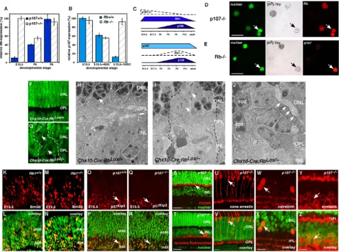
Reciprocal compensation after Rb or p107 inactivation Figure 3
Reciprocal compensation after Rb or p107 inactivation. (A) Real-time PCR analysis of Rb expression in retinas from three developmental stages of p107-deficient or wild-type mice. At E13.5, Rb was upregulated. Analysis was done in triplicate samples analyzed in duplicate. (B) In a similar experiment, p107 expression was analyzed in Rb-deficient retinas. Due to the embryonic lethality of deleting Rb, cultured retinas were used for the later stages; p107 was upregulated in a compensatory manner. (C) A summary of the changes in Rb family gene expression after Rb or p107 inactivation. (D) E13.5 p107-deficient retinas were labeled with [3H]thy for 4 h and immunostained for Rb expression (red). (E) In a similar experiment, cultured
Rb-deficient retinas were labeled for p107 expression. (F, G) In the absence of Rb in Chx10-Cre;RbLox/- P12 retinas, rod
photore-ceptors failed to form, and chromatin failed to condense in the ONL (arrow). (H) Chromatin (arrow) in rod photoreceptors normally condenses during differentiation. (I) In the absence of Rb, rods remained immature with diffuse chromatin (arrow).
(J) These defects in cell-fate specification also caused defects in synaptogenesis of horizontal cells (arrows); with processes extending apically into the ONL. (K-N) Wild-type and Rb-deficient E13.5 retinas were immunostained with an antibody against Brn3b (red), a ganglion cell marker. Brn3b was distributed across the retina (green nuclear stain overlay) and was indistinguish-able in the presence or absence of Rb. (O-R) Wild-type and p107-deficient E15.5 retinas were immunostained for p57Kip2
embryonic retinas. In the postnatal retina and in differen-tiated neurons and glia of the INL and GCL, Rb and p130 were expressed redundantly (Fig. 1V). Rb is the only fam-ily member expressed in rod photoreceptors, a finding that is consistent with its unique role in the development of this neuronal cell type [6]. Together, these data suggest that Rb and p107 are expressed in a largely nonoverlap-ping pattern during mouse retinal development, which raises the possibility of compensation by p107 when Rb is inactivated. Moreover, Rb and p130 expression overlaps, which supports the idea that these proteins play redun-dant roles in a subset of retinal neurons.
Expression of the Rb family during the cell cycle in retinal progenitor cells
Beyond determining when and where each Rb family member is expressed during retinal development, it is important to determine when during the cell cycle Rb, p107 and p130 are expressed. These data may help to elu-cidate their normal roles during development. For exam-ple, if a family member is only expressed in proliferating retinal progenitor cells, then that protein is unlikely to play a role in neuronal differentiation. It has been well established in a variety of culture systems that Rb, p107 and p130 are expressed during different phases of the cell cycle. To directly determine when during the cell cycle these proteins are expressed throughout retinal develop-ment, we scored the colocalization of [3H]thymidine
([3H]thy) with p107 (Fig. 2A–D), Rb (Fig. 2E–H) or p130
(Fig. 2I–L) at five time points during four stages of devel-opment [2,22,23]. The expression of p27Kip1 has been
characterized previously [22], and a parallel set of samples was analyzed for p27Kip1 expression to serve as an internal
control. The p27Kip1 protein is upregulated as cells exit the
cell cycle throughout retinal development, and the kinet-ics are different at E14.5, P0 and P2, because the length of the retinal progenitor cell cycle increases during retino-genesis [24]. At E14.5, 62% to 77% of proliferating cells expressed p107, and 4% to 6% expressed Rb (see Addi-tional file 1). The 36% reduction in the proportion of p107+ cells at the 24-h time point in E14.5 retinas was consistent with the previous finding that 20% to 25% of rat retinal progenitor cells exit the cell cycle at a similar stage [22,24,25].
At P0, 76% to 87% of proliferating retinal progenitor cells expressed Rb (see Additional file 1; Fig. 2E–H), and the remaining cells expressed p107 (data not shown). Unlike p107+ cells, Rb+ P0 retinal progenitor cells continued to express Rb after they exited the cell cycle (compare Fig. 2H and 2D). In the P2 retina, the level of p130 peaked in the late G2/early M phase of the cell cycle and persisted into G1/G0 (see Additional file 1; Fig. 2I–L). Many cells down-regulated p130 as they exited the cell cycle; this finding was consistent with the birth of rod photoreceptors (80%
of the total cell population in the retina), which do not express p130 (Fig. 1R–U). Results from the [3H]thy
pulse-labeling experiment combined with those from the immunostaining, real-time RT-PCR, in situ hybridization and immunoblot studies (Fig. 1) suggested that p107 is expressed primarily in retinal progenitor cells during embryonic retinal development; Rb is expressed in both retinal progenitor cells and postmitotic neurons and glia in the postnatal retina, and p130 is expressed in cells exit-ing the cell cycle durexit-ing the late stages of retinal develop-ment. These data help us to further refine the possible compensatory and redundant roles of Rb, p107 and p130 during retinal development. Specifically, Rb and p130 may play redundant roles in postnatal retinal progenitor cells and postmitotic differentiated neurons of the INL and GCL. Rb and p107 are both expressed in proliferating retinal progenitor cells, albeit at different stages of devel-opment. Inactivation of either Rb or p107 in proliferating retinal progenitor cells may lead to compensatory upreg-ulation of the other family member.
Reciprocal compensation between Rb and p107 in mouse retinal progenitor cells
To directly determine whether Rb family members com-pensate for each other in the developing retina, we iso-lated RNA from p107-/-, p107+/-, p107+/+, Rb-/-, Rb+/- and
Rb+/+ retinas at E13.5, P0 and P6 for real-time PCR
analy-sis. In E13.5 retinas lacking p107, Rb mRNA was upregu-lated in a compensatory manner (Fig. 3A). Similarly, in postnatal retinas lacking Rb, p107 mRNA was upregulated (Fig. 3B, C). The expression of p130 did not change signif-icantly in any of the samples (data not shown).
Next, we tested whether the Rb and p107 proteins were also upregulated in a compensatory manner in proliferat-ing retinal progenitor cells. Retinas were pulse-labeled with [3H]thy for 1 h, dissociated and immunostained for
Rb and p107 (Fig. 3D, E). The mean proportion of Rb+ cells in the E13.5 p107-/- retinas (54% ± 6%) was
signifi-cantly higher than that in stage-matched wild-type retinas (4% ± 1.2%; p < 0.001). The mean proportion of p107+ cells in the P2 Rb-/- retinas (7.3% ± 1.1%) was also
signif-icantly higher than that in age-matched wild-type retina (0.3% ± 0.2%; p < 0.005). The proportion of p107+ cells was lower than that of Rb+ cells, because there are fewer proliferating retinal progenitor cells at P2 than at E13.5 [24].
We have shown previously that Rb is required cell auton-omously for the development of postmitotic rod photore-ceptors [6,9]. To test whether p107 compensates for Rb in regulating the development of these postmitotic neurons, we performed electron microscopic (EM) analysis of
Chx10-Cre;RbLox/- retinas. One early morphologic feature
conden-BMC Biology 2006, 4:14 http://www.biomedcentral.com/1741-7007/4/14
Acute inactivation of RB1 in human fetal retinas Figure 4
Acute inactivation of RB1 in human fetal retinas. (A-D) Representative in situ hybridization of fetal week 16 human ret-inas with DIG-labeled probes (A, C) and radioactively labeled probes (B, D) shows that RB1 is the major RB family member expressed in proliferating retinal progenitor cells during development. (E-G) Immunofluorescent detection of RB1 (green) in fetal week 16 human retinas confirmed that the protein is expressed in the same pattern as the mRNA. (H) Real-time RT-PCR analysis using TaqMan® probes for RB1 demonstrated that the expression level does not dramatically change over the course of
retinal histogenesis. Data sets were analyzed twice, normalized to Gapdh expression and averaged. (I) The sclera and pig-mented epithelium were positive controls for p107 expression. (J) To inactivate RB1 in human fetal retinas, primary tissue was square-wave electroporated with a plasmid that encoded an siRNA to RB1 and a venus-YFP reporter gene. Retinas were then maintained in culture for several days and analyzed for compensation by p107. (K) COS cells transfected with the RB1 SiRNA vector shown in (J) showed a 21-fold reduction in the level of RB1 protein. Densitometry of normalized values for RB1 is shown in the lower portion of panel (K). (L, M) The venus-YFP+ retinal cells that were also [3H]thy+ downregulated RB1 but
sation (Fig. 3F–H). In the absence of Rb, p107 was upreg-ulated, but the cells fated to differentiate into rod photoreceptors, failed to condense their chromatin (Fig. 3I) and formed defective synapses in the outer plexiform layer (OPL) (Fig. 3J). These data suggest that the p107 compensation in the postnatal Rb-deficient mouse retina (Fig. 3B, C) helped to prevent deregulated proliferation but did not rescue the rod differentiation phenotype [6,9].
Rb is also expressed in a small subset of embryonic retinal progenitor cells (Fig. 1, 2; see Additional file 1), and sub-tle defects may occur in Rb-/- embryonic retinas if p107
does not compensate at this stage of development. We immunostained Rb-/-, Rb+/- and Rb+/+ retinas at E13.5 with
antibodies to several cell cycle proteins (p27Kip1, p57Kip2,
cyclin D1, cyclin D3) and markers of differentiated cell types (Brn3b, GAP43, syntaxin, calbindin, cone arrestin). There was no difference among the three genotypes, in either the distribution or proportion of cells expressing any of these proliferation or differentiation markers (Fig. 3K–R and data not shown).
To determine if the compensation by Rb in p107-deficient retinas (Fig. 3A, C) prevented any defects in retinal pro-genitor cell proliferation, differentiation or both, we ana-lyzed proliferation (BrdU labeling), apoptosis (TUNEL assay), cell-fate specification and neuronal differentiation (35 antibodies, see Methods) in p107-/-, p107+/- and p107+/ + retinas at E13.5, E15.5, P0, P3, P6 and P12. There was no
significant difference among the genotypes at any stage examined (Fig. 3E–H). Even in vivo lineage studies using a replication-incompetent retrovirus in p107-/-, p107+/- and
p107+/+ retinas revealed no defect in proliferation,
sur-vival, cell-fate specification or differentiation (Schweers and Dyer, in preparation). Together, these genetic analy-ses suggest that reciprocal compensation between Rb and p107 in retinal progenitor cells prevents deregulated pro-liferation during development. The p107 protein cannot compensate for Rb in postmitotic differentiating rods, and there was no evidence for p130 compensation in either Rb- or p107-deficient retinas at any stage of development.
p107 compensation does not occur in the human fetal retina after RB1 inactivation
RB1 inactivation is sufficient for retinoblastoma initiation in humans, which suggests that p107 does not compen-sate or play a redundant role when RB1 is lost in human fetal retinas. To begin to address this question, we charac-terized the expression of the Rb family during human fetal retinal development by performing immunostaining, in situ hybridization and real-time RT-PCR analyses (Fig. 4A–H). Primary human fetal retinas from fetal weeks 9, 10, 11, 12, 14, 16, 18, 20 and 23 were analyzed; these stages of human fetal development correspond to the entire period of retinal progenitor cell proliferation
(E14-P10) in the mouse [26]. RB1 is the primary family mem-ber expressed in proliferating retinal progenitor cells throughout human retinal development. Little p107 expression was detected by immunostaining, real-time RT-PCR or in situ hybridization analyses (Fig. 4C, D). An internal positive control for these expression studies was the surrounding sclera and pigmented epithelium that expressed high levels of p107 (Fig. 4I). During the late stages of retinal development, p130 was expressed in the retina in a pattern similar to that seen in mouse retinal development (data not shown).
To test if p107 compensation occurs when RB1 is inacti-vated, we performed square-wave electroporation to introduce the RB1 SiRNA and a venus-YFP reporter gene into human fetal week 11 retinas (Fig. 4J). The SiRNA knocked down RB1 protein expression by approximately 20 fold (Fig. 4K). An SiRNA to Gapdh and a scrambled RB1
SiRNA were used as negative controls. Retinas were main-tained in culture for several days by using a procedure we optimized for mouse and primate fetal retinas (see Meth-ods). Retinal progenitor cells proliferated normally with no appreciable cell death for at least 5 days while in this culture system (see Additional file 2). At several time points after electroporation, we labeled proliferating reti-nal progenitor cells with [3H]thy, dissociated the retinas,
plated the cells on glass slides and immunostained them for RB1, p107 and p130 (Fig. 4L and data not shown). By scoring the expression of the Rb family in venus-YFP+ cells
that were also [3H]thy+, we found that RB1 was
downreg-ulated by the SiRNA 48 h after electroporation, but there was no compensation by p107 or p130 (Fig. 4M). An identical experiment carried out on fetal week 14 retinas gave similar results (data not shown). As positive controls for RB1, p107 and p130 immunostaining, we electropo-rated parallel cultures with a plasmid expressing these proteins (Fig. 4M). The expression data combined with that from the RB1 gene inactivation studies in primary human fetal retinas suggested that p107 is not normally expressed in the developing human retina, and it cannot compensate for RB1 loss in this tissue.
Acute inactivation of Rb in the developing mouse retina
Compensation of p107 in MEFs depends on whether Rb is inactivated in an acute or chronic manner [20]. Our stud-ies on Rb-knockout retinas including Chx10-Cre;RbLox/-
ret-inas showed that p107 was upregulated in a compensatory manner after chronic Rb inactivation in the retina. To determine whether this result occurs after acute
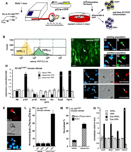
Rb gene inactivation in the developing retina in vivo, we introduced a Cre-expressing plasmid (pCre-vYFP) into the eyes of newborn RbLox/- mice (Fig. 5A). Real-time RT-PCR
BMC Biology 2006, 4:14 http://www.biomedcentral.com/1741-7007/4/14
Acute inactivation of Rb in the developing mouse retina Figure 5
Acute inactivation of Rb in the developing mouse retina. (A-G) In vivo or in vitro square-wave electroporation and purification of cells with acute Rb inactivation. (C) A retina after electroporation is shown. (D-G) Dissociated cells before and after FACS purification are shown. (H) Real-time PCR analysis of YFP+ purified cells after acute Rb inactivation; each sample was analyzed in duplicate and normalized to Gapdh and Gpi1. (I, J) Immunostaining of purified cells after Rb inactivation. (K-N)
To determine if retinal progenitor cells continue to divide after acute Rb inactivation, we scored the proportion of [3
H]thy-labeled BrdU+ cells. An example of a double-positive cell is shown in (K). (L) A significant proportion of progenitor cells was sensitive to deregulated proliferation after acute Rb inactivation, as indicated by the increase in double-positive cells. Data are normalized to account for the fraction of cells labeled with BrdU during a 1-h pulse. (M) The proportion of retinal progenitor cells that continue to divide after acute Rb inactivation is shown in the venus-Cre, YFP+ column. (N) Real-time PCR analysis of cell samples used in (L) and (M) revealed a significant increase in the expression of p107 and retinal progenitor cell markers
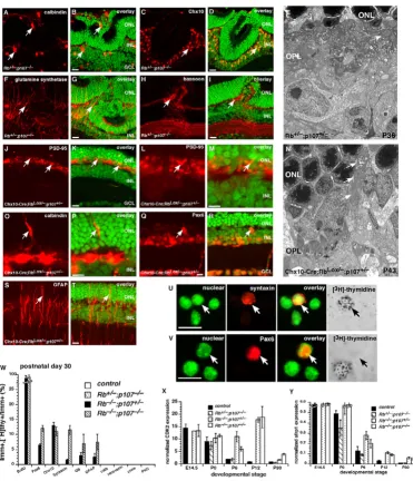
Haploinsufficiency of p107 in the developing mouse retina Figure 6
Haploinsufficiency of p107 in the developing mouse retina. Immunostaining of P30 Rb+/-; p107-/- retinas in the region of
the retinal dysplasia. (A, B) Antibodies against calbindin were used to identify horizontal cells; (C, D) those against Chx10, bipolar cells; (F, G) those against glutamine synthetase, Müller glia; and (H, I) those against bassoon, synapses in the OPL and IPL. (E) EM analysis of the P30 Rb+/-; p107-/- retinas showing the OPL with normal organization of synaptic connections in
con-trast to the Rb-deficient retinas. (J-T) Immunostaining of P14 Chx10-Cre;RbLox/-; p107+/- retinas in the region of Cre-mediated Rb
inactivation using antibodies to OPL synapses (PSD-95, J-M), horizontal cells (calbindin, O, P), amacrine/progenitor cells (Pax6,
Q, R), and reactive Müller glia (GFAP, S, T). (N) EM analysis of the P14 Chx10-Cre;RbLox/-; p107+/- retinas showing the OPL
with disrupted synaptic connections and an apical horizontal process (arrows). (U-Y) To determine if there are any ectopically dividing cells in Rb+/-; p107-/-, Rb-/-; p107+/- or Rb-/-; p107-/- retinas at E14.5, P0, P6, P12 and P30, we labeled retinas with [3H]thy
and BrdU for 1 h and then dissociated and immunostained the cells with antibodies against 12 markers of different retinal cell types. BrdU and [3H]thy always colocalized. (W) The only markers that colocalized with [3H]thy were amacrine/progenitor
cell markers Pax6 and syntaxin and bipolar/progenitor cell marker Chx10. There were also some reactive Müller glia, as indi-cated by GFAP, that incorporated [3H]thy. Ectopic proliferation was more prevalent in the double-knockout retinas than in the
other genotypes. Each bar is the average of triplicate samples scored in duplicate. (X, Y) Real-time PCR analysis demonstrated that throughout development there were more progenitor cell markers such as Cdk2 and Sfrp1 expressed in the double-knockout and Rb-/-; p107+/- retinas. Abbreviations: GCL, ganglion cell layer; INL, inner nuclear layer; ONL, outer nuclear
BMC Biology 2006, 4:14 http://www.biomedcentral.com/1741-7007/4/14
chronic Rb gene inactivation in the developing retina (Fig. 3). There was little change in p130 expression (Fig. 5H).
Next, we determined whether acute inactivation of Rb
altered rod development. Real-time RT-PCR analysis of RNA samples indicated that the early photoreceptor genes
Nrl and Nr2e3 were downregulated, as were several other photoreceptor-specific genes (Fig. 5H and data not shown). The cells that did not differentiate into photore-ceptors resembled progenitor cells, as indicated by the increased expression of Eya2 and Fgf15 (Fig. 5H) [27]. To further verify these developmental defects, we immunos-tained the purified cells that had undergone acute Rb inac-tivation and compared them to the control cells (Fig. 5I, J). Those data were consistent with real-time PCR data showing that acute inactivation of Rb not only leads to a compensatory increase in p107 expression, but also dis-rupts rod development. Results of experiments in which
p107 was acutely inactivated in wild-type E14.5 retinas by using an siRNA to p107 [20] were similar to those in the
p107-knockout retinas, i.e., Rb was upregulated in a com-pensatory manner (data not shown).
Proliferating cells and nondividing or quiescent MEFs exhibit differences in the timing of their compensatory upregulation of p107 after Rb inactivation [20]. To test for differences in compensation in proliferating retinal pro-genitor cells and transition cells [28] in the developing ret-ina, we acutely inactivated Rb at P3. Even though the majority (~ 95%) of cells in the P3 retina are postmitotic [24], a disproportionate number of proliferating retinal progenitor cells (42% ± 5.5%) underwent ectopic rounds of cell division (Fig. 5K, L; see Additional file 3). Retinal progenitor cells and transition cells were distinguished by BrdU labeling prior to electroporation (Fig. 5A). This find-ing suggests that retinal progenitor cells in which Rb has been acutely inactivated are susceptible to deregulated proliferation. A similar experiment in which all proliferat-ing retinal progenitor cells in the P3 retina were labeled with [3H]thy for 24 h prior to acute Rb inactivation
showed the same result (Fig. 5M). Specifically, even though retinal progenitor cells comprise only 7% of the total cell population at P3, they comprised 74% ± 4% of the cells that continued to divide after acute Rb inactiva-tion (Fig. 5M). Real-time RT-PCR analysis of the samples demonstrated that p107 was upregulated in a compensa-tory manner and that retinal progenitor cell genes such as
Sfrp1, Erdr1 and Chx10 were also upregulated (Fig. 5N).
To test if p107 compensation after acute Rb inactivation prevents deregulated proliferation, we acutely inactivated
p107 by using a previously characterized SiRNA [20] in retinal cells in which Rb was chronically inactivated. We found that proliferation significantly increased from 18%
vation (see Additional file 4). Moreover, the cells that con-tinued to divide expressed retinal progenitor cell markers Chx10, Pax6 and syntaxin and were BrdU+ retinal progen-itor cells at the time of p107 inactivation (see Additional file 4). Together, these data support the idea that p107 compensation is important for preventing deregulated proliferation in retinal progenitor cells whether Rb is inac-tivated in an acute or chronic manner.
Haploinsufficiency of p107 in the absence of Rb
Having shown that reciprocal compensation between Rb and p107 in the developing retina prevents deregulated proliferation of retinal progenitor cells, we tested whether a single copy of Rb was sufficient to compensate for p107 deficiency and whether a single copy of p107 was suffi-cient to compensate for Rb deficiency. Immunostaining of
Rb+/-; p107-/- retinas between P6 and P30 confirmed the
presence of minor focal retinal dysplasia, as reported pre-viously [13]. The laminar structure and synaptogenesis outside the regions of focal dysplasia were not substan-tially disrupted (Fig. 6A–I and data not shown), as con-firmed by EM analysis of the OPL (Fig. 6E). In addition, EM analysis confirmed that rod photoreceptors [6] and horizontal cells [9] form normally with just one copy of
Rb and no p107 (Fig. 6E). Even in the regions of the most severe dysplasia (Fig. 6A–I), cells were in their appropriate cellular layers, and synaptic markers were present in the corresponding plexiform layer sublaminae. EM analysis suggests that these focal dysplastic lesions are not formed by preneoplastic retinoblastoma cells. That is, there is no evidence for immature tumor cells localized to the lesions.
In contrast to the Rb+/-; p107-/- retinas, the Chx10-Cre;RbLox/ -; p107+/- retinas exhibited much more severe
developmen-tal defects. Immunostaining revealed deregulated prolifer-ation and laminprolifer-ation in an apical-basal mosaic pattern that was consistent with the expression of Cre from the
Chx10 promoter of the Chx10-Cre transgene [9,29]. Rod photoreceptors were not completely lost due to the mosaic pattern of gene inactivation by the Chx10-Cre
allele [29]. Defects in OPL synaptogenesis (Fig. 6J–T) were confirmed by EM analysis of P43 Chx10-Cre;RbLox/-; p107+/ - retinas (Fig. 6N). Müller glia showed upregulated
expres-sion of GFAP, which is a marker of reactive gliosis (Fig. 6S, T); however, GFAP expression was not detectable in Müller glia of the Rb+/-; p107-/- retinas.
To quantify the proportion and types of cells that were proliferating in Rb+/-; p107-/-, Rb-/-; p107+/- and Rb-/-; p107-/ - retinas during development, we labeled P0, P6, P12 and
P30 retinas with [3H]thy for 1 h, dissociated the retinas
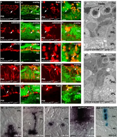
Retinal progenitor cells continue to proliferate throughout development in the absence of Rb and p107 Figure 7
Retinal progenitor cells continue to proliferate throughout development in the absence of Rb and p107. (A-T)
Immunostaining of P30 Chx10-Cre;RbLox/-; p107-/- retinas by using antibodies against (A-D) BrdU to identify ectopically dividing
cells; (E-H) calbindin, horizontal cells and Chx10, bipolar/progenitor cells; (M-P) GFAP, reactive Müller glia; (Q, R) cone arrestin, cone photoreceptors and (S, T) Pax6, amacrine/progenitor cells. (U, V) Electron micrographs showing that most retinal structures are disrupted in the absence of Rb and p107 and that continued proliferation eventually leads to retinoblast-oma. (W-Z) Newborn RbLox/Lox; p107-/- mice were injected with a retrovirus expressing Cre (LIA-Cre), and 3 weeks later, the
BMC Biology 2006, 4:14 http://www.biomedcentral.com/1741-7007/4/14
markers such as recoverin (photoreceptors), cone arrestin (cones) or PKCα (bipolar cells) incorporated [3H]thy (Fig.
6U–W; see Additional files 5,6,7,8). Primarily, cells expressing progenitor cell markers (see Discussion) such as syntaxin, Pax6 or Chx10 incorporated [3H]thy (Fig.
6U–W; see Additional files 5,6,7,8). A small proportion of Müller glia also incorporated [3H]thy, a finding that was
consistent with reactive gliosis-associated proliferation (Fig. 6S, T) [30]. The overall distribution of cell types was not significantly altered in Rb+/-; p107-/- retinas (see
Addi-tional files 5,6,7,8), but as expected, the number of rods was reduced in Chx10-Cre;RbLox/-; p107+/- retinas (see
Addi-tional files 5,6,7,8).
TUNEL assay results revealed no difference in the propor-tion of apoptotic cells in Rb+/-; p107-/- retinas and that of their wild-type littermates. The number of apoptotic cells in Chx10-Cre;RbLox/-; p107+/- retinas was slightly higher at P12 and P30, which corresponded to the stages of increased proliferation (see Additional files 5,6,7,8). Real-time RT-PCR analysis revealed increased expression of the retinal progenitor cell genes Cdk2 (Fig. 6X) and Sfrp2 (Fig. 6Y). Together, these data suggest that one copy of Rb in Rb+/-; p107-/- retinas is sufficient for much of retinal development (with the exception of minor focal dyspla-sia), but one copy of p107 in Chx10-Cre;RbLox/-; p107+/ - retinas is haploinsufficient to regulate proliferation. Spe-cifically, in retinal progenitor cells, two copies of the p107 gene are required to compensate for loss of Rb, but only one copy of Rb is required to compensate for loss of p107.
Deregulated proliferation in Rb;p107-deficient retinal progenitor cells
It has been shown previously that retinoblastoma forms in Rb;p107-deficient retinas [10,11,14,15]. Therefore, we propose that reciprocal compensation between Rb and p107 in retinal progenitor cells prevents retinoblastoma in mice. If this is true, then we predict that retinal progen-itor cells will continue to divide throughout development (E14-P30) in Rb;p107-deficient retinas. This is precisely what we found (Fig. 7A–D; see Additional files 5,6,7,8). The proliferating cells were detected in a mosaic pattern, which was consistent with Rb inactivation by the Chx10-Cre transgene (Fig. 7A–D) [9,29]. These immature prolif-erating cells disrupted synaptogenesis in the OPL (Fig. 7E–H) and expressed the progenitor cell markers syntaxin, Pax6 and Chx10 (Figs. 6W, 7I–L; see Additional files 5,6,7,8) [31-33]. With the exception of activated Müller glia (Fig. 7M–P; see Additional files 5,6,7,8), few of the proliferating cells expressed markers of differentiated reti-nal cells. It is important to point out that syntaxin, Pax6 and Chx10 are expressed in both retinal progenitor cells and postmitotic differentiated cells. However, the combi-nation of BrdU, Pax6, Chx10 and syntaxin
immunoreac-in electron micrographs lends support to their identifica-tion as retinal progenitor cells.
In contrast to findings from a recent study [10], we did not detect any defect in cone photoreceptor production in ret-inal sections (Fig. 7Q, R) or in dissociated cells scored at P0, P6, P12 or P30 (see Additional files 5,6,7,8). EM anal-ysis of Chx10-Cre;RbLox/-; p107-/- retinas revealed immature
cells spanning all cellular layers of the retina (Fig. 7U, V). These electron micrographs support the hypothesis that Chx10+, Pax6+, BrdU+ cells in the ONL and INL are reti-nal progenitor cells rather than differentiated cells. In addition, real-time PCR analysis demonstrated an increase in the expression of retinal progenitor cell mark-ers in Chx10-Cre;RbLox/-; p107-/- retinas at P6, P12 and P30
(Fig. 6X, Y).
Our analysis of the Rb;p107-deficient retinas over the course of postnatal retinal development showed that at all stages of development studied, there are ectopically divid-ing cells with morphologic features of retinal progenitor cells that express markers of retinal progenitor cells. Our data support the hypothesis that the role of reciprocal compensation between Rb and p107 is to prevent deregu-lated proliferation of retinal progenitor cells. If this is true, then we would expect that inactivation of Rb and p107 in a single proliferating retinal progenitor cell would lead to clonal expansion.
We infected proliferating retinal progenitor cells in new-born RbLox/-; p107-/- mice with the Cre-expressing
retrovi-rus LIA-Cre (Fig. 7W–Z). We observed significantly larger clone sizes and changes in cell-fate specification (Fig. 7Y, Z; see Additional file 9). To quantify the deregulated pro-liferation, we used the NIN-Cre retrovirus, which expresses nuclear lacZ and Cre, in E17.5 RbLox/Lox; p107
-/-retinas; NIN virus was used as a control. Clone size increased and was consistent with deregulated retinal pro-genitor cell proliferation. In control retinas infected with NIN, 248 of 313 (80%) clones were one cell, and 11 of 313 (3.5%) contained more than two cells. In retinas infected with NIN-Cre, 100 of 190 (52%) clones were one cell, and 42 of 190 (24%) contained more than two cells (Fig. 7AA). Similar results were seen in studies using the E1A 13S oncogene [6,15].
Proliferation of progenitor cells and postmitotic transition cells in Rb;p107-deficient retinas
TheRb and p107 prevents newly postmitotic cells from re-entering the cell cycle rather than preventing deregulated proliferation of retinal progenitor cells. To test this possi-bility directly, we labeled all proliferating retinal progeni-tor cells in P3 RbLox/Lox; p107-/- retinas with [3H]thy (Fig.
8A), inactivated Rb expression by electroporating a plas-mid that expressed Cre and vYFP and then incubated the retinas with BrdU. Cells that were BrdU+, vYFP+ and [3H]thy+ were retinal progenitor cells at the beginning of
the experiment that continued to divide after acute Rb
inactivation. In contrast, cells that were BrdU+, vYFP+ and [3H]thy- were postmitotic transition cells at the beginning
of the experiment that re-entered the cell cycle after acute
Rb inactivation. The time point P3 was selected because the vast majority (~ 93%) of cells in the retina at that stage are postmitotic [24]. If a postmitotic cell re-enters the cell cycle after acute Rb inactivation in RbLox/Lox; p107-/- retinas,
then the majority of BrdU+ cells would be [3H]thy-. If a
retinal progenitor cell is more susceptible to deregulated proliferation after acute Rb inactivation, then we would predict that the majority of BrdU+ cells would be [3H]thy+. If both retinal progenitor cells and postmitotic
cells divide after acute Rb inactivation, then the propor-tions of BrdU+, [3H]thy- cells and BrdU+, [3H]thy+ cells
will reflect the proportions of postmitotic cells and mitotic cells in the starting population, respectively.
Retinal progenitor cells are sensitive to deregulated proliferation after acute inactivation of Rb and p107
Figure 8
Retinal progenitor cells are sensitive to deregulated proliferation after acute inactivation of Rb and p107. (A)
To determine if retinal progenitor cells are sensitive to deregulated proliferation after acute Rb inactivation in a p107-deficient genetic background, we labeled RbLox/Lox; p107-/- P0 or P3 retinas for 24 h with [3H]thy and then electroporated them with a
Cre-venus-YFP plasmid. After 3 days in culture, the retinas were labeled for 1 h with BrdU and dissociated, and the YFP+ and YFP- cells were purified by FACS. (B-F) The cells were then analyzed by immunostaining, single-cell scoring and real-time PCR.
(B, C) Colocalization of BrdU and [3H]thy demonstrated that most cells that continued to proliferate after Rb inactivation
were retinal progenitor cells. (D) Real-time RT-PCR analysis confirmed that the proliferating cells expressed retinal progenitor cell markers. (E, F) Immunostaining and colocalization with [3H]thy further confirmed the retinal progenitor cell
BMC Biology 2006, 4:14 http://www.biomedcentral.com/1741-7007/4/14
Most (79.8% ± 6%) ectopically dividing cells were BrdU+, [3H]thy+ cells (Fig. 8B, C). Consistent with this finding,
results from real-time PCR and immunostaining analyses revealed that ectopically dividing cells expressed several retinal progenitor cell markers including Pax6, Chx10,
Syntaxin, Erdr1 and Sfrp1 (Fig. 8D–F). However, there was a small increase in the proportion of BrdU+, [3H]thy- cells
(Fig. 8C), which leaves open the possibility that either a retinal progenitor cell or a transition cell is the cell of ori-gin for retinoblastoma (discussed in [28]).
Discussion
We report here that in the developing mouse retina p107 is expressed in embryonic retinal progenitor cells, and Rb is expressed in postnatal retinal progenitor cells. When
p107 is inactivated, Rb is upregulated in a compensatory manner, and when Rb is inactivated, p107 compensates. The p130 protein is expressed during the late stages of retinogenesis and persists in the INL and GCL in the adult retina. Rb and p130 are expressed redundantly in these cells. There was no evidence of compensatory changes in p130 expression following either Rb or p107 inactivation. Compensation by p107 occurred when Rb was inactivated acutely or chronically in the developing mouse retina. Two copies of the p107 gene were required to prevent deregulated proliferation of Rb-deficient retinal progeni-tor cells, making p107 haploinsufficient for compensation in the developing retina. In contrast, only one copy of Rb
was required to regulate proliferation of p107-deficient retinal progenitor cells. We propose that reciprocal com-pensation between Rb and p107 prevents deregulated proliferation of retinal progenitor cells and retinoblast-oma in mice. Similarly, redundant expression of Rb and p130 may help to prevent retinoblastoma in mice.
In contrast to mice, human fetal retinal progenitor cells primarily express RB1 during development. When RB1
was acutely inactivated in human fetal retinas, there was little if any compensatory upregulation of p107. This may explain why humans are susceptible to retinoblastoma following RB1 gene mutations, but mice require inactiva-tion of Rb and p107 or Rb and p130.
Overlapping and unique functions of the Rb family in the developing mouse retina
Immunostaining, in situ hybridization, real-time RT-PCR and immunoblot analyses revealed that p107 is expressed primarily in proliferating retinal progenitor cells of the embryonic mouse retina. Pulse-labeling with [3H]thy
demonstrated that p107 is downregulated as cells exit the cell cycle. There were no defects in cell-fate specification or differentiation in p107-deficient retinas; thus, we propose that p107 primarily regulates retinal progenitor cell prolif-eration in the embryonic retina. Rb is expressed in
postna-glia. Consistent with a role in cell-fate specification or dif-ferentiation, Rb was expressed throughout the cell cycle and continued to be expressed as cells entered G0. In the absence of Rb, immature cells persist in the ONL and rod photoreceptors failed to form, a finding that indicates that Rb regulates the developmental processes in this specific sublineage.
Recent studies have extended these findings by focusing on the mechanism of Rb's role in rod development. Sun and colleagues generated an exon-specific knock-in allele of Rb with a single amino acid substitution (R654W) that reduces Rb binding to E2F1, E2F2 and E2F3 [34]. In
Chx10-Cre;Rb654/Lox retinas, rods still fail to form. This
finding led to a model in which Rb regulates a rod repres-sor gene through E2F1, E2F2 or E2F3, and in the absence of Rb, the rod lineage is blocked. Moreover, we predict that inactivation of the E2F that regulates this putative "rod repressor" would rescue the rod developmental defect. Preliminary data have shown that Rb;E2F1 -defi-cient retinas have normal rod development, a finding that suggests that E2F1 is involved in this important develop-mental process.
Our data suggest that Rb and p107 share the same role in regulating mouse retinal progenitor cell proliferation but the role of Rb is unique in rods. An extensive genetic anal-ysis of p130 is beyond the scope of this manuscript, but based on our expression studies and [3H]thy-labeling
experiments, we predict that Rb and p130 are redundant in newly postmitotic cells of the INL and GCL. It is not known if p130 is required for terminal cell cycle exit in the developing retina, for maintaining cells in the postmitotic state or both.
A recent study published by Spencer and colleagues char-acterized the expression of the Rb family in developing mouse retina and adult human retina [35]. Our data differ from theirs, and we have presented the most relevant dif-ferences in Figure 1. Our analysis combined several inde-pendent methods (real-time RT-PCR, in situ hybridization, immunostaining of tissue sections, immu-nostaining of [3H]thy-labeled dissociated cells and
gents being used. For example, it is possible that the Rb, p107 and p130 expression patterns in our study or that of Spencer and colleagues are broader than reported due to the limited sensitivity of the antibodies used. We have attempted to minimize such limitations by combining several independent approaches such as in situ hybridiza-tion, immunostaining, real time RT-PCR and dissociated cell staining. Nontheless, we anticipate that the retinal expression pattern of the Rb family will be further refined as new reagents become available.
Rb family redundancy and compensation in the developing mouse retina
The Rb family proteins are expressed in a dynamic manner during mouse retinal development. We show here that p107 is expressed in proliferating retinal progenitor cells in the embryonic retina and is downregulated as cells exit the cell cycle and undergo terminal differentiation. Around birth, the expression of p107 tapers off, and that of Rb takes it place in proliferating retinal progenitor cells. Rb expression persists in postmitotic differentiating neu-rons and glia of the mouse retina. The p130 protein is expressed in the late postnatal period of mouse retinal development. As cells exit the cell cycle, they upregulate p130, and its expression persists in postmitotic differenti-ating cells of the INL and GCL. Based on the expression pattern alone, one might predict that proliferation would be deregulated in p107-deficient embryonic retinas and Rb-deficient postnatal retinas. However, we report here that when p107 is inactivated, Rb is upregulated in a com-pensatory manner in the embryonic retina. Similarly, when Rb is inactivated, p107 is upregulated in a compen-satory manner in the postnatal retina. There is no evi-dence for compensation by p130 in either Rb- or p107-deficient retinas.
One of the interesting features of the compensation that we uncovered in this study is the haploinsufficiency of
p107 in this process. We show here that two copies of p107
are required for efficient compensation of Rb loss, but only one copy of Rb is required to compensate for p107
loss. This may indicate that there are different mecha-nisms for Rb and p107 gene compensation or that retinal progenitor cells exhibit different sensitivities to Rb and p107 protein levels. One intriguing possibility is that p107
haploinsufficiency for compensation may reflect the dif-ferential binding of Rb and p107 to different E2F family members.
It has been shown previously that p107 is an E2F-respon-sive gene; therefore, the mechanisms for p107 compensa-tion most likely involve a direct feedback loop [36]. However, the mechanism for Rb compensation in the absence of p107 has not been elucidated. We propose that Rb-deficient retinal progenitor cells fail to form
retino-blastoma because p107 is upregulated in a compensatory manner. However, it is possible that there is some over-lapping, redundant expression around P0 in the develop-ing mouse retina. Interestdevelop-ingly, Rb and p130 are expressed redundantly in the INL and GCL. The recent observation that retinoblastoma develops in Rb;p130-deficient retinas suggests that redundant expression of these two Rb family members prevents retinoblastoma in mice [11].
Previous studies using Rb-/- MEFs demonstrated that
inac-tivation of Rb in proliferating cells leads to compensatory upregulation of p107 and quiescence after serum starva-tion[20]. The p107 gene is persistently expressed in these quiescent Rb-deficient MEFs and is required for maintain-ing them in G0. When Rb was acutely inactivated in qui-escent RbLox/Lox MEFs, p107 was not immediately
upregulated in a compensatory manner, and the cells re-entered the cell cycle [20]. Therefore, chronic or acute inactivation of Rb can lead to very different outcomes in MEFs. The molecular changes that occur in MEFs as they become quiescent have been compared with those that accompany terminal cell cycle exit during development [20]. However, in the developing CNS, postmitotic neu-rons not only exit the cell cycle, but also migrate to their appropriate layer, extend axons and dendrites and form synaptic connections.
To test if there was any difference in p107 compensation following acute versus chronic Rb inactivation in the developing retina, we developed genetic tools [16] to acutely inactivate Rb in this context. Our data show that p107 compensation occurs when Rb is inactivated acutely or chronically in the developing retina. In addition, rod photoreceptors failed to form when Rb was acutely inacti-vated. These data suggest that p107 compensates for Rb in proliferating retinal progenitor cells, but it cannot take the place of Rb in differentiating rod photoreceptors.
We extended our acute inactivation studies to knock out
BMC Biology 2006, 4:14 http://www.biomedcentral.com/1741-7007/4/14
rare transition cells re-enter the cell cycle and expand to generate retinoblastoma or if only retinal progenitor cells continue to proliferate, as shown here [28].
Our data on the proliferation of Rb;p107-deficient retinal cells during development contrasts with previous studies by Chen and colleagues [10]. We show that ectopically dividing retinal cells are present at P0, P6, P12 and P30, whereas the previous study argued that proliferation ceased at P30. This difference at P30 may be due to the more sensitive and reproducible [3H]thy labeling we used
in our study compared to the BrdU labeling used by Chen et al. Alternatively, it could reflect the non-cell-autono-mous effect of broad Rb inactivation in Pax6-Cre;RbLox/Lox;
p107-/- retinas examined by Chen et al. compared with the
more restricted mosaic Rb inactivation in Chx10-Cre;RbLox/ -; p107-/- retinas that we investigated [16].
Another difference between our data and the study previ-ously published by Chen and colleagues involves the interpretation of the markers expressed in proliferating cells of the Rb;p107-deficient retinas. We found that at P0, the ectopically dividing cells were largely Pax6+ (see Addi-tional file 5). By P6, the proliferating cells were Pax6+, Chx10+ (see Additional file 6) and by P12, they were Pax6+, Chx10+ and syntaxin+ (see Additional file 7). Pax6
[32] and syntaxin [31] are expressed in both retinal pro-genitor cells and differentiating amacrine cells. Chx10 is expressed in retinal progenitor cells and in differentiated bipolar cells and Müller glia [29]. Interestingly, at P12 we noted that GFAP+ Müller glia were also [3H]thy+. This
finding is consistent with proliferation associated with reactive gliosis in Rb;p107-deficient retinas [30]. Chx10 immunopositivity may mark the proliferating Müller glia, bipolar cells or progenitor cells at this stage. However, we do not believe that the Chx10+ cells are bipolar cells, because there were no PKCα+ cells that incorporated [3H]thy at P12. At P30, most of the proliferating [3H]thy+
cells were syntaxin+. A smaller proportion were Pax6+, recoverin+ and PKCα+. Recoverin is expressed in a subset of bipolar cells as well as photoreceptors, so the PKCα+ and recoverin+ cells that incorporated [3H]thy are most
likely a small cohort of bipolar cells that re-entered the cell cycle.
Based on the timeline of marker colocalization with [3H]thy that we present here, we propose that a Pax6+,
Chx10+ and syntaxin+ retinal progenitor cell population that is biased toward the amacrine/horizontal cell fate continues to proliferate through development in Chx10-Cre;RbLox/-; p107-/- retinas and eventually gives rise to
retin-oblastoma. This model is consistent with the extensive differentiation of mouse retinoblastomas along amacrine/ horizontal cell lineage (Johnson and Dyer, in
prepara-tion in reactive Müller glia and possibly bipolar cells at P30, but based on the absence of glial and bipolar markers in mouse retinoblastomas, we propose that these cells do not contribute significantly to retinoblastoma in mice.
Chen and colleagues [10] showed that Pax6+, Chx10+, Prox1+, Math3+, Crx+ and Hes5+ cells incorporated BrdU. With the possible exception of Crx, each of these markers is expressed in both progenitor cells and differen-tiated cells [18,32,33,37-42]. We have found that several additional retinal progenitor cell markers (syntaxin 1, Fgf15, Sfrp1, Erdr1 and Eya2) are expressed in proliferat-ing cells of Rb;p107-deficient retinas. Overall, 10 of the 11 genes that are expressed in proliferating Rb;p107-deficient retinal cells have one feature in common; they are all expressed normally in retinal progenitor cells. The mor-phological features of the Rb;p107-deficient retinal cells in electron micrographs are also consistent with retinal pro-genitor cells. While these data support the hypothesis that retinal progenitor cells continue to divide during develop-ment of Rb;p107-deficient retinas and eventually give rise to retinoblastoma in mice, marker expression alone can-not distinguish between a progenitor cell and a transition cell as the cell of origin for retinoblastoma (discussed in [28]). Definitive proof of the cell of origin for retinoblas-toma will require clonal analysis of tumor formation from a single cell in vivo.
Retinoblastoma susceptibility in mice and humans
When the Rb-knockout mouse was generated over 13 years ago, it was expected that retinoblastoma would develop in Rb+/- mice, as it does in RB1+/- humans.
How-ever, that was not the case. We show here that the normal expression of Rb family members is dynamic during mouse retinal development and that reciprocal compen-sation between Rb and p107 may prevent retinoblastoma in mice.
In the human fetal retina, RB1 is the major family mem-ber expressed during much of retinal development. There is little or no expression of p107. We acutely inactivated
RB1 in primary human fetal retinas by electroporating an SiRNA to RB1. We demonstrated that although the RB1 protein was downregulated 20-fold, there was no com-pensation by p107 or p130. These data suggest that humans are susceptible to retinoblastoma formation after
RB1 inactivation, because they cannot upregulate p107 in a compensatory manner. It is possible that the 20-fold reduction in RB1 protein induced by the SiRNA used for these experiments is not sufficient to induce p107 com-pensation. However, we do not believe this is the case because another RB1 target gene (p14ARF) is induced by
explana-that the p107 promoter is sequestered in an inactive chro-matin conformation in human fetal retina, but it is in an active chromatin conformation in mouse retinal progeni-tor cells and is thus poised to respond to a loss of Rb. Indeed, it is believed that promoters and genes that have been recently transcribed remain in an active chromatin configuration. Therefore, this model is consistent with the expression data for mouse and human p107. That is, the embryonic expression of p107 in the mouse retina results in an open chromatin conformation, thereby making it readily accessible for compensatory upregulation if Rb is inactivated. Detailed promoter and transcription analyses will be required to test this hypothesis.
Conclusion
We have found that Rb and p107 are expressed in a largely non-overlapping pattern during mouse retinal develop-ment and that reciprocal compensation between these two Rb family members prevents retinoblastoma in mice. p107 compensation occurs whether Rb is inactivated chronically or acutely. In human retinal development, there is very little p107 expressed and compensation does not occur when RB1 is inactivated using an siRNA. We propose that this difference in compensation between mice and humans may explain why humans are suscepti-ble to retinoblastoma following RB1 gene inactivation but mice require simultaneous inactivation of the Rb and
p107 genes.
Our data also shed light on the retinoblastoma cell of ori-gin. Proliferating retinal progenitor cells are dispropor-tionately more susceptible to deregulated proliferation following Rb;p107 gene inactivation in comparison to newly postmitotic transition cells [28]. Consistent with this observation, 10 of 11 genes expressed in proliferating
Rb;p107-deficient retinal cells are normally expressed in retinal progenitor cells. Electron micrographs of Rb;p107 -deficient retinal cells provides further support for an immature cell with features of progenitor cells as the retin-oblastoma cell of origin. However, based on our data, it remains a formal possibility that a postmitotic amacrine or horizontal cell is the retinoblastoma cell of origin. Ongoing lineage studies should help to determine if a progenitor cell, a postmitotic neuron or both can give rise to retinoblastoma in mice.
Methods
Mouse strainsRb+/- mice were obtained from The Jackson Laboratory
(Bar Harbor, ME), and RbLox/Lox mice were obtained from
the National Cancer Institute. The p107-knockout mice were obtained from Dr. Tyler Jacks (MIT). Chx10-Cre mice were obtained from Dr. Connie Cepko (Harvard Medical School). All mice were crossed to C57Bl/6 mice purchased from Charles River Laboratories (Wilmington, MA). The
St. Jude Children's Research Hospital Institutional Animal Care and Use Committee approved all of the animal experiments.
Antibodies, immunostaining, BrdU, [3H]thymidine and
TUNEL studies
We immunolabeled retinal cryosections from mice of var-ious genotypes (p107-/-, p107+/-, p107+/+) at various
post-natal stages (P3, P6, P12 and adult) and dissociated retinas (500 cells per sample in triplicate) as previously described [22,43]. The list of antibodies used is provided (Supplementary Table 9). To label S-phase retinal progen-itor cells, we incubated freshly dissected retinas in 1 ml explant culture medium containing [3H]thy (5 µCi/ml; 89 µCi/mmol) or 10 µM BrdU for 1 h at 37°C. For these pulse-labeling experiments, retinas were dissected at four stages of development (E14.5, E17.5, P0, and P2) and exposed to the label for various durations (0, 4, 8, 16 and 24 h) to allow retinal progenitor cells to progress through the cell cycle. The p27Kip1 protein, which is upregulated at
G1/G0, was used as an internal control (Fig. 2D, H, L), because its expression during retinal development has been characterized previously [2,22,23].
Autoradiography and BrdU detection were carried out as described previously [22,43]. For apoptosis analysis, we sectioned (14-µm) retinas on a cryostat. We used the colorimetric TUNEL apoptosis system (Promega, Madi-son, WI) per the manufacturer's instructions; however, for detection, we used tyramide-Cy3 (Perkin-Elmer, Welles-ley, MA) rather than the colorimetric substrate.
Electroporation and FACS
Retinas were electroporated in vivo at P0 by injecting 0.5
µl CsCl preparation plasmid DNA (5 µg/µL) into the eye. Electroporation consisted of five 50-µsec pulses of 80 V each separated by 950-µsec recovery periods. As a control, retinas were electroporated with a plasmid lacking Cre. For explant cultures, the DNA was purified and resus-pended in HBSS at 1 µg/µL.
Electroporation consisted of five pulses of 25 V for 50 µsec each with 950-µsec recovery periods. For FACS purifica-tion of electroporated cells, retinas were dissociated as described previously [22,43], resuspended in explant cul-ture medium and sorted using vYFP fluorescence on a Bec-ton-Dickson FACS system (Rockville, MD).
Real-time RT-PCR
Real-time RT-PCR experiments were performed using the ABI 7900 HT Sequence Detection System (Applied Biosys-tems, Foster City, CA). Primers and probes were designed using Primer Express® software (Applied Biosystems).
Taq-Man® probes were synthesized with 5'-FAM and 3'-BHQ.