M E T H O D O L O G Y
Open Access
Detection of Epstein-Barr virus (EBV) in human
lymphoma tissue by a novel microbial detection
array
Joseph Tellez
1, Crystal Jaing
2, Jun Wang
3, Ralph Green
1and Mingyi Chen
1*Abstract
Background:Infectious agents are estimated to play a causative role in approximately 20% of cancers worldwide. Viruses, notably the Epstein-Barr virus (EBV), are associated with 10-15% of B-cell lymphomas and are found at a higher frequency in immunosuppressed patients. In this study, we screened human lymphoma tissues using a novel Lawrence Livermore Microbial Detection Array (LLMDA), a comprehensive detection system that contains probes for all sequenced viruses and bacteria. This technology has been applied to identify pathogen-associated diseases.
Results:We evaluated samples from 58 cases with various lymphoid tissue disorders using LLMDA. These included
30 B-cell lymphomas (9 indolent and 21 aggressive type), 2 T-cell lymphomas and 2 NK/T cell lymphomas, 4 plasmacytomas as well as 8 specimens of benign lymphoid tissue. Five of 21 high-grade B-cell lymphomas were positive for Epstein-Barr virus-encoded small RNA (EBER+), while all the indolent B-cell lymphomas were EBER-. Similarly, both NK/T cell lymphomas were EBER+, and the benign tissues were EBER-. We also screened 10 cases of post-transplant lymphoproliferative disorder (PTLD). Five of these cases (4 B-cell lymphomas and 1 NK/T cell lymphoma) were EBER+, and the remaining five cases were EBER-.
Conclusions: We have confirmed the reliability of the LLMDA methods by detecting EBV in EBV-positive
lymphomas while observing no false-positive results in EBV-negative lymphomas. The LLMDA technique provides a sensitive and alternative method for identifying known viral pathogen associated with tumors and may prove useful for future clinical identification of novel cancer-associated viral pathogens.
Keywords: Epstein-Barr virus (EBV), Lymphoma, Post-transplant lymphoproliferative disorder (PTLD), Lawrence
Livermore Microbial Detection Array (LLMDA)
Background
Infection associated cancers are on the rise worldwide due to shifting pathogen habitats, a more interconnected world and most importantly an aging population with a longer life-expectancy [1,2]. As humans age, the immune systems become dysregulated due to immunosenescence leaving them more susceptible to infection (comorbidi-ties and environmental exposures) [3-5]. It is estimated that at least 20% of the global cancer incidence is caused by infectious agents, with 10-15% of those caused by viruses [6]. Over the past three decades, research has
linked a number of cancers to infectious agents includ-ing viruses (Epstein-Barr virus (EBV), human papilloma virus (HPV), hepatitis B, human T-lymphotropic retro-virus, Kaposi’s sarcoma-associated herpesvirus (KSHV), and Merkel cell virus), bacteria (Helicobacter pylori), and parasites (Schistosoma haematobium, Clonorchis sinesis) [7]. Over 90% of the world’s population is in-fected with EBV [8]. The organ transplant patients are prone to viral infections due to immunosuppression which leaving them susceptible to post-transplant lym-phoproliferative disorder (PTLD) [9]. Immunocompetent individuals control EBV infection with EBV-specific cyto-toxic T lymphocytes (CTLs), while immunosuppressed transplant patients lack CTLs and allow EBV propagation that may lead to PTLD [8].
* Correspondence:[email protected] 1
Deptartment of Pathology and Laboratory Medicine, University of California, Davis Medical Center, PATH Bldg. 4400V Street, Sacramento, CA 95817, USA Full list of author information is available at the end of the article
EBV, or human herpesvirus 4, is one of the most com-mon viruses affecting humans. EBV is an episomal, double-stranded DNA virus that was discovered in a Burkitt lymphoma cell line by Epstein et al. [10-12]. EBV’s pro-miscuous tropism permits infection of a number of differ-ent cell types including B-cells, T-cells, NK-cells and epithelial cells [13-15]. Subsequently, EBV was found to be associated with various human malignancies, includ-ing nasopharyngeal carcinomas, PTLD, AIDS-associated lymphomas, T cell lymphomas, NK cell lymphomas and Hodgkin’s disease [11]. Importantly, EBV has been shown to induce B cell transformation [11]. EBV-positive lymph-omas can be divided into those occurring in immunodefi-cient individuals, which are virally driven lymphomas, such as PTLD and HIV-associated plasmablastic lymph-oma (PBL), and those occurring in immunocompetent in-dividuals. The latter group includes endemic and sporadic Burkitt lymphoma, and some T-and NK-cell malignancies. In the malignancies occurring in immunocompetent indi-viduals, EBV is a cofactor rather than the driving influence [10]. EBV likely exists as an episome with multiple copies in the host cell, making it easier to detect than viruses with one or few genomic copies per cell [12]. EBV epi-somes have been employed to determine the association of the virus with various aggressive types of lymphomas, indicating that it is likely involved in tumor progression but not tumor initiation, as might be assumed if present in indolent tumors [11].
While it is clear that EBV contributes to the progression of B-cell lymphoproliferative disease in immunosup-pressed patients, its role in lymphomagenesis is less clear in immunocompetent individuals [11]. However, the pres-ence of viral genomes in these lymphomas offers interven-tional targets and several approaches currently under evaluation which include adoptive immunotherapy, inter-feron, and small molecule targeting strategies in tumor virus biology [7]. A sensitive and comprehensive pathogen detection technology is critical to understand microbial profiles associated with lymphomagenesis and their contribution to the progression to high-grade lymphomas [16]. Sequencing produces the most comprehensive and unbiased information for microbial detection and dis-covery when analyzing nucleic acids from uncharac-terized samples, but high-throughput sequencing is time-consuming and expensive [17]. PCR is a cheap, fast and sensitive option, but it lacks the ability to detect the existence of large numbers of organisms simultaneously [18]. Microarray technology offers a reliable and sensitive alternative to sequencing and PCR when analyzing or screening tissue samples from patients with known or sus-pected pathological conditions [18]. Moreover, microar-rays are cheaper and faster than sequencing and permit detection of multiple microbes in the same sample [19]. In this study, we used the Lawrence Livermore Microbial
Detection Array (LLMDA), which is a pan-microbial detection array capable of detecting all sequenced virus, bacteria and plasmids, which uses a unique statistical method, Composite Likelihood Maximization Method for identifying multiple organisms in complex mixtures [18-21]. The family-specific probes selected were con-served enough to detect all known viral and bacterial organisms while containing sufficient sequence vari-ation for the detection of divergent species with homology to sequenced organisms [22,23]. Our previous studies have demonstrated the potential usage of the LLMDA for detection of a broad spectrum of pathogens in a di-verse set of clinical samples. We found that the microarray technique can detect both DNA and RNA viruses that are present in the same sample, as well as differentiate be-tween different virus subtypes [18-20].
In this study we demonstrated the reliability of the LLMDA technique in the detection of pathogen-associated lymphoproliferative diseases. The LLMDA accurately iden-tified EBV in EBV-positive samples and did not register false positive results in EBV-negative samples. We de-tected EBV in 5 out of 10 PTLD samples and showed that EBV-positivity is usually correlated with aggressive stages of lymphomas. We also demonstrated that the LLMDA can also be applied to detect EBV in Formalin Fixed Paraffin Embedded (FFPE) tissues which widens our sample pool and improves selection of relevant samples. Our results demonstrated that the LLMDA is a powerful tool for analyzing and detecting known microbial pathogens in lymphoproliferative disorders and potentially can be used as a diagnostic tool or to provide a basis for potential tar-geted and effective therapy regimens for these disorders.
Results
Detection of EBV in FFPE lymphoma samples
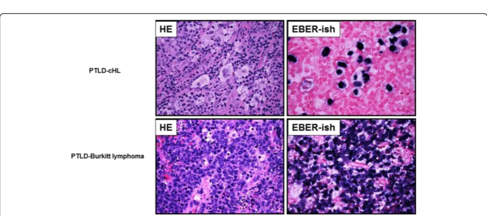
20 probes out of 26 expected to be specific to EBV were detected, or 83% of EBV probes detected. The EBV status was further confirmed by Epstein-Barr encoding region (EBER) in situ hybridization (Figure 2). The second sam-ple was a plasmablastic lymphoma (PBL) from an HIV+ patient where EBV was also detected by the LLMDA. The results demonstrated that FFPE samples could be used to reliably detect DNA sequences of known microbial origin and could therefore also be used to study micro-bial pathogen associations with lymphomas or other tumors in general, opening a new potential discovery tool for detection of previously unrecognized or novel associations linking pathogens with oncogenesis. Add-itionally, this technique can be used to assess for the per-sistence of known viral pathogens in tissue samples from patients who have undergone treatment to predict residual disease (See Figure 3) (See Table 2).
EBV screening of PTLD cases
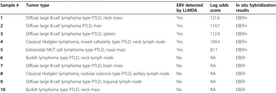
PTLD is a heterogeneous group of disorders that develop due to immunosuppressant regimens required for solid organ transplant or bone marrow allograft [9]. The risk for PTLD varies substantially depending on age, transplant type, immunosuppressant therapy, and EBV status. PTLDs are usually but not invariably associated with EBV. EBV can only be detected in half of PTLD cases that develop within a year of transplant [9,11,24]. The reported inci-dence, however, of EBV-negative PTLDs varies widely, and it is uncertain whether they should be considered analogous to EBV-positive PTLDs and whether they have any distinctive features [8]. We evaluated the accuracy of LLMDA for detecting EBV in 10 PTLD cases (Figure 3). DNA was isolated from the 10 frozen samples and applied to the LLMDA. Our system accurately detected the presence EBV in all the 5 EBV-positive cases which were confirmed by EBER in situ hybridization and PCR ana-lysis (data not shown), while no EBV sequence was de-tected in all the 5-EBV-negative cases. These results show that the LLMDA is capable of screening EBV-associated PTLD cases.
Detection of EBV from different lymphoma types
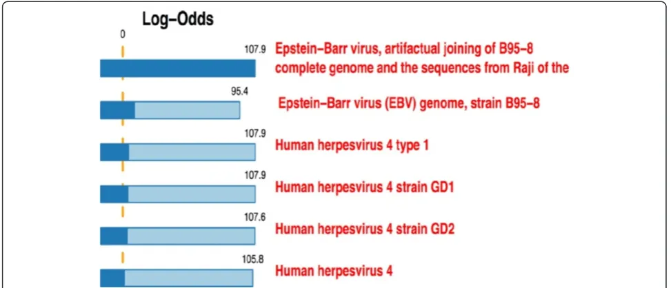
Out of the 58 cases of lymphoid tissues analyzed by the LLMDA, 5 out of the 30 B-cell lymphoma were positive for EBV, all 5 were from aggressive types (21 aggressive types vs 9 indolent types tested). All 9 of the indolent B-cell lymphomas were negative for EBV by LLMDA. Figure 1LLMDA’s detection of EBV in FFPE malignant lymphoma tissue from a 10μm section with a 150 mm2surface area.The
Epstein–Barr virus (EBV), also called human herpesvirus 4 (HHV-4). Array data was analyzed using the Composite Likelihood Maximization Method developed at LLNL. The lighter and darker-colored portions of the bars represent the unconditional and conditional log-odds scores, respectively. The conditional log-odds scores shows the contribution from a target that cannot be explained by another, more likely target above it, while the unconditional score illustrates that some very similar targets share a number of probes. 20 out of the 24 probes specific for EBV were detected on the LLMDA. The log odds score for EBV B95-8 genome is 107.9.
Table 1 Distribution of EBV-status of lymphoma cases in the study
Type of tissue EBER positive/total
Benign lymphoid tissues 0/8
B-cell lymphomas 5/30
T-cell lymphomas 1/2
NK/T cell lymphomas 2/2
PTLDs 5/10
Figure 2Representative cases EBV positive malignant lymphoma detected by LLMDA in FFPE tissues.The assay can detect the very high viral volume in plasmablastic lymphoma as well as relatively low viral volume in T-cell lymphoma.
One of two T-cell lymphoma samples (angioimmunoblas-tic T-cell lymphoma) was positive for EBV. Two out of two NK/T cell lymphoma samples were positive for EBV. Eight out of the eight specimens of control benign lymphoid tis-sues were tested negative for EBV by LLMDA. Addition-ally, only one of six classical Hodgkin lymphoma cases was detected for EBV by LLMDA (Table 1).
Discussion
The link between pathogen infections and tumorigenesis has been established in many cancers [2]. Virus-associated cancers are on the rise worldwide due primarily to the aging population as well as to the increasing use of solid organ transplantation and the associated use of immuno-suppressant drug regimens [7]. The identification of virus-associated malignancies will provide researchers and physicians with novel and effective therapeutic targets and treatment options. There is a need to be able to accur-ately and cost effectively screen increasing numbers of tu-mors in order to identify their cause and develop effective prevention and treatment options. The LLMDA is less ex-pensive than sequencing per sample and provides far greater flexibility than current sequencing and PCR diag-nostic techniques [23]. Although the current study focuses on EBV-associated cancers, the other viruses are also de-tectable when present providing a “one test detects all” technique.
Studying pathogen-induced tumorigenesis will require access to large sample pools [7]. Therefore, studies using frozen tissues come with limitations. Frozen tissues are often in short supply, and it is difficult to obtain specific tissues in significant numbers and in a timely fashion, as researchers need them. On the other hand, FFPE tissues are usually available in substantial numbers in many tissue banks. We have confirmed the ability of the LLMDA sys-tem to accurately detect EBV in FFPE samples. The option to use FFPE provides researchers with the opportunity to
screen large collections of relevant tumor samples in ar-chives worldwide, which is likely to reveal previously un-known pathogen-associated malignancies. The technique will also permit screening of samples from patients living in geographic locations from which collection and proper shipping of frozen tissues are prohibitive. The use of FFPE samples could hasten the identification of pathogen-induced malignancies and potentially help the develop-ment of therapies for these diseases.
We have previously demonstrated that the array de-tected viruses from a variety of human clinical samples such as urine, feces, serum, skin lesion, cerebral spinal fluid (CSF), tracheal aspirate, etc [18,19,21,25]. For ex-ample, the microarray detected BK polyomavirus posi-tive urine samples containing ≥1000 copies/mL (or an equivalent of 5 viral copies in a Phi29-amplification reac-tion) [18]. In another study, the array was used to detect Kaposi-sarcoma associated herpes virus, or human herpes virus 8 from bladder cancer samples. This further demon-strated the potential of the microbial detection array tech-nology to identify pathogens that might be linked to cancer and other diseases. The array detected viral DNA from as little as 20 fg or 100 genome copies input when combined with whole genome amplification [18,20]. Therefore, this microarray technique can be potentially used to detect EBV infection in clinical samples including blood and cerebral spinal fluid (CSF) [18-20,26].
Reliance on a technique as a clinical diagnostic tool re-quires that its accuracy is tested and confirmed at a reli-able level. In our study, the accuracy of the LLMDA has been demonstrated by correctly identifying EBV in 5 out of 10 PTLD clinically analyzed samples. Once the EBV’s initial lytic infection is brought under control, it can per-sist in latency. The EBV genome circularizes, resides in the cell nucleus as an episome, and is copied by cellular DNA polymerase. Each of the EBV latency programs leads to the production of a limited, distinct set of viral Table 2 Analysis of post-transplant lymphoproliferative disorder (PTLD) cases using the LLMDA
Sample # Tumor type EBV detected
by LLMDA
Log odds score
In situ hybridization results
1 Diffuse large B-cell lymphoma type PTLD, neck mass Yes 121.6 EBER+
2 Diffuse large B-cell lymphoma PTLD, liver Yes 114.7 EBER+
3 Diffuse large B-cell lymphoma type PTLD, spleen Yes 112.4 EBER+
4 Classical Hodgkin lymphoma, mixed cellularity type PTLD, neck lymph node Yes 104.4 EBER+
5 Extranodal NK/T cell lymphoma type PTLD, nasal mass Yes 87.1 EBER+
6 Burkitt lymphoma type PTLD, neck lymph node No NA
EBER-7 Diffuse large B-cell lymphoma type PTLD, brain mass No NA
EBER-8 Classical Hodgkin lymphoma, nodular sclerosis type PTLD, axillary lymph node No NA
EBER-9 Diffuse large B-cell lymphoma type PTLD, Inguinal lymph node No NA
EBER-10 Burkitt lymphoma type PTLD, neck mass No NA
proteins and viral RNAs [11]. For clinical diagnosis, EBER in situ hybridization is the standard methodology of choice for the detection of the EBV in tissue sections [16]. The large numbers of copies of EBERs can be de-tected in latently infected cells. Positive studies show stain-ing in the nuclei of the EBV-infected cells. False-negative results by EBER in situ hybridization are most often due to RNA degradation which still can be detected through the LLMDA with advantage of multiple probes. The LLMDA also possesses features unavailable to PCR and sequencing as it can more easily detect multiple pathogens in the same sample and this array also permits discovery of novel pathogens. In addition, we find that EBV-infection was de-tected from aggressive high grade lymphomas, but not the indolent low grade lymphomas. These results support the use of this system as a reliable clinical test for a variety of other tumor types.
Conclusions
In the present study, we were able to successfully screen FFPE lymphoma clinical cases, which will permit screening of a large volume of relevant samples, found in the ar-chives. We demonstrated the accuracy of the LLMDA sys-tem by detecting the presence or absence of EBV in PTLD cases by screening of clinical samples. Finally, we were able to show that the system can replicate clinical results by properly identifying EBV’s association with possible pro-gression from indolent to aggressive tumor stages. Taken together, our results support the use of the LLMDA as an important clinical tool in the screening of pathogen-associated lymphomas. In addition, this novel method has the ability to detect multiple broad-spectrum pathogen pathogens simultaneously [18].
In conclusion, using the LLMDA, we have developed an accurate and sensitive method for screening frozen and FFPE lymphoma samples. The technique performs well in the testing of most cancer types; however PCR and sequencing assays may be required to confirm re-sults produced by the LLMDA for tumors containing only a small percentage of transformed cells infected with pathogen. Nevertheless, the LLMDA system may provide a less expensive and/or more flexible clinical al-ternative to current techniques with FISH, PCR analysis and sequencing.
Methods
Patients and tissue specimens
A total of 58 patients (36 men and 22 women) diagnosed with lymphoma were included in the study. The tissues were collected at the Biorepository tissue bank, Department of Pathology, of University of California, Davis Medical Center during January 2010 to May 2014. The 10 PTLD samples were collected in Loma Linda University Medical Center. These cases previously had been diagnosed as
lymphoma, and subclassified using standard morphologic criteria, immunophenotyping, cytogenetic and selected fluorescence in situ hybridization (FISH) studies. Part of the fresh tissue samples was stored in liquid nitrogen until the extraction of DNA. The remaining tissues were fixed in formalin overnight. The study was approved by the IRB Ethics Committee of UC Davis medical center and in-formed consent was obtained.
DNA extraction
DNA was isolated from FFPE tissues using the AllPrep DNA/RNA FFPE Kit (Qiagen) following the manufacturer’s protocol with minor modifications. DNA was extracted from 1, 2 or 4, 10μm sections of 150 mm2surface area.
Microarray analysis
Genomic DNA (gDNA) was isolated from frozen and FFPE samples as described above. The gDNA was quantified using a Qubit 2.0 Fluorometer (Life Technologies). Then, 1μg gDNA was fluorescently labeled with Cy3-labeled ran-dom nonamers using the NimbleGen One-Color Labeling Kit (Roche). Labeled gDNA was re-quantitated with the Qubit 2.0 Fluorometer and 2μg of the labeled gDNA was hybridized to the 12×135K format of the LLMDA v.5 for 60 hr. The arrays were washed using a NimbleGen Wash Kit (Roche) and scanned at 532 nm (Cy3 chan-nel) using the NimbleGen MS 200 Microarray Scanner (Roche). The array was analyzed using the Composite Likelihood Maximization method at the 99% threshold (signal intensity of target probes were 99% above random control probes) [17,25].
EBER in situ hybridization for Epstein-Barr virus
All cases were subjected to in situ hybridization using oligonucleotides complementary to Epstein–Barr early RNA (EBER) transcripts in paraffin embedded tissues in an automated stainer (Integrated Oncology). The de-tail testing protocols using EBER riboprobes has been published [16].
Abbreviations
EBV:Epstein-Barr virus; HHV-4: Human herpesvirus 4; LLMDA: Lawrence livermore microbial detection array; PTLD: Post-transplant lymphoproliferative disorder; EBER: Epstein-Barr virus-encoded small RNA; HPV: Human papilloma virus; KSHV: Kaposi’s sarcoma-associated herpesvirus; FISH: Fluorescence in situ hybridization; PBL: Plasmablastic lymphoma; FFPE: Formalin fixed paraffin embedded; CSF: Cerebral spinal fluid.
Competing interests
The authors declare that they have no competing interests.
Authors’contributions
Acknowledgment
The study was supported by a UC Davis LLNL Fitpatrick Cancer Center Grant and UC Davis IRG grant. The authors thank Amelia Joslin from UC Davis for her technical assistance and Dr. Nicholas Be from LLNL for help with microarray experiments and review of the paper.
Author details
1
Deptartment of Pathology and Laboratory Medicine, University of California, Davis Medical Center, PATH Bldg. 4400V Street, Sacramento, CA 95817, USA. 2
Applied Genomics, Biosciences and Biotechnology Division, Lawrence Livermore National Laboratory, Livermore, CA 94551, USA.3Department of Pathology and Laboratory Medicine, Loma Linda University Medical Center, Loma Linda, CA 92354, USA.
Received: 13 October 2014 Accepted: 21 November 2014
References
1. Thun MJ, DeLancey JO, Center MM, Jemal A, Ward EM:The global burden of cancer: priorities for prevention.Carcinogenesis2010,31(1):100–110. 2. Zur Hausen H:Viruses in human cancers.Eur J Cancer1999,35(14):1878–1885. 3. Ginaldi L, Loreto MF, Corsi MP, Modesti M, De Martinis M:Immunosenescence
and infectious diseases.Microbes Infect2001,3(10):851–857.
4. Oyama T, Ichimura K, Suzuki R, Suzumiya J, Ohshima K, Yatabe Y, Yokoi T, Kojima M, Kamiya Y, Taji H, Kagami Y, Ogura M, Saito H, Morishima Y, Nakamura S:Senile EBV+ B-cell lymphoproliferative disorders: a clinicopathologic study of 22 patients.Am J Surg Pathol2003, 27(1):16–26.
5. Wong HH, Wang J:Epstein-Barr virus positive diffuse large B-cell lymphoma of the elderly.Leuk Lymphoma2009,50(3):335–340.
6. Mackay J:The Cancer Atlas.Atlanta: American Cancer Society Press; 2006:128. ISBN 9780944235621.
7. Moore PS, Chang Y:Why do viruses cause cancer? Highlights of the first century of human tumour virology.Nat Rev Cancer2010,10(12):878–889. 8. Taylor AL, Marcus R, Bradley JA:Post-transplant lymphoproliferative
disorders (PTLD) after solid organ transplantation.Crit Rev Oncol Hematol 2005,56(1):155–167.
9. Harris NL SS, Frizzera G, Knowles DM:Post Transplant Lymphoproliferative Disorders. Pathology and Genetics of Tumors of Hematopoietic and Lymphoid Tissues.Lyon: IARC Press; 2001:264–270.
10. Young LS, Rickinson AB:Epstein-Barr virus: 40 years on.Nat Rev Cancer 2004,4(10):757–768.
11. Rezk SA, Weiss LM:Epstein-Barr virus-associated lymphoproliferative disorders.Hum Pathol2007,38(9):1293–1304.
12. Epstein MA, Achong BG, Barr YM:Virus particles in cultured lymphoblasts from Burkitt’s Lymphoma.Lancet1964,1(7335):702–703.
13. Guerreiro-Cacais AO, Li L, Donati D, Bejarano MT, Morgan A, Masucci MG, Hutt-Fletcher L, Levitsky V:Capacity of Epstein-Barr virus to infect monocytes and inhibit their development into dendritic cells is affected by the cell type supporting virus replication.J Gen Virol 2004,85(Pt 10):2767–2778.
14. Savard M, Belanger C, Tardif M, Gourde P, Flamand L, Gosselin J:Infection of primary human monocytes by Epstein-Barr virus.J Virol2000, 74(6):2612–2619.
15. Yates JL, Warren N, Sugden B:Stable replication of plasmids derived from Epstein-Barr virus in various mammalian cells.Nature1985, 313(6005):812–815.
16. Weiss LM, Chen YY:EBER in situ hybridization for Epstein-Barr virus. Methods Mol Biol2013,999:223–230.
17. McLoughlin KS:Microarrays for pathogen detection and analysis.Brief Funct Genomics2011,10(6):342–353.
18. Erlandsson L, Rosenstierne MW, McLoughlin K, Jaing C, Fomsgaard A:The microbial detection array combined with random Phi29-amplification used as a diagnostic tool for virus detection in clinical samples.PLoS One 2011,6(8):e22631.
19. Gardner SN, Jaing CJ, McLoughlin KS, Slezak TR:A microbial detection array (MDA) for viral and bacterial detection.BMC Genomics2010,11:668. 20. Thissen JB, McLoughlin K, Gardner S, Gu P, Mabery S, Slezak T, Jaing C:
Analysis of sensitivity and rapid hybridization of a multiplexed Microbial Detection Microarray.J Virol Methods2014,201:73–78.
21. Rosenstierne MW, McLoughlin KS, Olesen ML, Papa A, Gardner SN, Engler O, Plumet S, Mirazimi A, Weidmann M, Niedrig M, Fomsgaard A, Erlandsson L: The microbial detection array for detection of emerging viruses in clinical samples–a useful panmicrobial diagnostic tool.PLoS One2014, 9(6):e100813.
22. Victoria JG, Wang C, Jones MS, Jaing C, McLoughlin K, Gardner S, Delwart EL: Viral nucleic acids in live-attenuated vaccines: detection of minority variants and an adventitious virus.J Virol2010,84(12):6033–6040. 23. Jaing C, Gardner S, McLoughlin K, Mulakken N, Alegria-Hartman M, Banda P,
Williams P, Gu P, Wagner M, Manohar C, Slezak T:A functional gene array for detection of bacterial virulence elements.PLoS One2008,3(5):e2163. 24. Thompson MP, Kurzrock R:Epstein-Barr virus and cancer.Clin Cancer Res
2004,10(3):803–821.
25. Paradzik M, Bucevic-Popovic V, Situm M, Jaing CJ, Degoricija M, McLoughlin KS, Ismail SI, Punda-Polic V, Terzic J:Association of Kaposi’s sarcoma-associated herpesvirus (KSHV) with bladder cancer in Croatian patients.Tumour Biol 2014,35(1):567–572.
26. Lin R, Liu Q:Diagnosis and treatment of viral diseases in recipients of allogeneic hematopoietic stem cell transplantation.J Hematol Oncol2013, 6:94.
doi:10.1186/s40364-014-0024-x
Cite this article as:Tellezet al.:Detection of Epstein-Barr virus (EBV) in human lymphoma tissue by a novel microbial detection array.
Biomarker Research20142:24.
Submit your next manuscript to BioMed Central and take full advantage of:
• Convenient online submission
• Thorough peer review
• No space constraints or color figure charges
• Immediate publication on acceptance
• Inclusion in PubMed, CAS, Scopus and Google Scholar
• Research which is freely available for redistribution