Original Article
Comparison of Three Staining Methods for the Detection of
Intesti-nal
Microspora
Spp.
Khadijeh KHANALIHA 1, Hamed MIRJALALI1, Mehdi MOHEBALI 1,2, Fatemeh TARIGHI 1,*Mostafa
REZAEIAN 1,2
1. Department of Medical Parasitology and Mycology, School of Public Health, Tehran University of Medical Sciences, Tehran, Iran 2. Center for Research of Endemic Parasites of Iran (CREPI), Tehran University of Medical Sciences, Tehran, Iran
Received 05 Apr 2014 Accepted 21 Sep 2014
Abstract
Background: This study aimed to compare three staining methods including: Calcofluor white, Chromotrope and Quick Hot Gram chromotrope used in diagnosis of intestinal mi-crosporidial spores.
Methods: One hundred and seventy five stool specimens were collected from patients re-ferred to Laboratory of Intestinal Protozoology at the School of Public Health, Tehran University of Medical Sciences during 2012-2013. All of specimens were evaluated by nest-ed PCR. The formalin–fixnest-ed stool samples were preparnest-ed from each specimen and drinest-ed at room temperature for 10 min, followed by 10 min methanol fixation. All the collected stool samples were evaluated blindly by calcofluor white, Chromotrope and Quick Hot Gram chromotrope staining methods separately.
Results: Microsporidial spores were recognized using Chromotrope, Quick Hot Gram chromotrope and Calcofluor white, in16 of 18 (88.8%), 17 of 18 (94.4%) and 18 of 18(100%) samples that were positive by nested PCR respectively. Regarding 14 stool sam-ples that were negative by nested PCR, 14 cases were negative by chromotrope and Quick hot Gram chromotrope and 13 samples were negative by Calcofluor white. One discordant sample interpreted as false positive.
Conclusion: Calcofluor white staining had the best performance for the detection of intes-tinal Microsprora spores and can be used as initial screen test for the detection of intestinal
Microspora spp.
Keywords: Microspora, Staining, Calcofluor, Chromotrope, Gram stain
*Correspondence
Email:
Introduction
icrospridia are obligate intracellular spore-forming protozoa. They have been recognized as human pathogen
particularly in immunodeficient patients (1). There are a number of methods available for diagnosing of microsporidial spores and
con-M
Iranian Society of Parasitology http:// isp.tums.ac.ir
Iranian J Parasitol
Open access Journal at http:// ijpa.tums.ac.ir Tehran University of Medical
firmation of microsporidiosis. Most of these methods were developed for diagnosing infec-tions in the immunocompromised population (2). The diagnosis of intestinal microsporidio-sis has traditionally depended on direct visual-ization of the parasites by light and or electron microscopy (1, 3). Although in some studies sensitivity of PCR particularly nested PCR have been greater than light microscopy for the diagnosis of intestinal microsporidia (3-5), but molecular methods have been some limi-tations. Several diagnostic methods may be needed for diagnosing of microsporidial infec-tion, especially when fecal specimens are
test-ed (2). The chromotrope staining method
us-ing modified trichrome was first described by Weber et al. for diagnosis of microsporidia (1). In this staining method, fast green was the counterstain and microscopical slides were stained for 2 hours.
Later Ryan et al. (6) modified chromotrope staining method by using aniline blue as the counterstain. Further Kokoskin et al. intro-duced an improved version of Weber standard staining technique by modifying the staining temperature to 50°C. The procedure is known as hot chromotrope (7). The staining method with calcofluor white that stain chitin in endo-spore layer of microsproridia endo-spores was de-scribed by Vavra, Chalupsky and Vicki (8-10). Another staining method for diagnosis of mi-crsporidia spores is Gram Chromotrope. This method is a combination of Gram staining and Weber’s staining methods. This method follows the steps of Gram staining method except safranin step, continuing with trope staining steps (11). The Gram chromo-trope was adapted by changing chromochromo-trope to hot chromotrope and short incubation time by Moura et al. (12).
In this study three different staining methods, calcofluor white, chromotrope and Quick hot Gram chromotrope were evaluated and com-pared for detection of microsporidial spores in the feces.
Materials and Methods
One hundred and seventy five stool speci-mens were collected from patients referred to laboratory of intestinal protozoology at school of Public Health, Tehran University of Medi-cal Science. All patients had gastro-intestinal signs and symptoms such as chronic or inter-mittent diarrhea. All of specimens were evalu-ated by nested PCR (13). The specimens were prepared to evaluate blindly by staining meth-ods. Conventional formalin-ether method was carried out for all samples. Thin smears were prepared from each specimen, dried at room temperature for 10 min, and followed by 10 minutes methanol fixation. All positive and negative stool samples were blindly evaluated by calcofluor white method, chromotrope and Quick Hot Gram chromotrope staining meth-ods.
Calcofluor white staining method
All microcopical slides were stained with 0.05% wt/vol calcofluor white M2R (Sigma chemical co, St Louis, Mo) as described by Vicki et al. (10).
Formalin- fixed positive and negative control stool samples that confirmed by nested PCR were examined in test series. The slides were cover slipped and screened under 100X oil– immersion using epiflourescence microscope fitted 455 nm.
Quick Hot Gram chromotrope staining method
decolor-ize solution and ethyl alcohol and mounted with cytosol. All slides were observed under light microscope with high magnification (1000X).
Chromotrope staining method
The trichrome was applied for this study as described by Weber et al. (1) with some modi-fication in the incubation time. The slides were prepared without coverslipping and viewed under a light microscope with high magnification (1000X).
Results
The chromotrope stain displayed transparent spores with pinkish wall of microsporidia and a background of faint green staining fecal bac-teria. Some of ovoid 1.0-1.5µm spores had a belt-like strip in the middle or at the end of body whereas fungal elements were much larger and stained red intensively (Fig.1, A). In the Quick Hot Gram chromotrope staining methods microsporidial spores appeared deep violet with ovoid structures. Yeasts were ob-served red in the background and easily rec-ognized from microsporidial spores. The equatorial belt–like stripes as a diagnostic fea-ture were seen in some spores (Fig.1, B). In the calcofluor white staining, microsporidial spores had oval shape with enhanced periph-eral fluorescence (Fig.1, C). Staining intensity was variable between fresh and old specimens. The fresh specimens fluoresced more brightly than old samples in our study. Internal
struc-tures were not visible in this method but pe-ripheral staining pattern and small size and unique shape of spores were very characteris-tic. Yeast cells also displayed fluorescence but were more round shaped and larger than mi-crosporidia spores.
Easy performance, cost benefit and stability of sample solution were advantages of using calcofluor in laboratory.
Microsporidial spores were recognized in16 of 18 (88.8%), 17of 18 (94.4%) samples that were positive by calcofluor white and nested PCR, using chromotrope and Quick Hot Gram chromotrope stain respectively. All 18 calco-fluor positive stool specimens confirmed by nested PCR .Two slides that were negative by chromotrope were stained again and subse-quent review on first slides and repeated ones, revealed few spores so these slides interpreted as false negative.
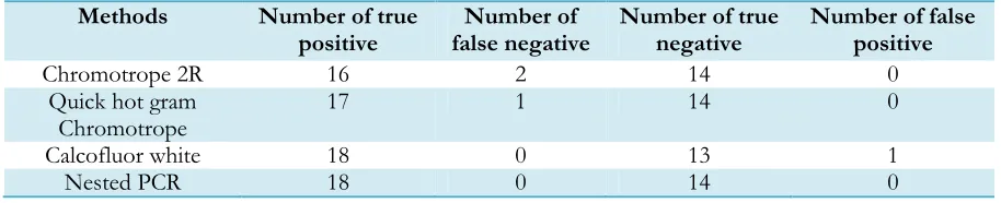
Fourteen stool samples that were negative by nested PCR were selected and analyzed to evaluate false positive in this study. All of the 14 specimens from these samples were nega-tive by chromotrope and Quick Hot Gram chromotrope staining methods whereas the results of calcofluor White method revealed 13 negative samples and 1discordant sample interpreted as false positive. Performance of three staining methods compared with PCR in 175 individuals for the detection of intestinal microsporidia spores and agreements degree between results obtained from three staining methods in diagnosing of microsporidia spores are summarized in Tables 1 and 2.
Table 1:Performance of three staining methods compared with PCR in 175 individuals for the detection of intestinal microsporidia spores
Methods Number of true
positive false negative Number of Number of true negative Number of false positive
Chromotrope 2R 16 2 14 0
Quick hot gram
Chromotrope 17 1 14 0
Calcofluor white 18 0 13 1
Table 2: Agreements among three staining methods in 175 individuals for the detection of intestinal micro-sporidia spores
Chromotrope Calcofluor white
Procedure
0.931
1.0 Calcofluor white
1.0
0.87 Chromotrope 2R
1.0
0.932 Quick-hot gram Chromotrope
*Agreement calculated by Cohen’s kappa test
A B
C
Discussion
In this study three staining methods, calcoflu-or white, chromotrope and Quick Hot Gram chromotrope stains were evaluated in the di-agnosis of microsporidial spores. The results indicate the sensitivity of Chromotrope, Quick Hot Gram chromotrope and calcofluor white staining methods in comparison with nested PCR were (88.8%), (94.4%) and (100%) re-spectively.
Microsporidia are reported in patients with acquired immunodeficiency syndrome (14-16).
The prevalence of intestinal microsporidiosis is reported to be 12-50% in European coun-tries and 2-64% in United States depending on the study population and methods of diagno-sis (17, 18). The chromotrope base stain is a routine diagnostic method (1, 19, 20), but be-cause of small size of microporidia species sometimes they are overlooked in this proce-dure, particularly in the cases with low con-centration of microsporidial spores (21). The Quick Hot Gram chromotrope technique is
Fig. 1: Appearance of microsporidial spores stained with chromotrope 2R (A), Quick Hot Gram chromotrope (B), Calcofluor white (C). Microspridia (white arrows), bacterial elements
another staining method that reported as a useful technique in diagnosis of microsporidial spores especially in cultured samples and tis-sue sections (11, 12). The calcofluor white de-scribed as a staining method with high sensi-tivity in some studies (10, 21).
Gram staining has been used occasionally to identify microsporidia spores in cell cultures and clinical samples such as urine that are ei-ther free of or have relatively low bacterial contamination (11).
There were some advantages found in Gram chromotrope staining method in this study. The microsporidia spores could be easily de-tected even they are present in small numbers; the staining time is short and could be photo-graphed easier in comparison with Chromo-trope.
The important advantages of calcofluor white were high sensitivity, speed, easiest perfor-mance between three staining method and stability of solution in the laboratory. Calco-fluor solution could be stored for long time in laboratory with little loss of fluorescence if solution stored in darkness. Another ad-vantage of this staining method is, individual microsporidia can be distinguished in thick area of smear that may be overlooked by chromotrope or Gram chromotrope staining methods. Chromotrope staining method is a method with high specificity but is time con-suming.
Sensitivity of chromotrope in comparison with calcofluor white and nested PCR was 88% in our study. In a study that were conducted by Vicki et al. sensitivity of chromotrope stain was (75%) in comparison with calcofluor white staining method and the calcofluor white is more sensitive than chromotrope that is consistent with our study (10).
Dider et al. compared modified trichrom blue, calcofluor white and Transmission Electron Microscopy (TEM) in detecting of microspor-idia. The sensitivity of calcofluor white, tri-chrom blue and TEM were 100%, 100% and 90.2% respectively. The calcofluor white and chromotrope stains are more sensitive than
TEM for detecting of microsporidia spores and the specificity of calcofluor white and modified trichrom blue and TEM were re-ported 90.5%, 100% and 100% respectively (21). These findings are similar to our results with high level of sensitivity and less specifici-ty for calcofluor white in comparison with chromotrope staining method.
The positive staining reaction of chitin con-taining objects in feces like yeasts and fungal elements and some parasites decrease specific-ity of calcofluor white staining that has been mentioned in previous studies (1, 12, 22) and false positive interpretations of calcofluor white reported in some studies (1, 9). Despite of these problems, the unique shape, small size, brightness of fluorescence and enhanced peripheral staining are very characteristic in diagnosing of microsporidial spores in this method that we observed in this study.
In fecal staining of Gram chromotrope micro-sporidial, spores appeared as deep violet to pink violet with ovoid structure and some-times spores have equatorial belt like stripes whereas yeasts were stained pink-red and easi-ly distinguished from microsporidial spores. Although in some studies the reported bacte-ria can be confused with microsporidia spores in fecal staining of Gram Chromotrope, they stained weakly with this technique in this study. The variability in some microsporidian spores with Gram stain is related to over-decolonization during staining or maturity of microporidian spores. Sporoblasts and imma-ture spores are Gram intermediate or Gram negative but mature forms tend to be Gram positive. This matter described in some previ-ous studies (11).
spores. The reason for this low intensity has been explained through late development of chitinous endospore layer during maturation (8, 9).
Conclusion
Our study shows that calcofluor white staining method has high sensitivity in comparison with other examined methods hence can be used as primary screen test in diagnosis of in-testinal microspridia. Further, we recommend Chromotrope and Gram chromotrope stain-ing methods for specificity improvement and result confirmation.
Acknowledgement
This study was self-funded. The authors de-clare that there is no conflict of interest.
References
1. Weber R, Bryan RT, Owen RL, Wilcox CM, et al. Improved light-microcopical detection of microsporidia spores in stool and duodenal as-pirates. N Engl J Med. 1992; 326: 161-6. 2. Garcia LS. Laboratory Identification of the
Microsporidia. J Clin Microbiol. 2002; 40(6): 1892-1901.
3. Muller A, Stellermann K, Hartmann P, Schrappe M, et al. A powerful DNA extraction method and PCR for detection of microsporid-ia in clinical stool specimens. Clin Diag Lab Immunol. 1999; 6:243-6.
4. Subrungruang I, Mungthin M, Chavalitshew-inkoon-Petmitr P, Rangsin R, et al. Evaluation of DNA Extraction and PCR Methods for Detection of Enterocytozoon bienuesi in Stool Specimens.J Clin Microbiol. 2004; 42(8): 3490-4.
5. Saigal K, Khurana S, Shama A, Sehgal R, Malla N. Comparison of staining techniques and multiplex nested PCR for diagnosis of intesti-nal microsporidiosis. Diagn Microbiol Infect Dis. 2013; 77(3): 248-9.
6. Ryan NJ, Sutherland G, Coughlan K, Globan M, et al. A new trichrome-blue stain for
detec-tion of microsporidial species in urine, stool, and nasopharyngeal specimens.J Clin Microbi-ol. 1993; 31: 3264-9.
7. Kokoskin E, Gyorkos T, Camus A, Celidotte L, Purtill T, Ward B. Modified technique for efficient detection of microsporidia.J Clin Mi-crobiol.1994; 32:1074-5.
8. Vavra J, Dahbiova R. Hollister WS, Canning EU. Staining of microporidian spores by opti-cal brighteners with remarks on the use of brighteners for the diagnosis of AIDS associat-ed human microsporidiosis. Folia Parasitol. 1993; 40:267-72.
9. Vavra J, Chalupsky J. Fluorescence staining of microporidian spores with the brightener calcofluor white M2R. J Protozool. 1982; 29(Suppl.):503.
10. Vicki AL, Stewart BK, Bergeron DL, et al. Use of the fluorochrome calcofluor white in the screening of stool specimens for spores of mi-crosporidia.Am J Clin Pathol. 1995; 103:656-9. 11. Moura H, Da Silva JL, Sodre FC, Brasil P, et al. Gram-Chromotrope: a New Technique that Enhances Detection of Microsporidial Spores in Clinical Samples.J Euk Microbiol. 1996; 43(5):94S-95S.
12. Moura H, Schwartz DA, Bornay-Llinares F, Sodré FC, et al. A new and improved "quick-hot Gram-Chromotrope" technique that dif-ferentially stains microporidian spores in clini-cal samples, including paraffin-embedded tissue sections. Arch Pathol Lab Med. 1997; 121(8): 888-93.
13. Mirjalali H, Mohebali M, Mirhendi H, Gholami R, et al. Emerging intestinal microsporidial in-fection in HIV+/AIDS patients in Iran:
micro-scopic and molecular detection. Iran J Parasitol. 2014; 9(2):149-54.
14. Orenstein JM. Microsporidiosis in the acquired immunodeficiency Syndrome. J Parasitol. 1991; 77: 843–64.
15. 15. Wuhib T, Silva TM, Newman RD, et al. Cryptosporidial and microsporidial infections in human immunodeficiency virus-infected pa-tients in northeastern Brazil. J Infect Dis. 1994; 170:494-7.
16. Shadduck JA. Human microsporidiosis in AIDS.Rev Infect Dis.1989; 11:203-7.
individ-uals referred for gastrointestinal evaluation.Am J Gastroenterol. 1994; 89:1998-2002.
18. Molina JM, Sarfati C, Beauvais B, Lemann M, et al. Intestinal microsporidiosis in human im-munodeficiency virus infected patients with chronic unexplained diarrhea: prevalence and clinical and biologic features. J Infect Dis.1993; 167:217- 21.
19. Isenberg HD. Clinical microbiology proce-dures handbook. American Society for Micro-biology Press: Washington, D.C; 1992.
20. Isenberg HD. Essential procedures for clinical microbiology. American Society for Microbiol-ogy Press: Washington, D.C; 1995.
21. Didier SD, Orenstein JM, Aldras A, et al. Comparison of three staining methods for de-tecting microsporidia in fluids.J Clin Microbiol. 1995; 33: 3138-45.
22. Harrington BJ, Hageage GJ. Calcofluor white: A Review of its Uses and Applications in Clini-cal Mycology and Parasitology. Lab Med.2003; 5(34):361-7.