R E S E A R C H
Open Access
Sex-associated differences in mitochondrial
function in human peripheral blood
mononuclear cells (PBMCs) and brain
C. Silaidos
1†, U. Pilatus
2†, R. Grewal
1, S. Matura
3,4, B. Lienerth
5, J. Pantel
3and G. P. Eckert
1*Abstract
Background:Alzheimer’s disease (AD) is the most common form of dementia, and it affects more women than men. Mitochondrial dysfunction (MD) plays a key role in AD, and it is detectable at an early stage of the degenerative process in peripheral tissues, such as peripheral mononuclear blood cells (PBMCs). However, whether these changes are also reflected in cerebral energy metabolism and whether sex-specific differences in mitochondrial function occur are not clear. Therefore, we estimated the correlation between mitochondrial function in PBMCs and brain energy metabolites and examined sex-specific differences in healthy participants to elucidate these issues.
Methods:The current pilot study included 9 male and 15 female healthy adults (mean age 30.8 ± 7.1 years).
Respiration and activity of mitochondrial respiratory complexes were measured using a Clarke-electrode (Oxygraph-2k system), and adenosine triphosphate (ATP) levels were determined using a bioluminescence-based assay in isolated PBMCs. Citrate synthase activity as a mitochondrial marker was measured using a photometric assay. Concentrations of brain energy metabolites were quantified in the same individuals using1H-magnetic resonance spectroscopy (MRS). Results:We detected sex-associated differences in mitochondrial function. Mitochondrial complexes I, I+II, and IV and uncoupled respiration and electron transport system (ETS) capacity in PBMCs isolated from blood samples of females were significantly (p< 0.05;p< 0.01) higher compared to males. ATP levels in the PBMCs of female participants were approximately 10% higher compared to males. Citrate synthase (CS) activity, a marker of mitochondrial content, was significantly (p< 0.05) higher in females compared to males. Sex-associated differences were also found for brain metabolites. TheN-acetylaspartate (NAA) concentration was significantly higher in female participants compared to males in targeted regions. This difference was observed in white matter (WM) and an area with a high percentage (> 50%) of gray matter (GM) (p< 0.05;p< 0.01). The effect sizes indicated a strong influence of sex on these parameters. Sex-associated differences were found in PBMCs and brain, but the determined parameters were not significantly correlated.
Conclusions:Our study revealed sex-associated differences in mitochondrial function in healthy participants. The underlying mechanisms must be elucidated in more detail, but our study suggests that mitochondrial function in PBMCs is a feasible surrogate marker to detect differences in mitochondrial function and energy metabolism in humans and it underscores the necessity of sex-specific approaches in therapies that target mitochondrial dysfunction.
Keywords:Sex differences, Blood cells, MR spectroscopy, Mitochondria, Mitochondrial respiration,N-Acetylaspartate
* Correspondence:[email protected]
†C. Silaidos and U. Pilatus contributed equally to this work.
1Nutrition in Prevention and Therapy, Institute for Nutritional Sciences, University of Giessen, Wilhelmstr. 20, 35392 Giessen, Germany Full list of author information is available at the end of the article
Background
Mitochondria are maternally inherited organelles of eukaryotic cells that are involved in numerous essential cell functions, e.g., energy metabolism, apoptotic pathways, and steroid hormone synthesis [1, 2]. The mitochondrial genome is optimized for functioning in women because the mitochondrial genome and mitogenome-nuclear genome interaction is effective in females only [3]. Recent studies report that female mito-chondria generate half the amount of hydrogen peroxide compared to mitochondria of males and contain higher levels of antioxidant enzymes and compounds [4–9]. Female sex may be a risk factor for Alzheimer’s disease (AD), which may be explained by the longer life expect-ancy of women [10,11]. However, women exhibit higher rates of AD than men even after adjusting for survival. Estimates from the Aging, Demographics, and Memory Study (ADAMS) revealed that 16% of women have Alzheimer’s disease or other dementias compared with 11% of men among people age 71 and older in the USA [12]. The prevalence of Alzheimer’s disease in Europe was estimated as 3.31% in men and 7.13% in women. The incidence of AD in Europe was 7.02 per 1000 per-son/years in men and 13.25 per 1000/years in women [13]. This disparity may be caused by differences in mitochondrial function between males and females. Sex-associated differences in the antioxidant capacity were examined [4–6, 14], and recent studies report that mitochondria in females of reproductive age generate half the amount of hydrogen peroxide [8]. Females also exhibit higher levels of antioxidant enzymes and com-pounds [4–9]. This sex difference decrease with age and following ovariectomy suggest a role of ovarian steroids [4, 6, 14, 15]. The decrease in gonadal hormone produc-tion during aging is gradual in men (testosterone), but es-trogen levels in women promptly decrease after menopause [16,17]. However, data of regarding sex-dependent differ-ences in mitochondrial function, such as respiratory activity and oxidative phosphorylation, are rare.
Mitochondria-related reactive oxygen species (ROS), including hydrogen peroxide play a key role in neurode-generative diseases, such as AD [8]. The brain of AD pa-tients is marked by severe synapse and neuronal loss, atrophy, and depletion of neurotransmitter systems in the hippocampus and cerebral cortex [18, 19]. Recent findings suggest that these changes are induced by mito-chondrial dysfunction (MD) and increased oxidative stress [20–24]. MD is detected at early stages of degen-erative processes [24–26], and it represents a promising target for nutrient-based preventive strategies. Brain tissue from living humans is not accessible, and cerebral mitochondrial function cannot be directly studied in clinical investigations. However, animal studies demonstrated that mitochondrial function of peripheral thymocytes reflected
mitochondrial function in brain cells [27]. Many observa-tions of substantial mitochondrial dysfunction in human peripheral tissues, especially fibroblasts and blood cells (primarily platelets and lymphocytes) indicate a relation be-tween peripheral and cerebral parameters, including age-related changes [28–32]. Increased oxidative stress during aging is not restricted to the brain; it is also present in peripheral cells, such as lymphocytes (a fractional part of peripheral mononuclear blood cells (PBMCs)). Leuner et al. observed mitochondrial dysfunction in peripheral blood cells isolated from Alzheimer’s patients that resulted in higher ROS production and oxidative-induced cell damage [32]. However, whether these changes reflect energy metab-olism in the brain is not clear. We investigated sex-specific differences and correlated mitochondrial function in peripheral blood cells with brain energy metabolites in healthy participants to elucidate these issues. Mitochondrial function and adenosine triphosphate (ATP) levels were determined in isolated PBMCs, and concentrations of brain energy metabolites were measured using 1H-magnetic resonance spectroscopy (MRS) in the same individuals. MRS offers a non-invasive method for the measurement of brain metabolites, and its diagnostic potential for many neurological diseases as dem-onstrated in numerous studies, particularly cerebral amino acid N-acetylaspartate (NAA). NAA is highly enriched in neurons [33], and its concentration is correlated with neur-onal density and damage. This metabolite is primarily syn-thesized in the mitochondria of neurons, and it is catalyzed by aspartate-N-acetyltransferase; moreover, the concentra-tion of NAA corresponds to neuronal energy consumpconcentra-tions [17,33,34], which supports NAA as a marker of mitochon-drial dysfunction in the brain [35–39].
The current pilot study examined possible sex-associated differences and correlated peripherally measured mitochon-drial function with cerebral energy metabolites in females and males.
Methods
Study design and participants
Thirty healthy volunteers were recruited for the study. Six men dropped out because of blood sampling fears or withdrew their agreement for participation. There-fore, a convenience sample of 24 healthy volunteers (9 males/15 females, mean age 30.8 ± 7.1 years) was included in the cross-sectional study. The Ethics Committee of the Goethe University of Frankfurt, Germany (reference no. 31/16) approved the study design, which was performed in agreement with the Declaration of Helsinki (Version Fortaleza 2012). All subjects declared that they understood the experimental procedure and signed a written informed consent.
brain structures and metabolites. Blood samples were collected in EDTA/K2-coated Sarstedt Monovetten
(#02.1333.001) from each person and transferred immediately to the Department of Pharmacology (Goethe University, Biozentrum, Niederursel, Frankfurt) for further analyses (e.g., determination of ATP levels and high-resolution respirometry). Weight, height, body mass index (BMI), waist-hip-ratio (WHR), and medical history were recorded (see Table 1). MRI exclusion criteria comprised cardiac pacemaker, neurostimulator, drug pump, metal parts in the body (metal clips, metal splinter), and claustrophobia. Exclusion criteria for blood sampling included hemophilia, hematophobia, or intake of anticoagulants.
Isolation of peripheral blood mononuclear cells (PBMCs)
Peripheral blood mononuclear cells were isolated from fresh blood from healthy participants within 1–2 h after collection using density medium centrifugation and Ficoll-Paque PLUS (GE Healthcare Bio-Science, Darmstadt, Germany). Blood samples were collected ei-ther before or after MRS in EDTA/K2-coated Sarstedt Monovetten. Briefly, blood was diluted 1:1 with phos-phate buffered saline (PBS), carefully layered onto Ficoll-Paque PLUS, and centrifuged at 400g for 40 min. Separated PBMCs were cautiously collected (2–4 ml), resuspended in 15 ml PBS, and centrifuged at 500g for 15 min. The supernatant was removed, and the pellet was resuspended in 15 ml PBS and centrifuged at 500g
for 10 min [GE Healthcare info]. The supernatant was removed, and the pellet was resuspended in 1 ml RPMI Glutamax™-1 medium (11.1 mM glucose, supplemented with 3% FBS, 50 units/ml penicillin, 50 g/ml strepto-mycin; #61870) for ATP-measurement or 1 ml MIRO 5 for high-resolution respirometry (see below).
Determination of ATP-levels in PBMC
To measure ATP concentrations, isolated PBMCs were resuspended in 1 ml RPMI medium, cultured in 96-well plate at a density of 1 × 10⁵ cells/100 μl/per well and
incubated for 3 h in humidified atmosphere supple-mented with 5% CO2at 37 °C.
ATP levels were assessed after 3 h using the ViaLight®-Plus bioluminescence kit (Lonza, Walkersville, USA), which is based on the production of light from ATP and Luciferin in the presence of the enzyme luciferase. A detailed description of the method was previously published [16]. The emitted light (bioluminescence) is linearly related to ATP concentration, and it was recorded using a luminometer (Victor21420 multilabel counter, Perkin Elmer, Rodgau-Jügesheim, Germany) [16]. The ATP concentration was normalized to cell number.
High-resolution respirometry in permeabilized PBMCs
For the high-resolution respirometry, isolated PBMCs were resuspended in 1 ml MiR05, which is a mitochon-drial respiration medium developed by Oroboros [40] containing EGTA (0.5 mM), magnesium dichloride (3 mM), lactobionic acid (60 mM), taurine (20 mM), potassium dihydrogenphosphate (10 mM), HEPES (20 mM), sucrose (110 mM), and essential fatty acid free bovine serum albumin (1 g/l). Cell density was adjusted to 10−6cells per ml. An Oxygraph-2k system (Oroboros Instruments, Innsbruck, Austria) and the DatLab soft-ware version 4.3.2.7 were used to analyze mitochondrial respiration.
A complex protocol (elaborated by Prof. Dr. Erich Gnaiger, University of Innsbruck, Austria) was used to investigate the function of the respiratory system, in-cluding different substrates, uncouplers, and inhibitors. The cell suspension (2 ml) was added to the two cham-bers of the Oxygraph-2k, and the chamcham-bers were closed to stabilize respiration (endogenous respiration). The plasma membrane of the cells was permeabilized with digitonin (1 μg/10−6 cells), which leaves the mitochon-drial outer and inner membranes intact. The capacity of oxidative phosphorylation was determined using com-plex I-related substrates (CI) glutamate (10 mM), malate (2 mM), and ADP (2 mM) followed by the addition of succinate (10 mM; OXPHOS). Leak respiration after the addition of glutamate/malate was labeled leak (G/M) and corresponded to state 4 respiration. Further addition of ADP induced state 3 respiration. The addition of oli-gomycin (2 μg/ml) allowed measurement of the state 2 respiration leak (omy). Uncoupling (ETS) was achieved with addition of carbonyl cyanide p-(trifluoromethoxy) phenylhydrazone (FCCP), injected stepwise up to 2.5 μM), and complex II respiration in the non-coupled state (CIIETS) was monitored after the addition of
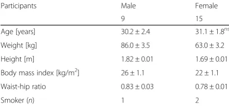
rote-none (0.5 μM) to the chambers. Residual oxygen con-sumption (ROX), which is oxygen concon-sumption caused by enzymes outside the electron transfer system, was de-termined after inhibition of complex III via the addition of antimycin A (2.5 μM) and was subtracted from all Table 1Demographic data
Participants Male Female
9 15
Age [years] 30.2 ± 2.4 31.1 ± 1.8ns
Weight [kg] 86.0 ± 3.5 63.0 ± 3.2
Height [m] 1.82 ± 0.01 1.69 ± 0.01
Body mass index [kg/m2] 26 ± 1.1 22 ± 1.1
Waist-hip ratio 0.83 ± 0.03 0.78 ± 0.01
Smoker (n) 1 2
respiratory parameters. COX activity (CIV) was mea-sured after ROX determination and application of 0.5 mM tetramethyl-phenylenediamine (TMPD) as an artificial substrate of complex IV and 2 mM ascorbate to maintain TMPD in the reduced state. The autoxidation rate was determined after the addition of sodium azide (≥100 mM), and COX respiration was corrected for autoxidation [16]. The data were normalized to citrate synthase (CS) activity [pmol/(s*IU CS)].
Citrate synthase (CS) activity
A subsample of isolated PBMCs was immediately frozen in liquid nitrogen and stored at−80 °C for photometric determination of citrate synthase activity. Measurements were performed in duplicate. A detailed description of the method was published previously [16]. CS activity was normalized to IU per 1 × 10⁶cells/ml.
Protein quantification
The Pierce TM BCA Protein Assay Kit (Thermo Fisher Scientific, Waltham, MA, USA) was used to measure protein content. Bovine serum albumin was used as the standard.
MR protocol
MRS of the brain was performed using a 3T whole body system (Magnetom Trio, Siemens Medical AG, Erlangen, Germany) equipped with a double tuned 1H/31P volume head coil (Rapid Biomedical, Rimpar, Germany). The proto-col was similar to a previously published study protoproto-col [41,42] including1H and31P magnetic resonance spectro-scopic imaging (MRSI) examinations. An axial slice at the level of the centrum semiovale partially including the trunk of the corpus callosum was recorded for 1H spectroscopy using 2D MRSI and an acquisition-weighted circular phase encoding scheme on a 20 × 20 matrix, field-of-view (FOV) of 240 × 240 mm2, 12-mm slice thickness, nominal voxel
size of 12 × 12 × 12 mm3, TR 1500 ms, TE 30 ms, and two acquisitions. The volume of interest (VOI) was selected using a combination of point-resolved spectroscopy (PRESS) and outer volume suppression, and it was adjusted to contain gray matter (GM) and white matter (WM) (Fig. 1). The matrix was extrapolated to 40 × 40 prior to Fourier transformation to produce a 6.0 × 6.0 mm2in-plane grid size (Fig.1).
We also obtained a B1 map [43] and an a 2D1H MRSI data set recording of the free induction decay (FID) sig-nal of unsuppressed water (25-mm slice thickness, FOV 240 mm2, matrix size 16 × 16 extrapolated to 32 × 32, delay before data acquisition 2.4 ms, 2° excitation pulse flip angle) for absolute quantification of metabolite con-centrations. The slice was aligned to the water sup-pressed PRESS MRSI slice. A T1-weighted MRI data set was recorded for tissue segmentation.
MR data processing
T1-weighted MRI data were segmented using the FAST tool in the FMRIB Software Library (FSL) toolbox [44]. B1 maps were calculated as described in Volz et al. 2010 [43] and registered to the T1-weighted data for each subject. Parameter maps (GM, WM, B1) were registered to the spectroscopic data.
The 1H data spectra were fitted in the frequency domain using a linear combination of a set of model spectra including the main metabolites Cho (choline-containing compounds), Cr (creatine/phosphocreatine), and NAA (N-acetyl-aspartate and N -acetyl-aspartate-glutamate), using the software tool LCModel (Version 6.3, http://s-provencher.com). Metabolite signal inten-sities were corrected for T1 and T2 relaxation assuming previously published relaxation times at 3 T [45]. B1 in-homogeneity was taken into account, and metabolite concentrations of tissue water were calculated as
described in the supplemental information (Additional file1) [45].
A set of voxels from right and left WM and central GM were selected for further data evaluation (Fig.1).
The31P data were not included in this study.
Chemicals
Chemicals were of the highest available purity and pur-chased from Sigma (St Louis, MO, USA) or Merck (Darmstadt, Germany) unless otherwise stated. Aqueous solutions were prepared using deionized, filtered water (Millipore, Billerica, MA, USA).
Statistics
Values are presented as the means ± standard error of the mean (SEM), unless otherwise stated. Group differ-ences were calculated using unpairedttest with Welch’s correction. Effect sizes were calculated using eta squared (η2) (Prism 7.03, GraphPad Software, San Diego, CA, USA). Statistical significance was defined forpvalues of *p< 0.05 and **p< 0.01.
Results
Sex-associated differences in peripheral mitochondrial parameters
Diversifications of mitochondrial efficacy and function are primarily dependent on alterations of the respiratory complex system. Mitochondrial respiration in isolated
PBMCs was used as an effective method to assess mito-chondrial efficacy and function in males and females.
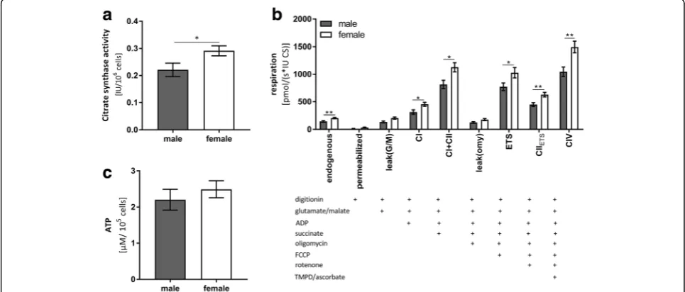
CS is an enzyme of the Krebs cycle, and it is located in the mitochondrial matrix. CS activity is a mitochondrial mass marker [46]. CS activity was significantly higher in females compared to males (see Fig.2a).
Mitochondrial complexes I, I+II, and IV and uncoupled respiration and ETS capacity in PBMCs iso-lated from males were significantly lower compared to females (see Fig.2b). Mitochondrial respiration was nor-malized to CS activity, and the results refer to maximal respiratory per mitochondrion [46].
The activity of the mitochondrial respiration chain complexes (CI–IV) creates a proton gradient at the inner mitochondrial membrane. The resulting mem-brane potential ultimately represents the driving force for complex V (CV; F1/F0-ATPase) to produce ATP.
ATP levels of female participants were approximately 10% higher compared to males (see Table2) (see Fig.2c). These differences were not significant, but the observed trends reflect lower mitochondrial respiration in PBMCs isolated from male volunteers (Fig.2c).
Sex-associated differences in cerebral parameters
NAA is a marker of neuronal energy consumptions [17,
33,34]. Figure 3shows the results from 1H-MR spectro-scopic data from the brain. The NAA concentration was significantly higher in female participants compared to male participants in the total WM region and GM region.
Correlation between mitochondrial function in PBMCs and the brain
Individual content of NAA in GM and WM did not cor-relate with the respective ATP levels or citrate synthase activity in PBMCs (see Table3and Fig.4).
Discussion
The present explorative pilot study measured peripheral markers of energy metabolism and compared these levels to the cerebral energy metabolite NAA. We ob-served sex-associated differences in mitochondrial func-tion, ATP levels and citrate synthase activity in isolated
PBMCs, and a significant effect of sex on the brain me-tabolite NAA.
Sex-associated differences in cerebral and peripheral markers of energy metabolism
Mitochondrial complexes I, I+II, and IV and uncoupled respiration and ETS capacity in PBMC isolated from females were significantly higher compared to males (see Fig.2b).
Activity of mitochondrial respiratory complexes CI– CIV is the driving force for ATP generation by complex V of the respiration chain. Therefore, the observed im-pairment in complex activities may account for the lower apparent ATP levels in males (see Fig. 2c). How-ever, ATP levels were not significantly different in PBMCs isolated from men and women. This result may indicate that all participants were adequately supplied with cellular energy at the time of measurement. Not-ably, the respiration measurements determined the max-imum possible oxygen consumption of the single respiratory complexes and thus, ATP levels may be much lower when damage (e.g., oxidative stress) occurs. CS activity was significantly higher in females compared to males (see Fig. 2a). These findings indicated that mitochondrial content is higher in females because CS activity, an enzyme of the mitochondrial Krebs cycle, is strongly associated with mitochondrial content [46]. Gaignard et al. detected no difference in CS activity be-tween intact male and female rats, which emphasizes the importance of measuring this parameter in humans [6].
Our study found lower ATP levels and reduced mito-chondrial function in PBMCs of males, which may indi-cate a generally lower mitochondrial function compared to women. This difference could produce the lower NAA concentrations in the brain. Mitochondria are the main source of NAA. Therefore, the significantly higher mitochondrial content may lead to significantly higher NAA concentrations in female brains. Maudsley et al. Table 2Measures of peripheral mitochondrial function and
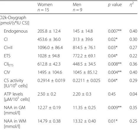
cerebral metabolism Women n= 15
Men n= 9
pvalue η2
O2k-Oxygraph [pmol/(s*IU CS)]
Endogenous 205.8 ± 12.4 145 ± 14.8 0.007** 0.40
CI 453.6 ± 36.0 313 ± 39.6 0.02* 0.30
CI+II 1096.0 ± 86.4 814.5 ± 76.1 0.03* 0.27
ETS 1028 ± 94.8 772.2 ± 69.1 0.04* 0.22
CIIETS 612.8 ± 42.3 448.5 ± 34.5 0.008** 0.36
CIV 1495 ± 104.6 1045 ± 85.12 0.004** 0.40
CS activity [IU/106cells]
0.2914 ± 0.019 0.2211 ± 0.025 0.04* 0.29
ATP levels
[μM/105cells] 2.50 ± 0.2 2.20 ± 0.3 0.45 0.04
NAA in GM [mmol/l]
12.27 ± 0.19 11.35 ± 0.25 0.009** 0.35
NAA in WM [mmol/l]
14.79 ± 0.38 13.32 ± 0.40 0.01* 0.25
Mean ± SEM, unpairedttest with Welch’s correction, significant differences are marked by asterisk, (*p<0.05; **p<0.01)
ATPadenosine triphosphate,CScitrate synthase,Ccomplex,ETSelectron transport system,GMgray matter,NAA N-acetylaspartate,WMwhite matter
also found significant sex-associated differences in NAA levels in some brain regions of 41 male and 47 female participants (mean age = 33 years) [47]. The sex differ-ences indicate increased NAA concentrations in brains of females relative to males, with an average of 4% in gray matter (GM) and white matter (WM).
Sex differences in mitochondrial function were mea-sured in other tissues.
Rutkai et al. observed higher mitochondrial respiration in freshly harvested cerebral arteries from adult female rats compared to males [48].
Genetic differences in females and males may contribute to the observed sex differences in energy metabolism. Basic sex differences in mitochondrial metabolic regula-tion may exist because of the maternal inheritance of mitochondria. Mitochondria from females exhibit better
coping with stressful conditions and are relatively resilient to DNA damage and mutations, which reduces the prob-ability of producing inheritable metabolic disorders [7].
Sex steroids are another factor that may be responsible for the observed sex differences. The influence of sex hormones would also explain why women are more likely to develop Alzheimer’s disease than men, despite exhibiting better mitochondrial function (see Figs.2and
3), lower levels of ROS [8], and better antioxidant cap-acity [4–9] at a reproductive age. The decrease in female sex hormones after menopause reverses sex differences. Previous studies demonstrated the reduced antioxidant capacity and the increased ROS production after meno-pause [5, 6]. Comparison of mitochondrial function in pre- and postmenopausal women and men of the same age should be compared in a future study to confirm this Table 3Correlations
Sex Correlated values Pearson r pvalue
Male n= 9
NAA GM [mmol] vs. CS [IU/106cells] 0.55 0.15
NAA WM [mmol] vs. CS [IU/106cells] 0.53 0.17
NAA GM [mmol] vs. ATP levels [μM/105cells] 0.46 0.26
NAA WM [mmol] vs. ATP levels [μM/105cells] 0.45 0.26
NAA GM [mmol] vs. CI activity [pmol/(s*IU CS)] 0.34 0.33
NAA WM [mmol] vs. CI activity [pmol/(s*IU CS)] 0.41 0.32
Female n= 15
NAA GM [mmol] vs. CS [IU/106cells] 0.06 0.82
NAA WM [mmol] vs. CS [IU/106cells] 0.25 0.40
NAA GM [mmol] vs. ATP levels [μM/105cells] 0.37 0.17
NAA WM [mmol] vs. ATP levels [μM/105cells] 0.45 0.28
NAA GM [mmol] vs. CI activity [pmol/(s*IU CS)] 0.30 0.38
NAA WM [mmol] vs. CI activity [pmol/(s*IU CS)] 0.04 0.90
ATPadenosine triphosphate,CIcomplex I,CScitrate synthase,GMgray matter,NAA N-acetylaspartate,WMwhite matter
mitochondrial respiration hypothesis. Pharmacological studies in young adult female rodents demonstrated that steroids influenced brain function. Ovariectomy de-creased brain mitochondrial oxidative phosphorylation and increased oxidative stress [49–53]. Gaignard et al. demonstrated a sex difference in brain mitochondrial respiration and oxidative stress that is suppressed with aging and ovariectomy. These findings in reproductive animals are consistent with our results.
A direct genomic effect of estradiol is unlikely, because neither the antioxidant enzyme superoxide dismutase (SOD) nor glutathione peroxidase (GPx) contain estrogen-responsive elements in their promotor region. However, estradiol may activate mitogen-activated pro-tein kinase (MAPK). MAPK activates the transcription factor NFκB, which upregulates the gene expression of antioxidant enzymes [54]. More antioxidant enzymes could protect the respiratory chain complexes against damage from ROS and explain the better mitochondrial respiration in females in reproductive age. Grimm et al. reviewed several studies that demonstrated an estrogen-induced upregulation of genes encoding for components of the mitochondrial electron transport chain, including CI, CIV, and the F1 subunit of ATP syn-thase, which is consistent with our detected significant differences in CI and CIV [15].
However, whether sex- or neurosteroids contributed to the differences in mitochondrial function in our study requires further investigation. The influence of the de-crease in estradiol during menopause on the mitochon-drial respiration should also be investigated. These findings emphasize the necessity to include males and females in experimental studies and particularly preven-tion strategies that target mitochondria.
Correlation between mitochondrial function in PBMCs and the brain
The higher level of peripheral markers of energy metab-olism in female subjects was paralleled by a higher con-centration of the metabolite NAA. Sex-associated differences were found in PBMCs and the brain, but statistical analyses revealed that the individual content of NAA in GM and WM did not correlate with the re-spective levels of ATP or citrate synthase activity in PBMCs (see Table 3). However, the effect size indicated a strong influence of the factor of sex on these parame-ters. The lack of significance may be due to the small number of subjects. Therefore, the lack of correlation does not necessarily indicate that energy metabolism in the brain and peripheral blood cells are not connected, especially because a tendency for positive correlations is observed on the graphs (Fig.3). The lack of controls for other confounding variables, such as blood sampling
times, smoking, and diet, is a further limitation of the study. Some recruited participants also dropped out, which resulted in an unequal number of women and men as another limitation of the study.
Several studies demonstrated mitochondrial dysfunc-tion and increased ROS levels and apoptosis in lympho-cytes of AD patients. Markers of mitochondrial function, such as NAA, also exhibited changes in the brains of AD patients compared to a control group. Mitochondrial function compromised in the peripheral cells and brains of AD patients, which is reflected in the alteredN -actey-laspartate levels.N-acetylaspartate has been proposed as a marker of neuronal health, viability, and number [55]. Many 1H-MRS studies demonstrated decreased NAA concentration in dementia and other neurological disor-ders [56–61] where the NAA reduction is frequently lo-cated in regions of gray matter volume reduction. This reduction may indicate reduced neurons [62, 63] be-cause NAA is highly concentrated in neurons. However, low NAA levels may reflect mitochondrial dysfunction because NAA concentration corresponds to mitochon-drial function [33,35–39,62–64].
To our knowledge, this study is the first report to dir-ectly compare markers of mitochondrial function in the brain and PBMCs. Bartolotti et al. 2016 investigated the CAMP response element-binding protein (CREB) pro-tein, which is important for the formation of memories [65]. CREB signaling is dysfunctional in mouse models of AD. These authors compared CREB expression in PBMCs and postmortem brain tissue. pCREB expression in PBMCs was positively correlated with pCREB expres-sion in the postmortem PFC, which indicates that pCREB expression in PBMCs may reflect pCREB expres-sion in the AD brain. Their results demonstrated im-paired pCREB in AD brain and PBMCs, which reinforces our hypothesis of a connection between the changes in the brain and peripheral tissues, such as PBMCs during aging and pathology. These data con-firms that AD is not a pure brain disease and empha-sizes that mitochondrial function in PBMCs may be a feasible surrogate marker to detect differences in mito-chondrial function in AD.
Conclusion
mitochondrial function. Future studies should confirm our findings using a larger sample size and investigate the sex-associated differences in males and females older than 65 years compared to young women and men. Fur-thermore, we would extend the range of methods to de-termination of mtDNA copy numbers to further strengthen the sex-specific data.
Additional file
Additional file 1:Calculation of B1 and coil-receive-profile-corrected metabolite concentrations normalized to tissue water. (PDF 58 kb)
Abbreviations
1H:Proton; 3 T: 3 Tesla; 31P: 31-Phosphor; AD: Alzheimer’s disease; ADAMS: Aging, Demographics, and Memory Study; ATP: Adenosine triphosphate; BMI: Body mass index; C: Complex; Ch: Choline-containing compounds; Cre: Creatine/phosphocreatine; CREB: CAMP response element-binding protein; CS: Citrate synthase; DNA: Deoxyribonucleic acid; ETS: Electron transport system; FCCP: Carbonyl cyanide p-(trifluoromethoxy) phenylhydrazone; FID: Free induction decay; FOV: Field-of-view; FSL: FMRIB Software Library; GM: Gray matter; GPx: Glutathione peroxidase;
IU: International unit; MAPK: Mitogen-activated protein kinase;
MD: Mitochondrial dysfunction; MiR05: Mitochondrial respiration media 05; MRS: Magnetic resonance spectroscopy; MRSI: Magnetic resonance spectroscopic imaging; mtDNA: Mitochondrial DNA; NAA:N-Acetylaspartate; NFκB: Nuclear factor“kappa-light-chain-enhancer”of activated B cells; OXPHOS: Oxidative phosphorylation; PBMCs: Peripheral blood mononuclear cells; PBS: Phosphate buffered saline; pCREB: Phosphorylated cAMP response element-binding protein; PRESS: Point-resolved spectroscopy; ROS: Reactive oxygen species; ROX: Residual oxygen consumption; RPMI: Roswell Park Memorial Institute; SEM: Standard error of the mean; SOD: Superoxide dismutase; TMPD: Tetramethyl-phenylenediamine; VOI: Volume of interest; WHR: Waist-hip-ratio; WM: White matter;η2: Eta squared
Acknowledgements
The assistance of Jan Köhler and Tanja Hölzinger is highly acknowledged.
Availability of data and materials
The datasets generated and/ or analyzed in the current study are available from the corresponding author on reasonable request.
Authors’contributions
CS, UP, SM, JP, and GPE conceived and designed the study and critically reviewed the manuscript. CS performed all analyses in PBMCs and primarily wrote the manuscript. UP performed all analyses of MRS. BL contributed to MRS data acquisition. RG contributed to analyses of mitochondrial respiration. CS and SM analyzed patient demographics and coordinated the study procedure. JP and GPE as PIs oversaw the entire study. All authors provided valuable feedback and approved the final manuscript.
Ethics approval and consent to participate
The Ethics Committee of the Goethe University of Frankfurt, Germany, approved the study design (reference no. 31/16), which is in agreement with the Declaration of Helsinki (Version Fortaleza 2012). All subjects declared that they understood the experimental procedure and signed a written informed consent.
Consent for publication Not applicable.
Competing interests
The authors declare that they have no competing interests.
Publisher’s Note
Springer Nature remains neutral with regard to jurisdictional claims in published maps and institutional affiliations.
Author details
1Nutrition in Prevention and Therapy, Institute for Nutritional Sciences, University of Giessen, Wilhelmstr. 20, 35392 Giessen, Germany.2Institute for Neuroradiology, Goethe University Frankfurt, Schleusenweg 2-16, 60528 Frankfurt/Main, Germany.3Institute of General Practice, Goethe University Frankfurt, Theodor-Stern-Kai 7, 60590 Frankfurt/Main, Germany.4Department of Psychiatry, Psychosomatic Medicine and Psychotherapy, University Hospital Frankfurt, Heinrich-Hoffmann Str. 10, 60528 Frankfurt/Main, Germany. 5Brain Imaging Centre, Schleusenweg 2-16, 60528 Frankfurt/Main, Germany.
Received: 6 March 2018 Accepted: 13 July 2018
References
1. Kuklinski B. Mitochondrien: Symptome, diagnose und therapie. 1st ed; 2015. Aurum. 2. Bakthavachalam P, Shanmugam PST. Mitochondrial dysfunction—silent killer
in cerebral ischemia. J Neurol Sci. 2017;375:417–23.
3. Tower J. Sex-specific regulation of aging and apoptosis [eng]. Mech Ageing Dev. 2006;127(9):705–18.
4. Viña J, Borrás C. Women live longer than men: understanding molecular mechanisms offers opportunities to intervene by using estrogenic compounds. Antioxid Redox Signal. 2010;13(3):269–78.
5. Mandal PK, Tripathi M, Sugunan S. Brain oxidative stress: detection and mapping of anti-oxidant marker‘glutathione’in different brain regions of healthy male/female, MCI and Alzheimer patients using non-invasive magnetic resonance spectroscopy. Biochem Biophys Res Commun. 2012;417:43–8. 6. Gaignard P, Savouroux S, Liere P, Pianos A, Thérond P, Schumacher M, Slama A, Guennoun R. Effect of sex differences on brain mitochondrial function and its suppression by ovariectomy and in aged mice [eng]. Endocrinol. 2015;156(8):2893–904.
7. Demarest TG, McCarthy MM. Sex differences in mitochondrial (dys)function: implications for neuroprotection. J Bioenerg Biomembr. 2015;47(1–2):173–88. 8. Ostan R, Monti D, Gueresi P, Bussolotto M, Franceschi C, Baggio G. Gender,
aging and longevity in humans: an update of an intriguing/neglected scenario paving the way to a gender-specific medicine [eng]. Clin Sci (London, England : 1979). 2016;130(19):1711–25.
9. Austad SN, Fischer KE. Sex differences in lifespan [eng]. Cell Metab. 2016; 23(6):1022–33.
10. Bachman DL, Wolf PA, Linn R, Knoefel JE, Cobb J, Belanger A, D'Agostino RB, White LR. Prevalence of dementia and probable senile dementia of the Alzheimer type in the Framingham study. Neurol. 1992;42(1):115–9. 11. Gabelli C, Cademo A. Gender diff erences in cognitive decline and
Alzheimer’s disease. Ital J Gender-Specific Med. 2015;1:21–8.
12. Wortmann M. World Alzheimer report 2014: dementia and risk reduction. Alzheimers Dement. 2015;11(7):P837.
13. Niu H, Álvarez-Álvarez I, Guillén-Grima F, Aguinaga-Ontoso I. Prevalence and incidence of Alzheimer’s disease in Europe: a meta-analysis. Neurolo (English Edition). 2017;32(8):523–32.
14. Borras C, Sastre J, Garcia-Sala D, Lloret A, Pallardo FV, Vina J. Mitochondria from females exhibit higher antioxidant gene expression and lower oxidative damage than males. Free Radic Biol Med. 2003;34:546–52. 15. Grimm A, Mensah-Nyagan AG, Eckert A. Alzheimer, mitochondria and
gender [eng]. Neurosci Biobehav Rev. 2016;67:89–101.
16. Hagl S, Grewal R, Ciobanu I, Helal A, Khayyal MT, Muller WE, Eckert GP. Rice bran extract compensates mitochondrial dysfunction in a cellular model of early Alzheimer’s disease. J Alzheimers Dis. 2015;43:927–38.
17. GOLDSTEIN FB. Biosynthesis ofN-acetyl-L-aspartic acid [eng]. Biochim Biophys Acta. 1959;33(2):583–4.
18. Querfurth HW, LaFerla FM. Alzheimer’s disease. N Engl J Med. 2010;362(4): 329–44.
19. Rhein V, Song X, Wiesner A, Ittner LM, Baysang G, Meier F, Ozmen L, Bluethmann H, Dröse S, Brandt U, Savaskan E, Czech C, et al. Amyloid-beta and tau synergistically impair the oxidative phosphorylation system in triple transgenic Alzheimer’s disease mice [eng]. Proc Natl Acad Sci U S A. 2009; 106(47):20057–62.
20. Leuner K, Schütt T, Kurz C, Eckert SH, Schiller C, Occhipinti A, Mai S, Jendrach M, Eckert GP, Kruse SE, Palmiter RD, Brandt U, et al.
Mitochondrion-derived reactive oxygen species lead to enhanced amyloid beta formation [eng]. Antioxid Redox Signal. 2012;16(12):1421–33. 21. Mattson MP, Magnus T. Aging and neuronal vulnerability. Nat Rev Neurosci.
22. Mattson MP, Gleichmann M, Cheng A. Mitochondria in neuroplasticity and neurological disorders. Neuron. 2008;60(5):748–66.
23. Eckert GP, Renner K, Eckert SH, Eckmann J, Hagl S, Abdel-Kader RM, Kurz C, Leuner K, Muller WE. Mitochondrial dysfunction—a pharmacological target in Alzheimer’s disease. Mol Neurobiol. 2012;46(1):136–50.
24. Schaffer S, Asseburg H, Kuntz S, Muller WE, Eckert GP. Effects of polyphenols on brain ageing and Alzheimer’s disease: focus on mitochondria. Mol Neurobiol. 2012;46(1):161–78.
25. Manczak M, Park BS, Jung Y, Reddy PH. Differential expression of oxidative phosphorylation genes in patients with Alzheimer’s disease: implications for early mitochondrial dysfunction and oxidative damage. NMM. 2004;5(2):147–62. 26. Hauptmann S, Scherping I, Dröse S, Brandt U, Schulz KL, Jendrach M, Leuner
K, Eckert A, Müller WE. Mitochondrial dysfunction: an early event in Alzheimer pathology accumulates with age in AD transgenic mice [eng]. Neurobiol Aging. 2009;30(10):1574–86.
27. Schuessel K, Frey C, Jourdan C, Keil U, Weber CC, Müller-Spahn F, Müller WE, Eckert A. Aging sensitizes toward ROS formation and lipid peroxidation in PS1M146L transgenic mice [eng]. Free Radic Biol Med. 2006;40(5):850–62. 28. Leutz S, Steiner B, Marques CA, Haass C, Muller WE. Reduction of trophic support
enhances apoptosis in PC12 cells expressing Alzheimer’s APP mutation and sensitizes cells to staurosporine-induced cell death. JMN. 2002;18(3):189–202. 29. Schindowski K, Kratzsch T, Peters J, Steiner B, Leutner S, Touchet N, Maurer
K, Czech C, Pradier L, Frölich L, Müller WE, Eckert A. Impact of aging. NeuroMolecular Med. 2003;4(3):161–77.
30. Migliore L, Fontana I, Colognato R, Coppede F, Siciliano G, Murri L. Searching for the role and the most suitable biomarkers of oxidative stress in Alzheimer’s disease and in other neurodegenerative diseases [eng]. Neurobiol Aging. 2005;26(5):587–95.
31. Leuner K, Pantel J, Frey C, Schindowski K, Schulz K, Wegat T, Maurer K, Eckert A, Müller WE. Enhanced apoptosis, oxidative stress and mitochondrial dysfunction in lymphocytes as potential biomarkers for Alzheimer’s disease. In: Gerlach M, Deckert J, Double K, Koutsilieri E, editors. Neuropsychiatric disorders an integrative approach. Vienna: Springer Vienna; 2007. p. 207–15. 32. Leuner K, Schulz K, Schütt T, Pantel J, Prvulovic D, Rhein V, Savaskan E,
Czech C, Eckert A, Müller WE. Peripheral mitochondrial dysfunction in Alzheimer’s disease: focus on lymphocytes. Mol Neurobiol. 2012;46:194–204. 33. Paslakis G, Träber F, Roberz J, Block W, Jessen F. N-acetyl-asparate (NAA) as a
correlate of pharmacological treatment in psychiatric disorders: a systematic review. Eur Neuropsychopharmacol. 2014;24:1659–75.
34. Truckenmiller ME, Namboodiri MA, Brownstein MJ, Neale JH. N-Acetylation of L-aspartate in the nervous system: differential distribution of a specific enzyme [eng]. J Neurochem. 1985;45(5):1658–62.
35. Heales R, Davies SEC, Bates TE, Clark JB. Deoletion of brain glutathione is accompanied by impaired micochondrial function and decreased N-acetyl aspartate concentration. Neurochem Res. 1995;20(1):31–8.
36. Bates TE, Strangward M, Keelan J, Davey GP, Munro PM, Clark JB. Inhibition of N-acetylaspartate production: implications for 1H MRS studies in vivo. Neuroreport. 1996;7:1397–400.
37. Dautry C, Vaufrey F, Brouillet E, Bizat N, Henry PG, Condé F, Bloch G, Hantraye P. Early N-acetylaspartate depletion is a marker of neuronal dysfunction in rats and primates chronically treated with the mitochondrial toxin 3-nitropropionic acid [eng]. J Cereb Blood flow Metab. 2000;20(5):789–99.
38. Signoretti S, Marmarou A, Tavazzi B, Lazzarino G, Beaumont A, Vagnozzi R. N-Acetylaspartate reduction as a measure of injury severity and mitochondrial dysfunction following diffuse traumatic brain injury [eng]. J Neurotrauma. 2001;18(10):977–91.
39. Vagnozzi R, Tavazzi B, Signoretti S, Amorini AM, Belli A, Cimatti M, Delfini R, Di Pietro V, Finocchiaro A, Lazzarino G. Temporal window of metabolic brain vulnerability to concussions: mitochondrial-related impairment—part I [eng]. Neurosurg. 2007;61(2):379–88. discussion 388-9
40. Stadlmann S, Renner K, Pollheimer J, Moser PL, Zeimet AG, Offner FA, Gnaiger E. Preserved coupling of oxidative phosphorylation but decreased mitochondrial respiratory capacity in IL-1beta-treated human peritoneal mesothelial cells. Cell Biochem Biophys. 2006;44:179–86.
41. Matura S, Fleckenstein J, Deichmann R, Engeroff T, Füzéki E, Hattingen E, Hellweg R, Lienerth B, Pilatus U, Schwarz S, Tesky VA, Vogt L, et al. Effects of aerobic exercise on brain metabolism and grey matter volume in older adults: results of the randomised controlled SMART trial [eng]. Transl Psychiatry. 2017;7(7):e1172.
42. Fleckenstein J, Matura S, Engeroff T, Füzéki E, Tesky VA, Pilatus U, Hattingen E, Deichmann R, Vogt L, Banzer W, Pantel JSMART. Physical activity and
cerebral metabolism in older people: study protocol for a randomised controlled trial [eng]. Trials. 2015;16:155.
43. Volz S, Nöth U, Rotarska-Jagiela A, Deichmann R. A fast B1-mapping method for the correction and normalization of magnetization transfer ratio maps at 3 T [eng]. NeuroImage. 2010;49(4):3015–26.
44. Zhang Y, Brady M, Smith S. Segmentation of brain MR images through a hidden Markov random field model and the expectation-maximization algorithm. IEEE Trans Med Imaging. 2001;20(1):45–57.
45. Hattingen E, Pilatus U, Franz K, Zanella FE, Lanfermann H. Evaluation of optimal echo time for 1H-spectroscopic imaging of brain tumors at 3 Tesla [eng]. J Magn Reson Imaging : JMRI. 2007;26(2):427–31.
46. Larsen S, Nielsen J, Hansen CN, Nielsen LB, Wibrand F, Stride N, Schroder HD, Boushel R, Helge JW, Dela F, Hey-Mogensen M. Biomarkers of mitochondrial content in skeletal muscle of healthy young human subjects. J Physiol. 2012;590:3349–60.
47. Maudsley AA, Domenig C, Govind V, Darkazanli A, Studholme C, Arheart K, Bloomer C. Mapping of brain metabolite distributions by volumetric proton MR spectroscopic imaging (MRSI). Magnetic resonance in medicine: official journal of the Society of Magnetic Resonance in medicine/society of Magn Reson Med. 2009;61(3):548–59.
48. Rutkai I, Dutta S, Katakam PV, Busija DW. Dynamics of enhanced mitochondrial respiration in female compared with male rat cerebral arteries [eng]. Am J Physiol Heart Circ Physiol. 2015;309(9):H1490–500. 49. Feng Z, Zhang J. Long-term melatonin or 17β–estradiol supplementation
alleviates oxidative stress in ovariectomized adult rats. Free Radic Biol Med. 2005;39:195–204.
50. Razmara A, Duckles SP, Krause DN, Procaccio V. Estrogen suppresses brain mitochondrial oxidative stress in female and male rats. Brain Res. 2007;1176:71–81. 51. Irwin RW, Yao J, Ahmed SS, Hamilton RT, Cadenas E, Brinton RD.
Medroxyprogesterone acetate antagonizes estrogen up-regulation of brain mitochondrial function. Endocrinol. 2011;152:556–67.
52. Yao J, Irwin R, Chen S, Hamilton R, Cadenas E, Brinton RD. Ovarian hormone loss induces bioenergetic deficits and mitochondrial-amyloid. Neurobiol Aging. 2012;38:1507–21.
53. Sandhir R, Sethi N, Aggarwal A, Khera A. Coenzyme Q10 treatment ameliorates cognitive deficits by modulating mitochondrial functions in surgically induced menopause. Neurochem Int. 2014;74:16–23. 54. Viña J, Sastre J, Pallardó FV, Gambini J, Borrás C. Modulation of
longevity-associated genes by estrogens or phytoestrogens [eng]. Biol Chem. 2008; 389(3):273–7.
55. Moffett JR, Ross B, Arun P, Madhavarao CN, Namboodiri MAA. N-Acetylaspartate in the CNS: from neurodiagnostics to neurobiology [eng]. Prog Neurobiol. 2007;81(2):89–131.
56. Ross AJ, Sachdev PS. Magnetic resonance spectroscopy in cognitive research [eng]. Brain Res Brain Res Rev. 2004;44(2–3):83–102.
57. Ferguson KJ, MacLullich AMJ, Marshall I, Deary IJ, Starr JM, Seckl JR, Wardlaw JM. Magnetic resonance spectroscopy and cognitive function in healthy elderly men. Brain. 2002;125(12):2743–9.
58. Clark JB. N-Acetyl aspartate: a marker for neuronal loss or mitochondrial dysfunction. Dev Neurosci. 1998;20(4–5):271–6.
59. Sibbitt WL, Haseler LJ, Griffey RR, Friedman SD, Brooks WM.
Neurometabolism of active neuropsychiatric lupus determined with proton MR spectroscopy. AJNR Am J Neuroradiol. 1997;18:1271–7.
60. Rajanayagam V, Grad J, Krivit W, Loes DJ, Lockman L, Shapiro E, Balthazor M, Aeppli D, Stillman AE. Proton MR spectroscopy of childhood
adrenoleukodystrophy. AJNR Am J Neuroradiol. 1996;17:1013–24. 61. Tsai G, Coyle JT. N-Acetylaspartate in neuropsychiatric disorders. Prog
Neurobiol. 1995;46(5):531–40.
62. Anglin RES, Mazurek MF, Tarnopolsky MA, Rosebush PI. The mitochondrial genome and psychiatric illness [eng]. Am J Med Genet B, Neuropsychiatr Genet. 2012;159B(7):749–59.
63. Manji H, Kato T, Di Prospero NA, Ness S, Beal MF, Krams M, Chen G. Impaired mitochondrial function in psychiatric disorders [eng]. Nat Rev Neurosci. 2012;13(5):293–307.
64. Signoretti S, Marmarou A, Tavazzi B, Dunbar J, Amorini AM, Lazzarino G, Vagnozzi R. The protective effect of cyclosporin A uponN-acetylaspartate and mitochondrial dysfunction following experimental diffuse traumatic brain injury [eng]. J Neurotrauma. 2004;21(9):1154–67.