Study of Apoptosis Induction of Hydatidiform Mole Trophoblastic Cell
by the Administration of Retinoic Acid
ANDRIJONO
W.L. HEFFEN*
Department of Obstetrics and Gynecology Faculty of Medicine University of Indonesia/
Dr. Cipto Mangunkusumo General Hospital
*Center for Research and Development of Dharmais Cancer Hospital Jakarta
INTRODUCTION
Hydatidiform mole is an abnormal pregnancy which at histological examination shows the prolif-eration of trophoblastic cells, avascular chorionic villi and hydrophic degeneration.1 The standard
management of hydatidiform mole was the evacu-ation of hydatidiform mole which was continued with clinical observation and the examination of hCG serum level.2
As high as 80% of patients underwent spontane-ous regression, and another 20% experienced ma-Pendahuluan: Molahidatidosa merupakan kehamilan abnormal
yang pada pemeriksaan histologi didapatkan proliferasi sel trofoblas. Sejumlah 80% penderita molahidatidosa akan mengalami regresi pasca-evakuasi. Regresi spontan pascaevakuasi disebabkan karena sel tro-foblas mempunyai aktivitas apoptosis. Sejumlah 20% penderita molahi-datidosa menderita degenerasi keganasan yang secara klinis disebut PTG (Penyakit Trofoblas Ganas). Degenerasi keganasan ini mungkin disebabkan karena aktivitas proliferasi yang dominan sehingga proli-ferasi terjadi berkelanjutan pascaevakuasi. Mekanisme apoptosis pada molahidatidosa belum diketahui sepenuhnya. Asam retinoat yang meru-pakan zat aktif retinol atau vitamin A mempunyai aktivitas merangsang arest siklus sel dan merangsang apoptosis. Menarik untuk diteliti, apa-kah pemberian asam retinoat pada sel trofoblas molahidatidosa juga menginduksi apoptosis. Penelitian ini bertujuan membuktikan pening-katan aktivitas apoptosis pada sel trofoblas molahidatidosa yang diberi-kan asam retinoat. Penelitian ini memberi manfaat sebagai dasar peneli-tian kemoprevensi vitamin A pada molahidatidosa.
Bahan dan cara kerja: Penelitian menggunakan spesimen kultur sel trofoblas molahidatidosa. Kultur sel trofoblas diperoleh dengan mengkultur sel trofoblas yang diperoleh dari gelembung molahidatidosa. Kultur dengan media RPMI. Pada usia 24 jam, dilakukan perlakuan dengan pemberian ATRA (all transretinoic acid) dengan dosis 50
μg/ml, 100 μg/ml, 150 μg/ml dan 200 μg/ml. Pelarut yang digunakan adalah DMSO (dimethyl sulfoxide). Dilakukan analisis aktivitas apop-tosis dengan flowcytometry pada 24 jam pascaperlakuan. Aktivitas apop-tosis tergambar pada sitogram di kwadran kanan bawah, sedangkan jum-lah sel hidup pada kwadran kiri bawah. Perhitungan sel dilakukan pada 1000 sel.
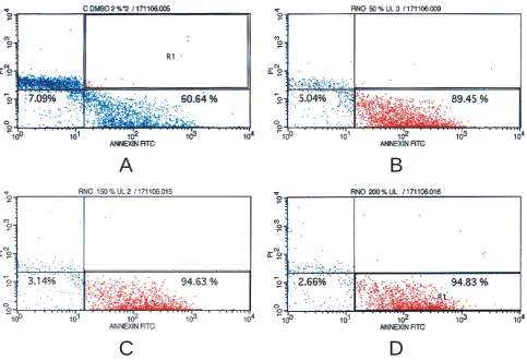
Hasil: Persentase apoptosis pada kontrol 60,64% sel hidup 7,09%. Persentase apoptosis pada 50 μg/ml 89,45%, 100 μg/ml sejumlah 87,23%, 150 μg/ml sejumlah 94.635 dan pada 200 μg/ml sejumlah 94,83%. Se-dangkan sel hidup pada 50 μg/ml sejumlah 5,04%, pada 100 μg/ml 5,71%, pada 150 μg/ml sejumlah 3,14% dan pada 200 μg/ml sejumlah 2,66%.
Kesimpulan: Terdapat peningkatan persentase jumlah sel yang apop-tosis dan penurunan sel yang hidup pada sel trofoblas yang diberikan ATRA.
[Maj Obstet Ginekol Indones 2008; 32-2: 99-104]
Kata kunci: apoptosis, molahidatidosa, sel trofoblas, asam retinoat
Introduction: Hydatidiform is an abnormal pregnancy which at histological examination shows the proliferation of trophoblastic cells. As high as 80% of hydatidiform mole patients would experience regres-sion after the evacuation. Spontaneous regresregres-sion after the evacuation occurred because trophoblastic cells had apoptosis activity. As high as 20% of hydatidiform mole patients suffered from malignancy degenera-tion which was clinically known as malignant trophoblastic disease (MTD). This malignancy degeneration may occur because of the domi-nant proliferation activity that took place continuously after the evacua-tion. Apoptosis mechanism in hydatidiform has not been completely un-derstood. Retinoic acid which is an retinol active substance or vitamin A plays a role in stimulating the arrest of cell cycle and stimulating apoptosis. It was interesting to study on whether or not the administra-tion of retinoic acid for hydatidiform mole trophoblastic cells would also induce apoptosis. The aim of this study was to demonstrate that the in-creased activity of apoptosis in hydatidiform mole trophoblastic cells re-ceiving retinoic acid. This study would be beneficial in providing the ba-sis for study of vitamin A chemoprevention in hydatidiform mole.
Material and methods: The study made use of culture specimens of hydatidiform mole trophoblastic cells. The culture of trophoblastic cells was obtained by cultivating trophoblastic cells taken from hydatidiform mole bubbles. Culture with RPMI media. At the age of 24 hours, treat-ment was made with the administration ATRA (all transretinoic acid) at a dose of 50 μg/ml, 100 μg/ml, 150 μg/ml and 200 μg/ml. The solution used was DMSO (dimethyl sulfoxide). We performed analysis of apop-tosis activity with flowcytometry in 24 hours after the treatment. The apoptosis activities were described in cytogram at the lower right quad-rant, while the number of life cells at the lower left quadrant. The cal-culation of cell was done at 1000 cells.
Results: The percentage of apoptosis in the control group was 60.64%, in the life cell 7.09%. The percentage of apoptosis at 50 μg/ml was 89.45%, at 100 μg/ml was 87.23%, at 150 μg/ml was 94.63%, at 200 μg/ml was 94.83%. On the other hand, the life cell at 50 μg/ml was 5.04%, at 100 μg/ml was 5.71%, at 150 μg/ml was 3.14%, and at 200
μg/ml was 2.66%.
Conclusion: There was an increased percentage of cells with apop-tosis and decreased percentage of life cells in trophoblastic cells receiv-ing ATRA.
[Indones J Obstet Gynecol 2008; 32-2: 99-104]
lignancy degeneration which was clinically known as malignant trophoblastic disease (MTD) or per-sistent hydatidiform mole. Regression after hyda-tidiform mole occurred because trophoblastic dis-ease had the activity of apoptosis.
It was suspected that persistent hydatidiform mole or MTD occurs due to some affecting factors, such as gene factor which regulates cell prolifera-tion, genes which regulate apoptosis and the factor of patient immunity. Cell proliferation is controlled by genes play a role as control in cell cycle.
The function of cell cycle occurs at the check point of G1 phase or S phase. The termination of cell cycle at the check point was known as G1 ar-rest and S phase arar-rest. Tumor suppressor genes (TSG) are the active genes which contribute to the arrest of cell cycle, such as p53, p21, p27, and other genes. If the function of TSG is disrupted, the con-trol mechanism will also be disrupted. Cell cycle takes place without control. The failure of control function may be a factor responsible for the occur-rence of persistent hydatidiform mole or MTD.3-6
The mechanism of apoptosis is a mechanism of body protection through cell suicide program. If the control function at the check point works, and the cell improvement does not occur, the cells that go through gene changes will be killed by apoptosis process, such that the mechanism of apoptosis takes place in conjunction with the control function, par-ticularly by p53.7,8,9
Various genes play a role in the activity of apop-tosis, the genes that stimulate apoptosis or the ge-nes that inhibit apoptosis.
Vitamin A or retinol, with retinoic as its active metabolite play a role in the various cells. The roles of retinol or retinoic include, among other things, controlling of cell proliferation through its activity which stimulates G1 phase arrest and S phase ar-rest. Such mechanism occurs because retinol or re-tinoic has plays a part in enhancing the expression of p53, activity of p21, and suppressing cylin ac-tivity.10
In addition, retinoic plays a part in enhancing apoptosis in several cancerous cells. The stimula-tion of apoptosis by retinoic occurs through the en-hancement of AP-1, stimulating caspase 7 and 9. Thus, retinol or retinoic has a suppressing effect on the growth of cancerous cells or has an impeding effect on the occurrence of cancer through two mechanisms, i.e. cell cycle and apoptosis.10,11
Retinol may enter the cell through the active mechanism or through retinol receptor. Inside the cell, the retinol is changed into retinoic active sub-stance. In order to understand the role of retinoic
in trophoblastic cells in the form of apoptosis ac-tivity, it is necessary to perform studies in tropho-blastic cells.
The aim of this study was to understand the in-duction of apoptosis activity in hydatidiform mole trophoblastic cells receiving retinoic acid. This stu-dy was expected to provide benefits that vitamin A can be use as chemoprevention post hydatidiform mole malignancy.
METHODS
This study was a laboratory trial. The samples used were the culture of hydatidiform mole. The process of trophoblastic cell culture was performed inde-pendently in laboratories. The method of culture was cell culture with RPMI media, with the ad-ministration of growth factor, antibiotics and anti fungus. The selection of culture specimen and ture method was done by try out, because the cul-ture of trophoblastic cells has not been reported yet. We conducted the treatment by administering ATRA (all transretinoic acid) in the growing cell culture. The doses of ATRA adminsitration were 50 μg/ml, 100 μg/ml, 150 μg/ml and 200 μg/ml. The evalua-tion of apoptosis activity was made by antibody Anexin V, and the examination of apoptosis made use of flowcytometry. The percentage of cell num-ber undergoing apoptosis was at the lower right quadrant, while the percentage of life cell number was at the lower left quadrant. The study was con-ducted with the permission from Ethical Committee of the Faculty of Medicine University of Indonesia.
RESULTS
Isolation/culture of hydatidiform mole tropho-blastic cells
The culture of trophoblastic cells was performed through various trials. After 7 trials, we obtained the method of sample collection and the appropriate processing of specimens. The samples were the fluid bubbles of hydatidiform mole. The fluid bub-bles of hydatidiform mole were taken by injection needle.
PBS. Culture for 24 hours at 37°C in 5% CO2 in-cubator. After 24 hours, the cells appeared to pro-liferate, the medium was disposed from tissue cul-ture flask and washed with PBS 2 times of 10 ml. ATRA was administered at doses of 50 μg/ml, 100 μg/ml, 150 μg/ml, 200 μg/ml within the well. In-cubation for 24 hours in CO2 incubator. Washed with cold PBS and centrifugation. Add 1 ml of the medium and calculate the cell number reaching 4-6 x 10⁷/ml with hematocytometry. Cells were ready to be analyzed with flow cytometry
Evaluation
The activity of apoptosis was examined using an-tibody Annexin-V. Annexin-V would bind phospha-ditilserin which was the residue of apoptosis cells on the surface of cell membrane.
The hCG examination was performed to demon-strate that the cells cultured were trophoblastic cells, and the results of beta hCG examination of culture media positively contained hCG. We performed re-peat culture up to day 10. Analysis of the medium on day 10 showed that it still contained hCG.
Examination of apoptosis activity (flowcytometry)
Identification of apoptosis was performed in 24 hours after treatment. The examination of apoptosis was made with the administration of Anexin-V ( An-nexin-V-fluorescein isothiocyanate/FITC). The cells that underwent apoptosis were calculated with flow-cytometry. The examination was performed in 1000 cells. The results of examination (cytogram) were presented at 4 quadrants. The examination of apop-tosis activity with flowcytometry constituted the better examination than any other methods.12,13
The cells that underwent apoptosis were presen-ted at the lower right quadrant, while the life cells at the lower left quadrant.12,13
Cytogram of control group and ATRA group
In the control (DMSO) of trophoblastic cell culture, we obtained 7.09% of life cells and 60.64% of the cells experienced apoptosis.
A
C
B
D
Figure 1. The results of flowcytometry cytogram examination (control)The life cells and the cells underwent apoptosis in the culture of trophoblastic cells receiving reti-noic of 50 μg/ml, 100 50 μg/ml, 150 μg/ml, 200 μg/ml were show at table 1.
Table 1. Percentage of cell life and apoptosis
CELL LIFE (%) APOPTOSIS (%)
CONTROL 7.09 60.64
50 μg ATRA 5.04 89.45
100 μg ATRA 5.71 87.23
150 μg ATRA 3.14 94.63
200 μg ATRA 2.66 94.83
DISCUSSION
Signal of apoptosis resulting from the complex of retinoic-receptor acid
With the presence of RBP receptor in the tropho-blastic cells (cytotrophoblast, syncytiotrophoblast, intermediate trophoblast) hydatidiform mole, it means that the retinol could enter the trophoblastic cells. The entry of retinol into trophoblastic cells would be continued with the metabolism of retinol into retinoic acid. Retinoic acid would enter the cell nucleus, and bind to retinoic receptor at the cell nucleus. The complex of retinoic-receptor acid would produce signals (see Figure 2).
The binding of retinoic-receptor complex would result in various signals, such as the signal of cell cycle arrest at G1 phase and S phase, and apoptosis signal.10,11
In our study, the administration of retinoic to the culture of trophoblastic cells caused an increase in the number of hydatidiform mole trophoblastic cells which underwent apoptosis.
In controls, in the observation after 24 hours, 7.09% of the cells were found to survive and 60.64% underwent apoptosis. This condition show-ed that the activity of apoptosis in the trophoblastic cells was reasonably high. This circumstance was consistent with the clinical condition, because the majority of hydatidiform mole cases experienced spontaneous regression. The spontaneous regres-sion of hydatidiform mole was caused by the ac-tivity of apoptosis in trophoblastic cells.
The administration of retinoic to the culture of trophoblastic cells caused an increase in the number of cell undergoing apoptosis. The increase in per-centage of apoptosis occurred with the increase in retinoic doses. With the administration of retinoic
at a dose of 50 μg/ml, 5.04% of the life cells were found, and the number of cells undergoing apop-tosis was 89.45%. On the other hand, with the ad-ministration of retinoic at a dose of 200 μg/ml, 3.18% of the life cells were found, and the number of trophoblastic cells undergoing apoptosis reached 93.81%. This finding showed that the administra-tion of retinoic acid increased the activity of apop-tosis in trophoblastic cells, and the percentage of apoptosis increased with the increase in the doses of retinoic administration.
The mechanism of apoptosis by retinoic acid oc-curred through the increase of TNF (tumor necros-ing factor) expression, inhibition of Bcl-2, and the expression induction of p53. The complex of reti-noic-receptor acid produced signals which directly stimulated caspase 9, and indirectly stimulated cas-pase 7.14,15 Retinoic acid also induced apoptosis through Apaf1 (apoptotic protease activating factor 1). Acid retinoic works as antagonist of transcrip-tion factor, activator protein-1 (AP-1) which played a role in the cell growth and cell differentiation.15 Retinoic acid causes the occurrence of G1 phase arrest, the termination of that cycle occurred be-cause of the decrease in G1 cyclin protein, E cyclin, and also prevents the phosphorilation of pRb. Thus, generally retinoic acid would cause G1 arrest.16
Arrest at G1 phase by retinoic acid could occur through several mechanisms, and one of the mecha-nisms taking place was the activation of p53 by re-tinoic acid. The increase of p53 activation caused the occurrence of arrest at G1 phase. This mechanism apparently constituted a direct mechanism.10,11
Arrest at S phase occurred because there was an inhibition of E2F phosphorilation, expression of clin A could be suppressed and the complex of cy-clin-cdk2 could not be formed. Therefore, the arrest at S phase could occur through two mechanisms, i.e. through p21 and through the inhibition of E2F phosphorilation.10,11
Retinoic acid also activated p53 and increased
p53 level. The increase of non-mutant p53 would cause the activation of control mechanism of cell cycle. Gene p53 caused the occurrence of G1 arrest, the termination of cell cycle at G1 phase would make it possible for the cell to be repaired. In ad-dition, p53 played a role in activating apoptosis mechanism (see Figure 2).12,13
Retinol inside the cytoplasm would be metabo-lized into retinoic acid, and the retinoic-receptor complex inside the cell nucleus would produce apoptosis signals and proliferation inhibition (see Figure 2).
In the study on the expression of retinoic-recep-tor in trophoblastic cells, it was demonstrated that trophoblastic cells had retinoic receptor. The pre-sence of retinoic receptor in trophoblastic cells sig-nified that retinoic could penetrate into hydatidi-form mole trophoblastic cells.
The study on the activation of retinoic in tro-phoblastic cells showed that retinoic acid could in-duce the activity of apoptosis in trophoblastic cells. In order to demonstrate the effects of vitamin A in hydatidiform mole trophoblastic cells as the ef-fort to prevent malignancy degeneration following hydatidiform mole, it is necessary to conduct fur-ther study with clinical trials.
CONCLUSIONS
The administration of retinoic acid in trophoblastic cells increased the activity of apoptosis in tropho-blastic cells.
REFERENCES
1. Tham KF, Ratnam SS. Current views on the management of trophoblastic tumors. Int J Gynecol Obstet 1995; 49: 77-89
2. Sasaki S. Clinical presentation and management of molar pregnancy. Best Practice and Research Clinical Obstetrics and Gynaecology 2003; 17(6): 885-92
3. MacDonald F, Ford CHJ, Casson AG. Molecular Biology of Cancer. Second edition, Garland Science/Bios Scientific Publishers, 2004: 2-4
4. MacDonald F, Ford CHJ, Casson AG. Molecular Biology of Cancer, second edition, Garland Science/BIOS Scientific Publisher, 2004: 61-72
RETINOIC + RAR
APOPTOSIS
CELL CYCLE
G
1S
G
2M
CASPASE-7 CASPASE-9
(+)
(+)
CRABP
Apaf-1
(+)
(+) (+)
Bcl-2
(-) (+)
(-)
CYCLIN-D1, A, E
P53
(+)
(-) (+)
(-)
P21
(+)
(-)
P27
E2F
pRb-E2F +
(-)
(-)
(+)
(+)
CYCLIN D3
(-)
Cmyc,Cjun
(+) (-)
GATA6 dab2
(+)
(+) (+)
Figure 2. Signals of retinoic-receptor complex
5. Zornig M, Baum W, Hueber AO, Evan G. Programmed Cell Death and Senescence, in The Molecular Basis of Cancer, 2nd edition, Philadelphia, WB Saunders company, 2001: 19-40
6. MacDonald F, Ford CHJ, Casson AG. Molecular Biology of Cancer, 2nd edition, Garland Science/BIOS Scientific Pu-blisher, 2004: 4-6
7. Tsao AS, Kim ES, Hong Wk. Chemoprevention of Cancer. CA Cancer J Clin 2004; 54: 150-80
8. Dragnev KH, Rigas JR, Dmitrovsky E. The Retinoids and Cancer Prevention Mechanisms. The Oncologist 2000; 5: 361-8
9. Greenwald P. Cancer chemoprevention. B Med J 2002; 324: 714-8
10. Budhu AS, Noy N. Direct Chanelling of Retinoic Acid be-tween Cellular retinoic Acid-Binding Protein II and Reti-noic Acid receptor Sensitizes Mammary Carcinoma Cells to Retinoic Acid-Induced Growth Arrest. Mol and Cell Bio-logy 2002; 22: 2632-41
11. Zhang Y, Rishi AK, Dawson MI, Tschang R, Farhana L. S-phase Arrest and Apoptosis Induced in Normal Mammary
Epithelial Cells by a Novel Retinoid. Cancer Res 2000; 60: 2025-32
12. Kravtsov VD, Daniel TO, Koury MJ. Comparative Analysis of Different Methodological Approaches to the in Vitro Study of Drug-Induced Apoptosis. Am J Pathol. 1999; 155: 1327-39
13. Yasuhara S, Zhu Y, Matsui T, Tipirneni N, Yasuhara Y, Kaneki M, Rosenzweig A, Martyn JAJ. Comparison of Comet Assay, Electron Microscopy, and Flow Cytometry for Detection of Apoptosis. J Histochem Cytochem 2003; 51: 873-85
14. Farias EF, Ong DE, Ghyselinck Nb, Nakajo S, Kuppumbatti Ys, Lopez RM. Cellular Retinol-Binding Protein I, a Regu-lator of Breast Epithelial Retinoic Acid Receptor Activity, Cell Differentiation, and Tumorigenicity. J J Nat Can Inst 2005; 97: 21-9