Journal of Applied Pharmaceutical Science Vol. 2 (9), pp. 122-126, September, 2012 Available online at http://www.japsonline.com
DOI: 10.7324/JAPS.2012.2924 ISSN 2231-3354
Effect of Saccharum spontaneum Linn. on Lysosomal enzymes of
Uro-lithiatic rats
M. Sathya and R. Kokilavani
Department of Biochemistry, Kongunadu Arts and Science College, Coimbatore-29, India.
ARTICLE INFO ABSTRACT
Article history: Received on: 22/08/2012 Revised on: 05/09/2012 Accepted on: 15/09/2012
Available online: 28/09/2012
The ethanolic root extract of Saccharum spontaneum of family Poaceae was used to treatthe urolithiasis induced by glycolic acid On this course, the extract also repairs the changesthat happened in the lysosomal enzymes like β-D-glucuronidase, xanthine oxidase in liver and kidney and n-acetyl _-d-glucosaminidase in serum, liver, kidney and urine of the urolithiatic rats. The ethanolic root extract (200 and 300 / kg b.w.) elevated the levels of reduced β-D-glucuronidasein liver and n-acetyl _-d-glucosaminidase in liver and kidney and reduced the level of xanthineoxidase in liver and kidney and n-acetyl _-d-glucosaminidase in serum and urine significantly (p<0.05) when compared with the toxic groups. The results shown by the ethanolic root extract (200 and 300 mg / kg b.w.) was compared to standard thiazide drug treated group, showing no significantdifference (p<0.05) and thus it proves that the ethanolic root extract of S.spontaneum exhibits potent antiurolithiatic activity.
Key words:
Saccharum spontaneum, Lysosomal Enzymes, Thiazide, Glycolic acid
INTRODUCTION
Urinary stone disease continues to occupy an important place in everyday urological practice. The average life time risk of stone formation has been reported in the range of 5-10 %. A predominance of men over women can be observed with an incidence peak between the fourth and fifth decade of life. Recurrent stone formation is a common part of the medical care of patients with stone disease (Tiselius et al., 2001).Urolithiasis is a recurrent renal disease affects 4-8% in UK, 15% in US, 20% in Gulf countries and 11% population in India. Stone formation tends to recur at very high rate; without preventative measures after a first stone. After 3 years this is about 40%, by 10 years up to 75% and by 25 years virtually every patient has formed at least one more stone (Leye et al., 2007).
There are several types, most commonly consisting of calcium phosphates and calcium oxalates; others are composed of magnesium ammonium phosphate (struvite), uric acid or cystine (Sellaturay and Fry, 2008). Epidemiological data suggests that 60-80% of stone is composed mainly of calcium oxalate (CaOx). Stones formation occurs when urinary concentrations of stone forming salts, exceed the limit of metastability for that salt in solution. This most often reflects excessive excretion of one or more stone constituents, deficient inhibitory activity in urine, or simply a low urine volume resulting in excessively concentrated urine (Steven, 2003). The pathogenesis of calcium oxalate stone formation is a multi-step process, which includes-nucleation, crystal growth, crystal aggregation and crystal retention (Pareta et al., 2011). Various substances in the body have an effect on one or more of the above stone forming processes, thereby influencing a person’s ability to promote or prevent stone formation. Promoters of stone formation facilitate stone formation whilst inhibitors * Corresponding Author
Research scholar, Department of Biochemistry,
prevent it. Low urine volume, low urine pH, calcium, sodium, oxalate, and urate are known to promote stone formation (Basavaraj et al., 2007).
The recent resurgence of plant remedies results from several factors like effectiveness of plant medicines and no side effects compared to modern medicines. Plant medicine was commonly used for traditional treatment of some significant diuretic activity. Many investigators have demonstrated that studies of herbal plant used in traditional medicine as diuretic have increased recent years (Maghrani et al., 2005) and might be a useful tool in the treatment of urolithiasis. A large number of Indian medicinal plants have been used in the treatment of urolithiasis and they have been reported to be effective with fewer side effects (Selvam et al., 2001). Even today, plants provide a cheap source of drugs for majority of world’s population. Several pharmacological in vitro and in vivo investigations on the medicinal plants used in traditional antiurolithiatic therapy revealed their therapeutic potential (Bashir and Gilani, 2009).
Saccharum spontaneum L. known as Kasa (Family:
Poaceae) is a traditional herb, it has excellence medicinal value; has been advocated in the treatment gynaecological troubles, respiratory disease. Roots are used as galactagogue and diuretic and in ayurveda system roots are also used as astringent, emollient, refrigerant, diuretic, purgative, tonic, and aphrodisiac and useful in treatment of dyspepsia, burning sensation, piles and sexual weakness (Khalid and Siddiqui, 2011). The stems (culm) are useful in vitiated conditions of pitta and vata burning sensation strongly, renal and vesicol calculi dyspepsia, haemorrhoids, menorrhagia dysentery, agalactia phthisis and general debility (Suresh kumar et al., 2009).
MATERIALS AND METHODS
Collection of plant material
Saccharum spontaneum Linn. was collected from Koorappalayam, Erode district, Tamil Nadu, India during the month of September to November, 2008. The plant was identified and authenticated by taxonomist Dr.K. Arumugasamy, Assistant Professor, Department of Botany, Kongunadu Arts and Science College, Coimbatore, Tamilnadu, India. Voucher specimen was deposited in herbarium centre, Department of Botany, Kongunadu Arts and Science College, Coimbatore.
Preparation of the ethanolic root extract for in vivo studies Roots of the plants were washed, shade dried, powdered and stored in tight containers under refrigeration. 100g of S.spontaneum powder was taken in a conical flask. To this 500ml of 99% ethanol was added. The content of the flask was kept in the shaker for 48 hr. and the suspension was filtered and residue was resuspended in an equal volume of 99% ethanol for 48hr. and filtered again. The two filtrates were pooled and the solvents were dried in an oven at 37ºC and a crude residue was obtained. The yield was 21.8 g, and the residue was suspended in water and administered orally to the experimental rats.
Selection of animals for In vivo studies
For the purpose of sub acute toxicity, diuretic, pharmacological screening of anti urolithiatic and In vivo biological evaluation of urolithiatic studiesin in adult male wistar albino rats weighing about 150 to 200 g were collected from animal breeding centre, Kerala Agricultural University, Mannuthy, Thrissur, Kerala, India. The ethical committee permission license number is 659/02/a/CPCSEA. The rats were kept in properly numbered large polypropylene cages with stainless steel top grill having facilities for pelleted food. The animals were maintained in 12 hr. light and dark cycle at 28oC ± 2o C in a well ventilated animal house under natural conditions in large polypropylene cages and they were acclimatized to laboratory conditions for 10 days prior to the commencement of the experiment. The animals were fed with standard pelleted diet supplied by AVM foods, Coimbatore, Tamilnadu, India.All animal experiments were performed according to the ethical guidelines suggested by the Institutional Animal Ethics Committee (IAEC). Paddy husk was used as beding material and changed twice a week.
Experimental design for in vivo biological evaluation studies The rats were divided into 5 groups of six animals in each group and the experimental design of animals is given in table1 for in vivo studies.
Group I: Control rats - received normal pelleted diet.
Group II: Glycolic acid intoxicated rats - Urolithiasis induced by fed with a calculi-producing diet (CPD: commercial diet mixed with 3% glycolic acid) for 28 days.
Group III: Root extract treated rats - Urolithiasis induced rats received ethanolic root extract of S.spontaneum (200 mg / kg b.w.) by oral administration for 28 days at a rate of 1.0 ml / rat / day.
Group IV: Root extract treated rats - Urolithiasis induced rats received ethanolic root extract of S.spontaneum (300 mg / kg b.w.) by oral administration for 28 days at a rate of 1.0 ml / rat / day.
Group V: Standard drug thiazide treated rats - Urolithiasis
induced rats received thiazide (150μg/ kg b.w.) by oral
administration for 28 days at the rate of 1.0 ml / rat / day.
Collection of urine sample
Before the day of sacrifice the rats were placed in metabolic cages and urine was collected for 24 hours. Urine was freed from faecal contamination. Rats were provided with water but no feed. Urine collected in 50 ml beaker maintained at 0oC in an ice bath. The collected urine samples were centrifuged for 10 minutes and any sediment present was discarded. The urine was used for further analysis.
Collection of serum sample
ratio of 1:10. Aliquots of the diluted serum were then used for the determination of serum constituents and serum enzymic activities.
Collection of liver and kidney samples
The experimental animals were sacrificed, liver and kidney were removed immediately, washed with ice cold saline10% tissue homogenate was prepared by homogenizing 1.0g of chopped liver or kidney tissue in 10ml of 0.1M tris HCl homogenizing buffer at pH 7.5. The homogenate was used for assaying the enzyme activities
Estimation of β-D-glucuronidase
β -D-glucuronidase is estimated by using themethod as in (Kawai and Anno, 1971). 0.5 ml of substrate, 0.05mlof acetate buffer, 0.3ml of homogenate was incubated at 37oC for 1 hr.. The reaction was arrested by the addition of 3.9ml of glycine buffer. Standards were also run simultaneously along with a blank. The colour developed was read at 420nm using acolorimeter. The
enzyme activity is expressed as μmoles of p-nitrophenol
liberated/L.
Estimation of Xanthine Oxidase
Xanthine oxidase estimation is done as per the method given in(Bergmeyer et al.,1974).To the test added 0.6ml of phosphate buffer, 0.4ml EDTA, 0.4ml gelatin, 0.3ml NBT,0.1ml PMS and 0.1ml of enzyme mixture. The tubes wee incubated at 37°C for 15min.Then add 0.6ml of phosphate buffer and 0.5ml of xanthine. To the blank instead of enzyme mixture add the enzyme buffer and repeat the same, the enzyme activity was measured at 432nm in aspectrophotometer for every 2sec for 10min. The
enzyme activity was expressed as μm of xanthine
oxidized/min./mg protein.
Estimation of n-acetyl _-d-glucosaminidase
The method of (Marhun, 1976). was followed for the determination of NAG activity. To 0.2 ml ofdialysed urine, 0.2ml of buffered substrate.
Chemicals
All the chemicals used in the present study were of analytical reagent grade.
Statistical analysis
The results of the biochemical estimations were reported as mean ± SD of six animals in each group. Total variations, present in a set of data were estimated by one way Analysis Of Variance (ANOVA) followed by the analysis of level of significance between different groups based on ANOVA using SPSS statistical package (Version 15.0). Difference among means were analysed by least significant difference (LSD) at 5% level (p<0.05).
RESULTS AND DISCUSSION
Lysosomal Enzymes in Liver and Kidney
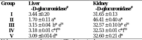
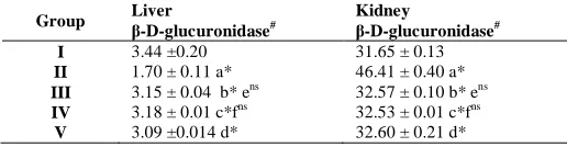
From the table1, it is evident that the levels of lysosomal enzymes in liver homogenate was significantly decreased (P<0.05)
whereas in the kidney homogenate it was significantly increased in glycolic acid intoxicated rats (Group II) comparing to control rats
(Group I). β-D-glucuronidase a renal tissue enzyme was found in
kidney but low levels in liver during hyperoxaluric condition.
Group comaprison
‘a’ represents comparison between group II and I ‘b’ represents comparison between group III and II ‘c’ represents comparison between group IV and II ‘d’ represents comparison between group V and II ‘e’ represents comparison between group III and V ‘f’ represents comparison between group IV and V
The symbols represent statistical significance p* < 0.05; ns - not significant
Units
#
µmoles of phenol liberated / mg protein.
Group III and IV rats treated with the S.spontaneum root extract showed a significant restoration of lysosomal enzymes in liver and kidney when compared to glycolic acid treated rats (group II), which might be an indication of recovery due to the antiurolithiatic property of ethanolic extract of S. spontaneum.
When S.spontaneum root extract treated rats (Group III and IV) were compared with thiazide treated rats (Group V), there was no significant difference between these groups of rats. This result gives a supportive evidence of the antiurolithiatic activity of ethanolic root extract which is similar to standard drug thiazide.
Our results coincides with that of Veena et al. (2005) who showed that the sulfated polysaccharides from edible sea weed Fucus vesiculosus restore this enzyme level in hyperoxaluric rats. Subha et al. (1992) reported that administration of sodium pentosan phosphate restored the levels of this enzyme in urolithiatic rats. Experimental studies reveal that the ethanolic extract from S. spontaneum root (200 and 300 mg/kg) orally administered for 28 days normalized the levels of the levels of lysosomal enzymes in liver and kidney.
Xanthine oxidase levels in liver and kidney
Table 2 represents the effect of ethanolic root extract of S.spontaneum on xanthine oxidase in liver and kidney of control and experimental rats. From the table 2, it is evident that the levels of xanthine oxidase in liver and kidney homogenate was significantly increased (P<0.05) in calculi induced animals (Group II) comparing to control rats (Group I).
The enzymatic reaction converting hypoxanthine to xanthine and uric acid is usually linked to the reduction of NAD+
Table. 8: Effect S.spontaneumroot extract of on β-D-glucuronidase in liver and kidney of control and experimental rats.
Group Liver
β-D-glucuronidase#
Kidney
β-D-glucuronidase# I
II III IV V
3.44 ±0.20 1.70 ± 0.11 a*
3.15 ± 0.04 b* ens
3.18 ± 0.01 c*fns
3.09 ±0.014 d*
31.65 ± 0.13 46.41 ± 0.40 a*
32.57 ± 0.10 b* ens
32.53 ± 0.01 c*fns
to NADH and the protein involved is termed as xanthine dehydrogenase. The oxygen-reducing activity of this enzyme or xanthine oxidase results when the protein undergoes oxidation of certain thiol or proteolysis and a 2-fold significant increase in the activity of xanthine oxidase was observed in EG-treated rats (Hille and Nishino, 1995).
The most common kidney stone is the calcium oxalate stone; however, uric acid stones can be formed as a mixture of uric acid and calcium oxalate stones. Uric acid production is catalysed by xanthine oxidase. This enzyme causes gout and is responsible for oxidative damage of living tissues. glycoic acid treated rats showed significant loss in body weight and increase in the activities of oxalate synthesizing enzymes and free radical producing enzyme xanthine oxidase.
Table. 2: Effect ethanolic root extract of S.spontaneum on xanthine oxidase in liver and kidney of control and experimental rats.
Values are expressed as mean ± SD of six animals
Experimental design and comparison between the groups are as in table 1 The symbols represent statistical significance p* < 0.05, ns – not significant
Units
##
µ moles of xanthine oxidised / min. / mg protein The ethanolic root extract of S.spontaneum. treated rats showed a significant restoration of xanthine oxidase in liver and kidney when compared to glycolic acid treated rats (group III and IV ), which might be an indication of recovery due to the antiurolithiatic property ethanolic root extract of S.spontaneum. When S.spontaneum. root extract treated rats (Group III and IV) were compared with thiazide treated rats (Group V), there was no significant difference between these groups of rats in xanthine oxidase activity.
Our results coincides with that of Santhosh kumar and Selvam (2003)who showed that supplementation of vitamin E and selenium decreased the level of oxalate synthesizing enzymes with a concomitant increase in the activities of enzymatic antioxidants and non- enzymatic antioxidant. The antioxidant vitamin E+ selenium thereby protected from hyperoxaluria.
Pragasam et al. (2005) reported that L-arginine co-supplementation to EG-treated rats maintained the activities of the oxalate synthesizing enzymes and free radical producing enzyme xanthine oxidase with in the normal range.
Moriyama et al. (2007) reported an increase in NADPH-induced O2
−
production, or NADPH oxidase activity, in the homogenate of cells injured by oxalate exposure. These findings suggest that the reduction in oxalate-induced O2
−
production contributes to the cytoprotective effect of Quercus salicina extract.
Levels of N-acetyl β -D glucosaminidase in serum, urine kidney and liver
Table 3 represents the levels of N-acetyl β-D glucosaminidase in urine of control and experimental rats.
Units
*µ moles of P-nitrophenol liberated / L
** µ moles of P-nitrophenol liberated /24 hr..urine
ψ
µ moles of P-nitrophenol liberated /min./mg protein
From the table 3, it is evident that the levels of NAG was significantly increased (p<0.05) in serum and urine whereas in liver and kidney the levels were significantly decreased in urolithiatic rats (Group II) when compared to control rats (Group I).Urinary excretion of the lysosomal enzyme NAG was measured as a specific indicator of tubular cell damage. NAG is renal tubule specific and is a valid indicator of tubular damage. It has been found to correlate with the corresponding morphological tubule lesions arising through other mechanisms. To prevent falsification due to individual difference in baseline NAG secretion, the individual post therapeutic NAG increase was determined in the pooled urine of each animal and set in relation to creatinine clearance to avoid dilution artifacts (Jacobsen et al., 1999).
Damage to the tubules was indicated by increased excretion N-acetyl β -D glucosaminidase (NAG). Fibrinolytic activity was found to be reduced. Administration of ethanolic root extract of S.spontaneum reduced the tubular damage and decreased the markers of crystal deposition markedly, which was substantiated by the reduction in weight of bladder stone formed.
However treatment with the extract ethanolic root extract of S.spontaneum root extract showed a significant restoration of N-acetyl glucosaminidase levels in serum, urine, kidney and liver when compared to glycolic acid treated rats (group III and IV), which might be an indication of recovery due to the healing property of the ethanolic root extract of S.spontaneum which possess antiurolithiatic property.
When S.spontaneum root extract treated rats (Group III and IV) were compared with thiazide treated rats (Group V), there was no significant difference between these groups of rats, proving the antiurolithiatic activity of S.spontaneum which is similar to standard drug thiazide. From the above results, it is prevalent that the ethanolic root extract of S.spontaneum normalized the levels of
N - acetyl β -D glucosaminidase in urine, serum, kidney and liver
of experimental animals.
Our results are in accordance with that of Lenin et al. (2001) who showed attenuation of oxalate-induced nephrotoxicity by eicosapentaenoate-lipoate (EPA-LA) derivative in experimental rats. Jeong et al. (2006) reported that green tea supplementation decreased the excretion of urinary oxalate and the activities of urinary gamma glutamyl transpeptidase and N-acetyl glucosaminidase in ethylene glycol treated rats.
Group Liver
β-D-glucuronidase#
Kidney
β-D-glucuronidase# I
II III IV V
3.44 ±0.20 1.70 ± 0.11 a*
3.15 ± 0.04 b* ens
3.18 ± 0.01 c*fns
3.09 ±0.014 d*
31.65 ± 0.13 46.41 ± 0.40 a*
32.57 ± 0.10 b* ens
32.53 ± 0.01 c*fns
CONCLUSION
Our results highlight that Saccharum spontaneum is the most effective drug in inhibiting stone formation and healing renal damage caused by oxalate toxicity, thus confirming its antiurolithiatic property.
REFERENCES
Basavaraj, D.R., C.S. Biyani, A.J. Browning and J.J. Cartledge, The role of urinary kidney stone inhibitors and promoters in the pathogenesis of calcium containing renal stones. EAU-EBU Update Series. 2007; 5: 126-136.
Bashir, S., Gilani, A.H., Antiurolithiatic effect of Berenia ligulata rhizomes: an explation of the underlying mechanisms. J Ethnopharmacol., 2009;122, 106-116.
Bergmeyer, H.U., Gawehn, K. and Grassl, M. Methods of Enzymatic Analysis.2nd edn., Academic Press Inc., New York,1974; (1): 521-522.
Hille, R. and Nishino, T. Xanthine oxidase and xanthine dehydrogenase. The federation of American societies for experimental biology, 1995; 9:995-1003.
Jacobsen, K.J.W., Bannowski, A., Kuppers, F., Loch, T. and Stockle, M. Direct amifostine effect on renal tubule cells in rats. Cancer. Res., 1999; 59: 3451-3453.
Jeong, B.C., Sub Kim, B., In Kim, J. and Hoe Kim, H. Effects of green tea on urinary stone formation: An in vivo and in vitro Study. J. endourol., 2006; 20(5): 356-361.
Kawai, Y. and Anno, K. Mucopolysaccharide - degrading enzymes from the liver of the squid, ommastrephes Sloani pacificus. I. Hyaluronidase. Biochem. et Biophys.Acta., 1971; 242(2): 428-436.
Lenin, M., Thiagarajan, A., Nagaraj, M. and Varalakshmi, P. Attenuation of oxalate-induced nephrotoxicity by eicosapentaenoate– lipoate (EPA-LA) derivative in experimental rat model. Prostaglandins, Leukotrienes and Essential Fatty Acids, 2001; 65(5-6): 265-270.
Leye A, Jaeger P, Robertson W and Unwin R; Renal stone disease. Medicine, 2007; 35: 415-419.
M. Maghrani, N.-A. Zeggwagh , M. Haloui M. Eddouks Acute diuretic effect of aqueous extract of Retama raetam in normal rats Journal of Ethnopharmacology., 2005;99(1):31–35.
Maruhn, D. Rapid colorimetric assay of β- galactosidase and N-acetyl- β- D- glucosaminidase in human urine, Analytical Chemical Acta,1976; 73: 453-461.
Mohammad Khalid, Hefazat H. Siddiqui Pharmacognostical Evaluation and Qualitative Analysis of Saccharumspontaneum (L.) Root International Journal of Pharmaceutical Sciences and Drug Research., 2001;3(4): 338-341.
Moriyama, M.T., Miyazawa, K., Noda, K., Oka, M., Tanaka, M. and Suzuki, K. Reduction in oxalate-induced renal tubular epithelial cell injury by an extract from Quercus salicina Blume/ Quercus stenophylla Makino. Urol. Res., 2007; 35 (6):295-300.
Pareta, S.K., K.C. Patra and R. Harwansh, In-vitro calcium oxalate crystallization inhibition by Achyranthes indica linn. Hydroalcoholic extract: An approach to antilithiasis. Int. J. Pharm. Bio. Sci., 2011 ; 2: 432-437.
Pragasam, V. and Kalaiselvi, P. Counteraction of oxalate induced nitrosative stress by supplementation of L-arginine, a potent antilithic agent. Clin. Chem. Acta., 2005; 354 (1-2):159-166.
Santhosh kumar, M. and Selvam, R. Supplementation of vitamin E and selenium prevents hyperoxaluria in experimental urolithic rats. J. Nutri. Biochem., 2003; 14(6): 306-313.
Selvam, R., Kalaiselvi, P., Govindaraj, A., Murugan, V.B. and Sathish kumar, A.S. Effect of A. lanata leaf extract and Vediuppu chunnam on the risk factor of calcium oxalate urolithiasis during experimental hyperoxaluria. Pharmacol Res., 2001; 43 (1), 89-93.
Sellaturay, S. and C. Fry. The metabolic basis for urolithiasis. Surgery, 2008; 26: 136-139.
Steven, J. Urinary calculi. Medicine, 2003 ; 32: 77-80.
Subha, K., Siva Murugesan, A. and Varalakshmi, P. Changes in serum lipids and lipoproteins in calcium oxalate stone forming rats treated with sodium pentosan phosphate. Biochem. Mol. Biol. Inter.1992; , 27(6): 1011-1018.
Suresh kumar , C.A., R.Varadharajan , P.Muthumani1, R. Meera1,P.Devi , B.Kameswari Pharmacognostic and Preliminary Phytochemical Investigations on the stemof Saccharum spontaneum J. Pharm. Sci. & Res.. 2009; 1(3): 129-136.
Veena, C.K., Josephine, A., Preetha, S.P. and Varalakshmi, P. Beneficial role of sulphated polysaccharides from edible seaweed Fucus vesiculosus in experimental hyperoxaluria. Food Chem., 2005; 100: 1552-1559.
Tiselius HG, Ackermann D, Alken P, Buck C, Conort P, Gallucci : Guidelines on urolithiasis 2001; 40: 362-371.
Table. 3: Effect S.spontaneum root extract of on N-acetyl β-D glucosaminidase levels in serum urine, kidney and liver of control and experimental rats.
Group Serum * Urine * * Kidneyψ Liverψ
I II III IV V
16.53 ± 0.026 26.50 ± 0.12 a*
17.25 ± 0.02 b* ens
17.20 ± 0.021 c*fns
17.31 ± 0.24 d*
220.53 ± 0.23 410.44 ± 0.13 a*
222.32 ± 0.018 b* ens
222.27 ± 0.12 c*fns
222.38 ± 0.13 d*
16.82 ± 0.12 12.53 ± 0.06 a*
14.07 ± 0.14 b* ens
14.02 ± 0.01 c*fns
14.11 ±0 .02 d*
14.38 ± 0.13 8.71 ± 0.16 a*
14.07 ± 0.14 b* ens
14.05 ± 0.12 c*fns
14.12 ± 0.24 d*
Values are expressed as mean ± SD of six animals .
Experimental design and comparison between the groups are as in table 1. The symbols represent statistical significance p* < 0.05, ns – not significant.
How to cite this article: