Cite as
Bednarz-Misa I, Berdowska I, Zboch M, et al. Paraoxonase 1 decline and lipid peroxidation rise reflect a degree of brain atrophy and vascular impairment in dementia. Adv Clin Exp Med. 2020;29(1):71–78. doi:10.17219/acem/111377
DOI
10.17219/acem/111377
Copyright
© 2020 by Wroclaw Medical University This is an article distributed under the terms of the Creative Commons Attribution 3.0 Unported (CC BY 3.0) (https://creativecommons.org/licenses/by/3.0/)
Address for correspondence
Izabela Berdowska
E-mail: [email protected]
Funding sources
None declared
Conflict of interest
None declared
* Iwona Bednarz-Misa and Izabela Berdowska contributed equally to this work.
Received on February 18, 2019 Reviewed on March 30, 2019 Accepted on July 28, 2019
Published online on January 22, 2020
Abstract
Background. Paraoxonase 1 (PON1) is an enzyme with the capability to protect against lipid oxidation and atherosclerotic lesions formation. Impaired antioxidative capacity and enhanced lipid peroxidation (reflected by malondialdehyde rise) accompany dementias.
Objectives. The aim of this study was to discern the possible differences in the activity and phenotype distribution of PON1, and lipid peroxidation level in dementias of neurodegenerative and vascular pathology, to assess whether they reflect structural changes in the brain, and to evaluate their potential as dementia markers.
Material and methods. Paraoxonase 1 arylesterase activity and polymorphisms (dual-substrate method), and malondialdehyde/thiobarbituric acid reactive substances (MDA/TBARS) levels were determined spec-trophotometrically in 257 serum samples derived from 136 dementive patients (with Alzheimer’s disease (AD; n = 63), vascular dementia (VaD; n = 40) and mixed-type dementia (MD; n = 33), as well as from 121 non-dementive individuals. The results were analyzed with reference to dementia type and severity (assessed with Mini Mental State Examination (MMSE) and Clinical Dementia Rating (CDR) scales), structural brain changes (estimated with magnetic resonance imaging (MRI) – Global Cortical Atrophy (GCA), Medial Temporal lobe Atrophy (MTA) and Fazekas scales)) and brain ischemia (Hachinski Ischemic Scale (HIS) index), and evaluated using receiver operating characteristic (ROC) analysis.
Results. Malondialdehyde/thiobarbituric acid reactive substances were increased in dementia (more in VaD than AD). In patients with vascular involvement, MDA/TBARS elevation reflected a degree of global cortical atrophy. Paraoxonase 1 activity was decreased in patients with dementia, especially in patients with severe cognitive deficits. In VaD, a drop in PON1 reflected a degree of MTA and brain ischemia. MDA/TBARS displayed 75% accuracy as a general dementia marker, but, similarly to PON1, were a poor differential marker. Conclusions. Both indices were more associated with vascular involvement and the severity of brain atrophy or ischemia rather than with degree of cognitive decline.
Key words: Alzheimer’s disease, vascular dementia, paraoxonase 1, mixed-type dementia, MDA/TBARS
Paraoxonase 1 decline and lipid peroxidation rise reflect a degree
of brain atrophy and vascular impairment in dementia
*Iwona Bednarz-Misa
1,A,B,D,F, *Izabela Berdowska
1,B,D–F, Marzena Zboch
2,B,C,F, Błażej Misiak
3,4,B,E,F, Bogdan Zieliński
1,B,F,
Sylwia Płaczkowska
5,B,F, Mariusz Fleszar
1,B,F, Jerzy Wiśniewski
1,B,F, Andrzej Gamian
1,A,E,F, Małgorzata Krzystek-Korpacka
1,A–F1 Department of Medical Biochemistry, Wroclaw Medical University, Poland 2 Alzheimer Center, Wroclaw Medical University, Ścinawa, Poland 3 Department of Psychiatry, Wroclaw Medical University, Poland 4 Department of Genetics, Wroclaw Medical University, Poland
5 Department of Professional Training in Clinical Chemistry, Wroclaw Medical University, Poland
A – research concept and design; B – collection and/or assembly of data; C – data analysis and interpretation; D – writing the article; E – critical revision of the article; F – final approval of the article
Introduction
Progressive degeneration of neurons, a hallmark of neu-rodegenerative disorders, leads to serious deficits in cogni-tive performance and general functioning.1 Due
to the pro-gressing aging of societies, the number of people suffering from various forms of dementia is projected to double every 20 years and will become a serious burden for public healthcare in the near future. Alzheimer’s disease (AD) is the main cause of dementia in the elderly population, followed by vascular dementia (VaD). Alzheimer’s disease is estimated to account for 60–80% of cases and is char-acterized by the accumulation of extracellular β-amyloid plaques and intracellular neurofibrillary tangles.1 Vascular
dementia is the result of the blockage or damage of the cere-bral blood vessels, leading to infarcts, bleeding and/or isch-emia and ultimately to brain injury. It was initially consid-ered to be the sole form of dementia attributable to vascular lesions; however, several studies have demonstrated that the development of cardiovascular risk factors also play a substantial role in AD. Indeed, neuropathological indices of brain infarcts can be found in up to half of AD patients. The co-existence of AD and VaD pathologies is observed in the vast majority of mixed dementia (MD) cases.1
Epidemiological studies have revealed that lipid peroxi-dation caused by oxidative imbalance is a common etiol-ogy of both cardiovascular2 and neurocognitive diseases.3
Among others, oxidative stress (OS) alters the permeability of vascular and cerebral endothelium and promotes in-flammatory responses.3 Brain tissue is particularly
vulner-able to OS due to high content of oxidative damage-prone polyunsaturated fatty acids and intense oxidative metabo-lism combined with low antioxidant capacity.3
Accord-ingly, the decline in antioxidants as well as the accumula-tion of oxidative damage markers, e.g., malondialdehyde (MDA), considered as the most abundant aldehyde derived from lipid peroxidation, have been reported in both AD and VaD.3,4
Paraoxonase 1 (PON1) is a liver-synthesized enzyme of multiple biological functions, which has gained particu-lar attention as an atherogenic agent. It displays anti-oxidative properties and protects low-density lipoprotein (LDL) and high-density lipoprotein (HDL) lipids from oxi-dation, decreases oxidative status of macrophages and in-creases cholesterol efflux, thus contributing to the preven-tion and attenuato the preven-tion of the development of atherosclerotic lesions.5 Moreover, PON1 participates
in the detoxifica-tion of homocysteine thiolactone, another pro-atherogenic compound, and plays an anti-inflammatory role by inhibit-ing the expression of monocyte chemoattractant protein (MCP)-1 and activity of myeloperoxidase, which serve as key players in vascular inflammation and OS. It has also been reported to stabilize lipid membranes and enhance their integrity under the conditions of oxidative imbalance. Accordingly, decreased enzyme activity has been repeat-edly linked with an increased atherogenic risk.5
The study evaluated the potential relationship between oxidative imbalance and the degree of structural and func-tional impairment of the brain in patients with dementia of neurodegenerative and vascular pathology.
Material and methods
Study population
The study population consisted of 257 individuals: 136 with dementia and 121 without dementia who served as a control group. Among patients with dementia, 63 were diagnosed with AD, 33 with MD and 40 with VaD. The following diagnostic criteria were used: 1) DSM-IV6
and NINCDS-ADRDA7 for AD; 2) ICD-108 and
NINDS-AIREN9 for VaD, and 3) ICD-10 with the Hachinski
Isch-emic Scale (HIS)10 for MD. Patients with dementia were
recruited from the Alzheimer Center, Wroclaw Medi-cal University, Ścinawa, Poland (48 AD patients and all patients with MD and VaD) and from the Department of Psychiatry, Wroclaw Medical University (15 AD pa-tients). All of the patients underwent a routine medical examination. Global cognitive function was assessed using the Mini Mental State Examination (MMSE) and Clinical Dementia Rating (CDR) scales. The presence of vascular involvement and global or focal atrophy were evaluated us-ing magnetic resonance imagus-ing (MRI) and estimated with the application of the following scales: the Global Cortical Atrophy (GCA) scale, the Medial Temporal lobe Atrophy (MTA) scale, and the Fazekas scale for white matter le-sions.11 Computed tomography (CT) was used in patients
in whom contraindications for MRI were observed (e.g., pacemaker or other metal elements in the body or fear of staying in confined spaces). The MRI scans were as-sessed by 2 trained persons (one of the authors (MZ) and an independent blinded radiologist), whose scores were averaged. Nutritional status of patients with dementia was evaluated using the body mass index (BMI) and the Mini Nutritional Assessment (MNA).12 Clinical characteristics
of patients with dementia are presented in Table 1. Control group consisted of the following:
1) BD group – 68 apparently healthy blood donors (age >45 years, no significant health history, no active inflammation, no pregnancy, no complaints concerning memory and cognitive function), recruited from the Re-gional Center for Blood Donation and Therapeutics in Wrocław, Poland;
2) HA group – 38 otherwise healthy individuals suffer-ing from headaches, dizziness and/or complainsuffer-ing about weak memory in a degree justifying neuroimaging, but in whom neither loss of cognitive function nor any sig-nificant somatic or mental illnesses were diagnosed. They were recruited at the Alzheimer Center;
to Petersen’s criteria13 were recruited from the Department
of Psychiatry.
Basic demographic data of the control groups, including clinical characteristics of HA patients, are summarized in Table 1.
Ethical considerations
The study protocol was approved by the Medical Ethics Committee of Wroclaw Medical University (approval No. KB-679/2011 and KB-367/2017). The study was conducted in accordance with the Helsinki Declaration of 1975, as re-vised in 2013, and informed consent was obtained from all study participants. In the case of patients with severe dementia, legal guardians were consented.
Analytical methods
Blood samples were obtained following overnight fasting through venipuncture, clotted for 30 min, and centrifuged (15 min, 720 × g). Serum was collected, aliquoted and kept frozen at −80°C until examination.
Malondialdehyde
and malondialdehyde-like substances
Serum concentrations of MDA/TBARS were determined spectrophotometrically with thiobarbituric acid (TBA) assay.14 To increase the specificity of the reaction, MDA/
TBARS were assessed in the presence of butylated hy-droxytoluene (BHT) (Fluka Chemie, Buchs, Switzerland).15
Measurement of PON1 activities
and PON1 phenotyping
To express PON1 activity, we determined its arylesterase activity against phenyl acetate as a substrate. Since phenyl acetate is equally metabolized by PON1 alloforms, which result from the most frequent Q192R polymorphism, the obtained enzymatic activity is considered as a sur-rogate for the enzyme concentration.16
To evaluate PON1 phenotype distribution, we used a dual substrate method, employing a determination of arylester-ase and paraoxonof arylester-ase activities of the enzyme.17 Subsequent
plotting of arylesterase against paraoxonase activity re-sults in the separation of 3 forms of PON1, representing individuals homozygous for the Q alloenzyme (A pheno-type of PON1 with Q at 192), individuals homozygous for the R alloenzyme (B phenotype of PON1 with R at 192), and heterozygous individuals (AB phenotype of PON1). Due to the very low occurrence of B phenotype, for the purpose of the present study, phenotypes AB and B were combined and are further described as phenotype B.
Paraoxonase 1 arylesterase and paraoxonase activities were determined spectrophotometrically by measuring the rates of hydrolysis of respective substrates: phenyl ace-tate (Sigma-Aldrich, St. Louis, USA) according to the Aryl-esterase/Paraoxonase assay kit protocol (ZeptoMetrix
Table 1. Characteristics of the study population
Variables Patients with dementia (D) Not demented controls Pall
AD MD VaD Pdementia BD HA MCI
Number of cases 63 33 40 68 38 15
Demographics
Age [years] 75.5 ±8.0 75.6 ±7.0 72.8 ±8.3 0.1851 55.1 ±7.0 61.6 ±8.8 66.5 ±10 <0.0011
Sex, F/M 40/23 22/11 22/18 0.5512 42/26 28/10 9/6 0.5592
Nutritional status
BMI [kg/m2] 27.1 ±4.1 27.7 ±4.2 28.7 ±5.4 0.3001 27.7 ±5.4 0.5231
MNA 12.8 ±0.6 12.9 ±0.4 12.9 ±0.3 0.8111 13.8 ±0.74 <0.0011
Mental deficits
CDR 1.48 ±0.58 1.42 ±0.50 1.48 ±0.55 0.8951 04 <0.00013
MMSE 17.5 ±4.2 19.4 ±4.3 18.7 ±4.0 0.1371 29.1 ±0.94 <0.0011
Neuroimaging and Hachinski Ischemic Scale
MTA 2.5 (2.4–3.0) 2.5 (2.0–3.5) 2.63 (2.3–3.0) 0.7253 0.5 (0.0–0.5)4 <0.00013
GCA 2.3 ±0.5 2.3 ±0.5 2.5 ±0.5 0.1331 0.9 ±0.54 <0.0011
Fazekas 1.3 (0.5–1.5)5,6 2 (1.5–2.5)6,7 2.5 (2–3)5,7 <0.000013 0.5 (0.5–1.0)4 <0.00013
HIS 3 (2–3)5,6 5 (5–6)6,7 7 (7–8)5,7 <0.000013 2 (1–3)4 <0.00013
Co., Buffalo, USA) (CV = 3.0%) and paraoxon (ChemSer-vice Inc., West Chester, USA) with the method designed by Charlton-Menys et al.18 One unit (U) of enzyme activity
was defined as 1 mmol of released phenol (arylesterase activity) or 1 μmol of released p-nitrophenol (paraox-onase activity) per 1 L of serum per 1 min. Intra-assay coefficients of variation (CV) for these methods were 1.1% (paraoxonase activity) and 3% (arylesterase activity). All measurements were conducted in duplicates and technical replicates were averaged.
Statistical analysis
Normality of data distribution was tested using the χ2
test and homogeneity of variances using Levene’s test. Log-transformation was used, if appropriate. If not otherwise stated, data is presented as medians or means with 95% confidence interval (95% CI). Continuous variables were analyzed using the Kruskal–Wallis H test or one-way analysis of variance (ANOVA) with Bonferroni correc-tion for multiple testing and the t-test for independent samples with the Welch correction, if required. Correlation analysis was conducted using either the Spearman test (ρ) or the Pearson’s test (r). Frequency analysis and compari-son of 2 proportions were conducted using the χ2 test. Age-
and sex-adjusted analyses were conducted using the analy-sis of covariance (ANCOVA). The discriminative power of PON1 and MDA/TBARS was evaluated using the ROC analysis. Overall accuracy was expressed as an area under the ROC curve (AUC). Additionally, an optimal cut-off was determined and corresponding sensitivities and specifici-ties were calculated. Backward method of multiple regres-sion was used to discern independent predictors of PON1 activity and MDA/TBARS concentrations with p < 0.05 as inclusion and p > 0.1 as exclusion criteria. A two-tailed probability <0.05 was considered significant. The analy-ses were performed using MedCalc Statistical Software v. 17.4.4 (MedCalc Software bvba, Ostend, Belgium; https:// www.medcalc.org; 2017).
Results
PON1 and MDA/TBARS in dementia
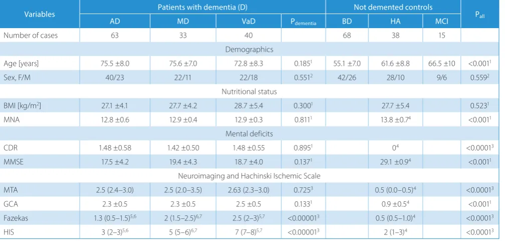
Patients with dementia had significantly higher concen-trations of MDA/TBARS and lower levels of arylesterase activity of PON1 (Fig. 1).
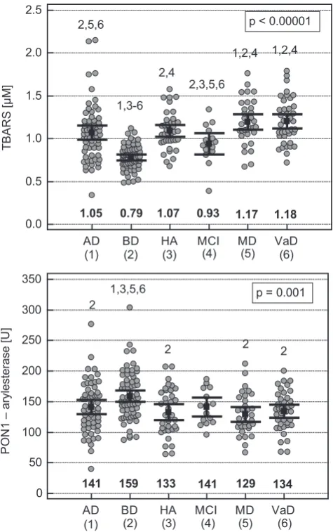
A detailed analysis revealed differences in MDA/TBARS in both control and dementia groups (Fig. 2). Among in-dividuals recruited as controls, the BD group had sig-nificantly lower concentrations of MDA/TBARS than the HA or MCI group. Among patients with dementia, the presence of vascular impairment was responsible for significant upregulation of MDA/TBARS concentrations. Arylesterase activity of PON1 was significantly higher
in BD as compared to other groups, with an exception of MCI, but did not show significant differences with re-spect to the type of dementia.
There was a significant age discrepancy between the con-trol and dementia groups (Table 1). MDA/TBARS concentra-tions and PON1 arylesterase activities did not correlate with age in any of the studied groups (MDA/TBARS: p = 0.790 for AD group, p = 0.405 for MD group, p = 0.526 for VaD group, p = 0.811 for BD group, p = 0.418 for HA group, and p = 0.720 for MCI group; PON1: p = 0.515 for AD group, p = 0.435 for MD group, p = 0.543 for VaD group, p = 0.935 for BD group, p = 0.498 for HA group, and p = 0.562 for MCI group). However, there were significant correlations with age when the study population was analyzed as a whole (r = 0.43, p < 0.001 for MDA/TBARS and r = −0.24, p < 0.001 for PON1). Therefore, for the whole cohort evaluation, age- and sex-adjusted analysis was employed. Analysis of covariance revealed a significant effect of health status (p < 0.001) and insignificant effects of age (p = 0.931) and sex (p = 0.339) on MDA/TBARS. Similarly, health status (p = 0.041), but not age (p = 0.163) or sex (p = 0.071), was a significant predictor of PON1 arylesterase activity.
Fig. 1. Comparison of MDA/TBARS concentrations and PON1 activity between non-demented and demented individuals. Circles represent individual MDA/TBARS or PON1 values. Solid squares with whiskers represent means with 95% confidence intervals (95% CI)
0.0 0.5 1.0 1.5 2.0 2.5
TBARS
[µM]
dementia controls
1.14 (1.1–1.2) 0.9 (0.86–0.94) p < 0.0001
0 50 100 150 200 250 300 350
PO
N1
– arylesterase [U
]
dementia controls
136.1 (130–143) 148.8 (142–156)
MDA/TBARS and PON1 as dementia
and dementia differential markers
The ROC analysis was employed to evaluate MDA/ TBARS and PON1 power in discriminating demented from non-demented individuals (dementia markers) as well as in differentiating dementia with and without vascular component (differential markers).
The AUCs for MDA/TBARS and PON1 as dementia markers were 0.75 (p < 0.0001) and 0.59 (p = 0.011), re-spectively. At the cut-off >1.0 µM and ≤142.2 U for MDA/ TBARS and PON1, the sensitivities and specificities were as follows: 71% and 75% for MDA/TBARS and 60% and 59% for PON1.
The AUCs for MDA/TBARS and PON1 as differential markers were 0.58 (p = 0.150) and 0.57 (p = 0.269), respec-tively. At the cut-off >1.061 µM and ≤153.5 U for MDA/ TBARS and PON1, the sensitivities and specificities were as follows: 73% and 46% for MDA/TBARS and 75% and 48% for PON1.
PON1 phenotype distribution
The prevalence of the A phenotype of PON1 was 63% in AD group, 59% in VaD group, 50% in MD group, 47% in MCI group, 61% in HA group, and 48% in BD group. There was a 15% difference in the prevalence be-tween AD and BD groups at the trend level significance (p = 0.095).
In patients with vascular component (MD and VaD) stratified by the presence of strategic infarcts, the preva-lence of the A phenotype was 61.5% in patients without and 55.5% in patients with strategic infarcts (p = 0.671).
Interrelationship between PON1 alloforms
and MDA/TBARS
Controls with the phenotype A of the enzyme had sig-nificantly lower concentrations of MDA/TBARS than those with the phenotype B (0.85 µM (0.8–0.9) vs 0.94 µM (0.87–1.0), p = 0.044). Similarly, MD patients with the phe-notype A had significantly lower concentrations of MDA/ TBARS than those with the phenotype B of the enzyme (1.1 µM (0.97–1.23) vs 1.29 µM (1.15–1.42), p = 0.045). In the other studied groups, the phenotype-related dif-ference in MDA/TBARS was not significant.
The correlation of PON1 and MDA/TBARS
with the degree of cognitive deficits
The MDA/TBARS were positively correlated with MMSE in demented patients with vascular involvement (MD and VaD groups: ρ = 0.28, p = 0.017). Paraoxonase 1 did not exhibit any association with MMSE, but it corre-lated with CDR scale in the VaD group; the patients with CDR-1 grade had significantly higher PON1 activity than those with CDR-2 and CDR-3 grades (140.6 U (132–149) vs 127.4 U (118–137), p = 0.040).
PON1 and MDA/TBARS and the level
of brain atrophy and vascular changes
MDA/TBARS positively correlated with GCA scores in all patients who underwent neuroimaging (ρ = 0.21, p = 0.020, n = 127). The association was stronger in pa-tients with dementia and vascular involvement (ρ = 0.30, p = 0.022, n = 57), particularly those with strategic infarcts (ρ = 0.58, p = 0.015, n = 17). Arylesterase activity of PON1 negatively correlated with the MTA score in patients with dementia and with vascular involvement (ρ = −0.27,
Fig. 2. Comparison of MDA/TBARS concentrations and PON1 activity between healthy individuals, individuals with benign headaches and memory deficits, patients with mild cognitive impairment, and patients with dementia of different pathology. Circles represent individual MDA/ TBARS or PON1 values. Solid squares with whiskers represent means with 95% confidence intervals (95% CI). The numbers above the data indicate significance of differences between groups in: Alzheimer’s disease (1); blood donors (2); individuals with headaches, dizziness and/or complaining about weak memory but without dementia or MCI (3); mild cognitive impairment (4); mixed-type dementia (5); and vascular dementia (6)
0.0 0.5 1.0 1.5 2.0 2.5
TBAR
S [µ
M]
AD BD HA MCI MD VaD
1.05 0.79 1.07 0.93 1.17 1.18
2,5,6
1,3-6 2,4
2,3,5,6
1,2,4 1,2,4 p < 0.00001
(1) (2) (3) (4) (5) (6)
0 50 100 150 200 250 300 350
PO
N1
– arylesterase [U
]
AD BD HA MCI MD VaD
p = 0.001 2
2 2 2
1,3,5,6
141 159 133 141 129 134
p = 0.049, n = 57), particularly those with strategic infarcts (ρ = −0.55, p = 0.024, n = 17). None of the parameters cor-related with the Fazekas score.
PON1 and MDA/TBARS and ischemia
Arylesterase activity of PON1 negatively correlated with the HIS in VaD patients (ρ = −0.46, p = 0.004) and in pa-tients with strategic infarcts (ρ = −0.56, p = 0.015). A sim-ilar tendency was observed in MD patients (ρ = −0.34, p = 0.061). MDA/TBARS displayed only a tendency towards a weak correlation with the HIS score in a whole studied population (ρ = 0.14, p = 0.073).
Multivariate analysis
The effect of mental deficits on MDA/TBARS and PON-1 was co-examined with the effect of brain abnormalities detected with neuroimaging and with the HIS. The GCA score was the only variable retained in the multiple re-gression model explaining the MDA/TBARS variability with partial correlation coefficient r = 0.23. The HIS was the only variable retained in the multiple regression model explaining PON1 variability with partial correlation coef-ficient r = −0.44.
Discussion
In this study, we demonstrated an increase in MDA/ TBARS accompanied by a drop in PON1 activity in pa-tients with dementia, particularly these with vascular involvement, which was related to the severity of brain atrophy or ischemia rather than to the degree of cogni-tive decline.
Oxidative stress has been shown to contribute to the pathogenesis of AD with the markers of oxidative imbalance evident both locally and systemically.19
Accord-ingly, the accumulation of MDA has been consistently reported in AD patients.4 There is a scarcity of studies
comparing MDA levels between AD and VaD patients. In addition, these studies have yielded conflicting results, showing either equally high MDA levels in both types of dementia20 or significantly higher MDA accumulation
in AD21 or, contrarily, in VaD.22 Taking into account
dif-ficulties in strict AD and VaD differentiation, a need for easily accessible biomarkers of vascular involvement has been stressed, and possible application of MDA has been suggested but not tested.22 In this study, we corroborated
previous reports on elevated levels of MDA/TBARS in de-mentia. By showing MDA/TBARS to be significantly higher in VaD/MD than AD, we confirmed its particular associa-tion with vascular involvement. Our finding is in line with close cause-and-effect relationship between OS and vascu-lar diseases supporting its involvement in the pathogenesis of VaD.3 Adding to existing knowledge, we assessed MDA
suitability as a differential dementia marker. However, de-spite its higher levels in dementia with vascular involve-ment, MDA/TBARS failed to display significant diagnostic power to be applied as a differential biomarker. Yet, MDA/ TBARS showed a moderate accuracy as a general dementia marker in our mixed cohort with AUC identical to previ-ously reported ones for AD.23
Similarly to many other authors,24–28 we demonstrated
a significant decrease in PON1 arylesterase activity in AD. However, studies comparing PON1 activity between dis-tinct subtypes of dementia have yielded conflicting results. Bednarska-Makaruk et al.24 observed that a drop in PON1
activity was particularly expressed in the case of AD-relat-ed pathology, whereas Paragh et al.28 showed no
differenc-es. Others, like us, have found it to be lowered in demen-tias with vascular involvement.26,27,29 Moreover, Cervellati
et al.30 reported low PON1 activity to be associated with
an increased risk of MCI progression to VaD rather than AD. Even though the difference in PON1 activity between patients with dementia with and without vascular involve-ment in our cohort did not reach statistical significance, the enzyme association with vascular involvement seems to be confirmed by the negative correlation between PON1 activity and HIS index, reflecting the degree of ischemia. This finding is in agreement with the biological activity of PON15 and the results of epidemiological studies
show-ing that the PON1 activity is inversely correlated with ca-rotid and cerebral atherosclerosis.31
A direct correlation between MDA accumulation and cognitive decline is not unanimously observed. While some studies32 have reported erythrocyte and serum MDA
to be inversely related to MMSE scores in AD patients, this relationship has not been mentioned by others.22,23,33 Liu
et al.34 found this correlation in the general stroke patients
but not in the subgroup suffering from post-stroke cogni-tive impairment. Similarly, the correlation between PON1 and MMSE has been reported only by 1 group of research-ers.25 We, in turn, found MDA/TBARS to correlate with
MMSE, but this association was counterintuitive, implying an improvement of cognitive function with the exacer-bation of oxidative status. As expected, the association of PON1 with CDR suggested impaired protection against OS with the deterioration of cognitive functions. However, both correlations were weak and might have resulted from an interference with confounding factors. Indeed, when co-examined with the degree of brain alterations and HIS, the correlations between MDA/TBARS levels with MMSE, as well as between the PON1 activity and the CDR, lost their significance.
differential diagnosis, has been postulated.11 The issue
of possible correlation between biochemical markers of OS and neuroimaging has been addressed only recently. Bulboacă et al.35 demonstrated a positive correlation
be-tween markers of nitro-OS and posterior cortical atrophy. To our knowledge, neither MDA nor PON1 have been pre-viously analyzed in the context of presence and the degree of brain alterations accompanying dementia. We found that in patients with dementia, particularly those with stra-tegic infarcts (within VaD group), systemic MDA/TBARS accumulation was proportional to the degree of structural brain changes expressed in terms of GCA rating scale. Also, PON1 activity negatively correlated with the degree of medial temporal lobe atrophy (MTA rating scale). How-ever, structural changes in the brain were independently associated only with MDA/TBARS, whereas they lost sig-nificance when their effect was co-evaluated with ischemia in the case of PON1.
Results from animal studies have shown OS to pre-cede plaque formation characteristic for AD.36 The MCI
is viewed as a transitional condition between physiological aging and AD and, as such, has gained increased attention. However, reports on OS in MCI are scarce and incon-clusive.4 Although some authors have reported the lack
of a significant difference in MDA between controls and MCI patients37 or its gradual increase along the sequence
from healthy individuals to MCI and AD,32 we and others
found serum MDA/TBARS to accumulate in MCI and AD patients to a similar extent,33 substantiating
the no-tion that lipid peroxidathe no-tion is a phenomenon that occurs early in the progression of dementia.34 Likewise, in the case
of MDA/TBARS, there is no consensus in the literature concerning PON1 in MCI, the activity of which has been reported to resemble that in healthy controls29 or,
con-trarily, to be decreased similarly to patients with demen-tia.26,27,38 Also, our results are inconclusive – the PON1
activity in MCI was similar to that observed in AD; yet, MCI was the only group for which the difference, as com-pared to the healthy controls, did not reach statistical significance.
An interesting observation of our study was an evident oxidative imbalance in HA subgroup of controls com-prising individuals admitted to the Alzheimer Center due to persistent headaches, dizziness and/or memory loss complaints. The etiology of their problems was not elucidated; neither the loss of cognitive function nor any significant somatic or mental illnesses were diagnosed. Nevertheless, these patients had significantly lower PON1 activity than BD controls recruited through the Blood Do-nation Center. Moreover, their concentration of lipid per-oxidation markers (MDA/TBARS) exceeded that observed in both BD group and MCI patients. Therefore, our results implicate OS also in the pathogenesis of seemingly benign conditions.
Genetic studies addressing variations in the PON1 gene have revealed a lower frequency of the 192R allele among
AD patients than in the general population, suggesting its role as a protective factor against disease development.38
Although not consistently observed, this finding seems to corroborate our observations on the PON1 pheno-type B that tended to be less prevalent in AD patients compared to healthy blood donors. The protecting effect attributed to the 192R allele might be associated with the PON1 activity as a natural inhibitor of cholinesterase. Cholinesterase inhibitors are employed as the first-line treatment for AD to prevent further loss of cognitive function via an increase in the availability of acetylcho-line.39 On the other hand, the alloenzyme Q of PON1
is believed to be more efficient in counteracting lipid peroxidation.40 Accordingly, the concentrations of MDA/
TBARS were lower in the control group and MD pa-tients with phenotype A of the enzyme at the trend level significance.
Our study has certain limitations that need to be dis-cussed. Due to a natural history of neurocognitive disor-ders and the fact that the age over 65 years is considered a contraindication for blood donation, there was a signifi-cant difference in age distribution between controls and patients. However, the PON1 activity and MDA/TBARS levels correlated with age exclusively in the whole cohort and the issue was addressed in age- and sex-adjusted analysis. The ANCOVA confirmed that observed dif-ferences in the PON1 activity and MDA/TBARS levels could be attributed solely to a diagnosis of dementia and not to age or sex. The low number of patients with MCI as well as the lack of neuroimaging data or the assessment of degree of cognitive impairment is yet another limitation, negatively affecting the relevance of our findings concern-ing this particular group.
Conclusions
In this study, we confirmed an impaired oxidative status in patients with dementia and, for the first time, showed that an increase in MDA/TBARS level and a decrease in the PON1 activity reflects the severity of brain atrophy established with neuroimaging. We found ischemia, as-sessed with the application of HIS index, to be an inde-pendent predictor of the PON1 activity. Additionally, our findings suggest that OS also accompanies pathological changes in benign conditions.
ORCID iDs
Iwona Bednarz-Misa https://orcid.org/0000-0001-7244-2017 Izabela Berdowska https://orcid.org/0000-0002-0275-4522 Marzena Zboch https://orcid.org/0000-0002-6853-0433 Błażej Misiak https://orcid.org/0000-0002-5392-6398 Bogdan Zieliński https://orcid.org/0000-0002-5330-9904 Sylwia Płaczkowska https://orcid.org/0000-0002-1466-3820 Mariusz Fleszar https://orcid.org/0000-0001-7857-327X Jerzy Wiśniewski https://orcid.org/0000-0003-2831-7643 Andrzej Gamian https://orcid.org/0000-0002-2206-6591
References
1. Alzheimer’s Association. 2016 Alzheimer’s disease facts and figures.
Alzheimers Dement. 2016;12(4):459–509.
2. Ahotupa M. Oxidized lipoprotein lipids and atherosclerosis. Free Radic Res. 2017;51(4):439–447. doi:10.1080/10715762.2017.1319944 3. Luca M, Luca A, Calandra C. The role of oxidative damage
in the patho-genesis and progression of Alzheimer’s disease and vascular dementia.
Oxid Med Cell Longev. 2015;2015:504678. doi:10.1155/2015/504678 4. Schrag M, Mueller C, Zabel M, et al. Oxidative stress in blood in
Alzheimer’s disease and mild cognitive impairment: A meta-analysis.
Neurobiol Dis. 2013;59:100–110. doi:10.1016/j.nbd.2013.07.005 5. Chistiakov DA, Melnichenko AA, Orekhov AN, Bobryshev YV.
Paraox-onase and atherosclerosis-related cardiovascular diseases. Biochimie. 2017;132:19–27. doi:10.1016/j.biochi.2016.10.010
6. American Psychiatric Association. Diagnostic and Statistical Manu-al of MentManu-al Disorders. 4th ed. Washington, DC: American Psychiatric
Association; 2000. doi:10.1002/jps.3080051129
7. McKhann G, Drachman D, Folstein M, Katzman R, Price D, Stadlan EM. Clinical diagnosis of Alzheimer’s disease: Report of the NINCDS– ADRDA Work Group under the auspices of Department of Health and Human Services Task Force on Alzheimer’s Disease. Neurology. 1984; 34(7):939–944. doi:10.1037/0894-4105.19.4.520
8. World Health Organization. International Statistical Classification of Diseases and Related Health Problems, 10th Revision. Vol 41. Geneva,
Switzerland: World Health Organization; 1992. http://www.who.int/ classifications/icd/ICD-10_2nd_ed_volume2.pdf.
9. Erkinjuntti T. Clinical criteria for vascular dementia: The NINDS-AIREN criteria. Dement Geriatr Cogn Disord. 1994;5(3–4):189–192. doi:10.1159/ 000106721
10. Hachinski V, Iliff L, Zilhka E, et al. Cerebral blood flow in dementia.
Arch Neurol. 1975;32(9):632–637. doi:10.1001/archneur.1975.00490510 088009
11. Wahlund LO, Westman E, van Westen D, et al. Imaging biomarkers of dementia: Recommended visual rating scales with teaching cases.
Insights Imaging. 2017;8(1):79–90. doi:10.1007/s13244-016-0521-6 12. Vellas B, Villars H, Abellan G, et al. Overview of the MNA: Its history
and challenges. J Nutr Heal Aging. 2006;10(6):456–463.
13. Petersen RC. Mild cognitive impairment as a diagnostic entity. J Intern Med. 2004;256(3):183–194. doi:10.1111/j.1365-2796.2004.01388.x 14. Rice-Evans CA, Diplock AT, Symons MCR. Techniques in free
radi-cal research. In: Burdon RH, van Knippenberg PH, eds. Laboratory Techniques in Biochemistry and Molecular Biology. Vol. 22. Amster-dam, the Netherlands: Elsevier Science Publishers BV; 1991:1–291. doi:10.1016/S0075-7535(08)70046-9
15. Bartosz G. The Other Face of Oxygen. Free Radicals in the Environment
[in Polish]. Warszawa, Poland: PWN; 2004.
16. Costa LG, Vitalone A, Cole TB, Furlong CE. Modulation of paraox-onase (PON1) activity. Biochem Pharmacol. 2005;69(4):541–550. doi:10. 1016/j.bcp.2004.08.027
17. La Du BN, Billecke S, Hsu C, Haley RW, Broomfield CA. Serum paraox-onase (PON1) isozymes: The quantitative analysis of isozymes affect-ing individual sensitivity to environmental chemicals. Drug Metab Dispos. 2001;29(4 Pt 2):566–569.
18. Charlton-Menys V, Liu Y, Durrington PN. Semiautomated method for determination of serum paraoxonase activity using paraoxon as sub-strate. Clin Chem. 2006;52(3):453–457. doi:10.1373/clinchem.2005. 063412
19. Massaad CA. Neuronal and vascular oxidative stress in Alzheimer’s disease. Curr Neuropharmacol. 2011;9(4):662–673. doi:10.2174/1570 15911798376244
20. Polidori MC, Mattioli P, Aldred S, et al. Plasma antioxidant status, immu-noglobulin G oxidation and lipid peroxidation in demented patients: Relevance to Alzheimer disease and vascular dementia. Dement Geriatr Cogn Disord. 2004;18(3–4):265–270. doi:10.1159/000080027 21. Casado A, Encarnación López-Fernández M, Concepción Casado M,
de La Torre R. Lipid peroxidation and antioxidant enzyme activities in vascular and Alzheimer dementias. Neurochem Res. 2008;33(3): 450–458. doi:10.1007/s11064-007-9453-3
22. Gustaw-Rothenberg K, Kowalczuk K, Stryjecka-Zimmer M. Lipids’ peroxidation markers in Alzheimer’s disease and vascular dementia.
Geriatr Gerontol Int. 2010;10(2):161–166. doi:10.1111/j.1447-0594.2009. 00571.x
23. Lopez-Riquelme N, Alom-Poveda J, Viciano-Morote N, Llinares-Ibor I, Tormo-Diaz C. Apolipoprotein E ε4 allele and malondialdehyde level are independent risk factors for Alzheimer’s disease. SAGE Open Med. 2016;4:2050312115626731. doi:10.1177/2050312115626731
24. Bednarska-Makaruk M, Graban A, Lipczyńska-Łojkowska W, et al. Positive correlation of paraoxonase 1 (PON1) activity with serum insulin level and HOMA-IR in dementia: A possible advantageous role of PON1 in dementia development. J Neurol Sci. 2013;324(1–2): 172–175. doi:10.1016/j.jns.2012.11.003
25. Wehr H, Bednarska-Makaruk M, Graban A, et al. Paraoxonase activity and dementia. J Neurol Sci. 2009;283(1–2):107–108. doi:10.1016/j.jns. 2009.02.317
26. Cervellati C, Romani A, Bergamini CM, et al. PON-1 and ferroxidase activities in older patients with mild cognitive impairment, late onset Alzheimer’s disease or vascular dementia. Clin Chem Lab Med. 2015; 53(7):1049–1056. doi:10.1515/cclm-2014-0803
27. Castellazzi M, Trentini A, Romani A, et al. Decreased arylesterase activity of paraoxonase-1 (PON-1) might be a common denominator of neuroinflammatory and neurodegenerative diseases. Int J Biochem Cell Biol. 2016;81:356–363. doi:10.1016/j.biocel.2016.06.008 28. Paragh G, Balla P, Katona E, Seres I, Égerházi A, Degrell I. Serum
paraox-onase activity changes in patients with Alzheimer’s disease and vas-cular dementia. Eur Arch Psychiatry Clin Neurosci. 2002;252(2):63–67. doi:10.1007/s004060200013
29. Dantoine TF, Debord J, Merle L, Lacroix-Ramiandrisoa H, Bourzeix L, Charmes JP. Paraoxonase 1 activity: A new vascular marker of demen-tia? Ann N Y Acad Sci. 2002;977:96–101. doi:10.1111/j.1749-6632.2002. tb04802.x
30. Cervellati C, Trentini A, Romani A, et al. Serum paraoxonase and aryles-terase activities of paraoxonase-1 (PON-1), mild cognitive impair-ment, and 2-year conversion to dementia: A pilot study. J Neuro chem. 2015;135(2):395–401. doi:10.1111/jnc.13240
31. Arslan A, Tüzün FA, Arslan H, et al. The relationship between serum paraoxonase levels and carotid atherosclerotic plaque formation in Alzheimer’s patients. Neurol Neurochir Pol. 2016;50(6):403–409. doi:10.1016/j.pjnns.2016.07.002
32. Torres LL, Quaglio NB, De Souza GT, et al. Peripheral oxidative stress biomarkers in mild cognitive impairment and Alzheimer’s disease.
J Alzheimer’s Dis. 2011;26(1):59–68. doi:10.3233/JAD-2011-110284 33. Balmuș I-M, Strungaru S-A, Ciobica A, et al. Preliminary data on the
interaction between some biometals and oxidative stress status in mild cognitive impairment and Alzheimer’s disease patients. Oxid Med Cell Longev. 2017;2017:1–7. doi:10.1155/2017/7156928
34. Liu Z, Liu Y, Tu X, et al. High serum levels of malondialdehyde and 8-OHdG are both associated with early cognitive impairment in patients with acute ischaemic stroke. Sci Rep. 2017;7:9493. doi:10.1038/ s41598-017-09988-3
35. Bulboacă AE, Bulboacă SD, Bulboacă AC, Prodan CI. Association between low thyroid-stimulating hormone, posterior cortical atro-phy and nitro-oxidative stress in elderly patients with cognitive dys-function. Arch Med Sci. 2017;13(5):1160–1167. doi:10.5114/aoms.2016. 60129
36. Praticò D, Uryu K, Leight S, Trojanoswki JQ, Lee VM-Y. Increased lipid peroxidation precedes amyloid plaque formation in an animal model of Alzheimer amyloidosis. J Neurosci. 2001;21(12):4183–4187. doi:21/ 12/4183 [pii]
37. Martín-Aragón S, Bermejo-Bescós P, Benedí J, et al. Metalloprotein-ase’s activity and oxidative stress in mild cognitive impairment and Alzheimer’s disease. Neurochem Res. 2009;34(2):373–378. doi:10.1007/ s11064-008-9789-3
38. Marsillach J, Parra S, Coll B, Joven J, Camps J. Paraoxonase-1 in chron-ic liver diseases, neurologin chron-ical diseases and HIV infection. In: Mack-ness B, MackMack-ness M, Aviram M, Paragh G, eds. The Paraoxonases: Their Role in Disease Development and Xenobiotic Metabolism. Dordrecht, the Netherlands: Springer; 2008:187–198.
39. Wilkinson D, Francis P, Schwam E, Payne-Parrish J. Cholinesterase inhibitors used in the treatment of Alzheimer’s disease. Drugs Aging. 2004;21(7):453–478. doi:10.2165/00002512-200421070-00004 40. Draganov DI, La Du BN. Pharmacogenetics of paraoxonases: A brief