http: // www.ijrtsm.com© International Journal of Recent Technology Science & Management 36
ISSN : 2455-9679
[Anshu Singh et al. , 3(9), Sept 2018 Impact Factor : 2.865
IJRTSM
INTERNATIONAL JOURNAL OF RECENT TECHNOLOGY SCIENCE & MANAGEMENT
“Improvement In Kirsch’s Template Algorithm By Introducing Adaptive
Histogram Equalisation And Image Matting Structure Element Algorithm To
Create Trimap of Blood Vessels For Diabetic Retinopathy Diagnostic”
Anshu Singh
1, Gaurav Bhardwaj
21,PG, Scholar, Dept. of Electronics & Communication Engineering, RJIT BSF Tekanpur,Gwalior, MP, India 2Assistant Professor, Dept. of Electronics & Communication Engineering, RJIT BSF Tekanpur,Gwalior, MP, India
ABSTRACT
Diabetic Retinopathy (DR) can be detected by analysing tri-map of blood vessels in human eye but an expert is required to create it properly as well as manually creation of tri-map is time consuming task. This problem can be solved by using computer algorithms based on image processing. Kirsch’s template algorithm is popular in creating a tri-map of blood vessels using retinal image. In our proposed work, adaptive histogram equalisation and image matting structure element algorithm has been used to further improve the results of kirsch’s template.
Keyword: blood vessel,Diabetic Retinopathy (DR),Computer Aided Diagnosis (CAD), image matting,adaptive histogram equalisation, image pre-processing, image enhancement, RGB (Red, Green & Blue), Kirsch’s Template
algorithm, MATLAB, Gray scale image.
I. INTRODUCTION
Diabetic Retinopathy can be detected by diagnostic trimap of blood vessels out of retinal images. In past days, trimap has been created by an expert and only an expert can analyse it. But with the development in computer based algorithms, creation of trimap is quite easy and accurate. Kirsch’s template is one of such computer algorithm which can take out the blood vessels out of retinal image and create a trimap. Image matting is also one of the methods of extracting the features from the retinal image. These segmentation techniques give out the image into two regions. First is called as foreground (shown in white) and another is background (shown in black)[1].If the retinopathy detects in early stage, the probability of visual loss resulting from diabetic retinopathy can
be prevented. The filteringbased method which is used for the effective extraction the blood vessels of retinal images are described in [5].
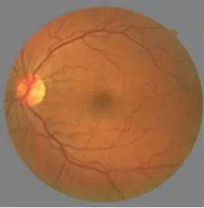
Blood vessels in retinal image appear like a wire mesh-like structure or tree-like structure as shown in figure 1. Diseases like stroke, vein occlusions, diabetes and arteriosclerosis can be detected from the morphological features like length, width and branching of blood vessels [4].
In our proposed work, kirsch’s template algorithm has been improved by using histogram based contrast enhancement technique which improves the detection better and image matting structure element algorithm which helps in removing unwanted part like eye boundary and centre oval removal.
http: // www.ijrtsm.com© International Journal of Recent Technology Science & Management 37
ISSN : 2455-9679
[Anshu Singh et al. , 3(9), Sept 2018 Impact Factor : 2.865
II. KIRSCH’S TEMPLATE BLOOD VESSEL EXTRACTION ALGORITHM
Kirsch templates of window size 3x3 are used for the extraction of blood vessels from retinal image. Edge detection is a process of finding edges in the form of frequently and abrupt changes. In this algorithm, edge is determined by comparing the intensity of neighbouring pixels around a center pixel. The detection of edge can be taken as the major difference in the brightness level of neighbouring pixel else it is taken as smooth change or no edge. The described procedure is most common and fundamental approach among all the available edge detection algorithms such as, Prewitt, Sobel etc. The Kirsch edge detection algorithm uses a single mask of size 3x3 and rotates it in 45 degree increments through all 8 directions as shown in figure 2 [9].
The edge magnitude of the Kirsch operator is calculated as the maximum magnitude across all direction. The matrix contains the information of a pixel and its neighbours. The Kirsch algorithm detects direction of the edge as well as an edge. Accordingly, there are eight possible directions south, east, north, west, northeast, southeast, southwest and northwest as shown in figure 5. Out of the several templates the biggest one is considered for the output value and later the edges are extracted. Threshold value can be changed to get the best result [5]. Mathematically it can be represented as shown in equation 3.
Where, h(n,m) is the edge magnitude of the Kirsch operator in all direction and n and m are the coordinates of center pixel. The figure 5 shows the different values of g (total 8 values). Image neighbouring pixel intensities are represented by f. where z enumerates the compass direction kernels g [6].
III
.
HISTOGRAM EQUALISATIONHistogram equalization is used for enhancing the gray level contrast of the image [2]. The contrast of the image is enhanced by transforming these values using contrast limited adaptive histogram equalization, which operates on small regions in the image called tiles. Each tile's contrast is enhanced in such a way that the histogram of the output region approximately matches the histogram specified by the distribution parameters. While combining the neighbouring tiles the problem of artificially induced boundaries are found and it is eliminated using bilinear interpolation [3].
IV
.
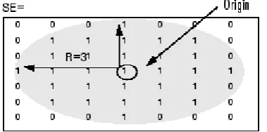
STRUCTURE ELEMENT (MORPHOLOGICAL OPENING) ALGORITHMA strel object represents a flat morphological structuring element, which is an essential part of morphological dilation and erosion operations.
Figure 3: Image enhancement Figure 2: Kirsch’s convolution
http: // www.ijrtsm.com© International Journal of Recent Technology Science & Management 38
ISSN : 2455-9679
[Anshu Singh et al. , 3(9), Sept 2018 Impact Factor : 2.865
A flat structuring element is a binary valued neighbourhood,either 2-D or multidimensional, in which the true pixels are included in the morphological computation, and the false pixels are not. The center pixel of the structuring element, called the origin, identifies the pixel in the image being processed. Use the strel function (described below) to create a flat structuring element. You can use flat structuring elements with both binary and grayscale images. The following figure illustrates a flat structuring element.
V. PROPOSED METHODOLOGY
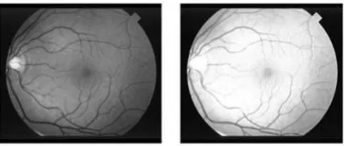
In proposed work, kirsch’s template technique is taken as base algorithm. MATLAB is used for simulation for the work. First image is imported in MATLAB by using MATLAB’s function. The extracted image is then passed through histogram based image enhancement technique which enhances the contrast of the image which helps in better extraction of blood vessels. The dataset of images used for experimentation is shown in figure 6 below.
.
Figure 4:Strel algorithm effect on matrix
Figure 5: Dataset of Retinal image
Figure 6:Proposed Algorithm
http: // www.ijrtsm.com© International Journal of Recent Technology Science & Management 39
ISSN : 2455-9679
[Anshu Singh et al. , 3(9), Sept 2018 Impact Factor : 2.865
Table 1: Output of image segmentation from base algorithm and from proposed algorithmhttp: // www.ijrtsm.com© International Journal of Recent Technology Science & Management 40
ISSN : 2455-9679
[Anshu Singh et al. , 3(9), Sept 2018 Impact Factor : 2.865
VI. CONCLUSION
Blood vessels trimap was the way to find DR in early ages. But first it is required to be expert to make it accurately as well as it takes too much of time. Image processing algorithms are successfully solving this issue by creating computerized trimap of blood vessels. Image matting can successfully find the blood vessels out of retinal image. While other two algorithms based on kirsch’s template and hessian matrix can find blood vessels upto satisfactory extent. In future work, combination of all three can be studied to increase the accuracy of detection.
REFERENCES
[1] Zhun Fan, Jiewei Lu, Wenji Li, Caimin Wei, Han Huang, XinyeCai, Xinjian Chen, “A Hierarchical Image Matting Model for Blood Vessel Segmentation in Fundus images”, IEEE Submitted on 4 Jan 2017 (v1), last
revised 9 Oct 2017 (this version, v3).
[2] Minal B. Wankhade, Dr. A. A. Gurjar, “Analysis of Disease using Retinal Blood Vessels Detection”,
International Journal Of Engineering And Computer Science, Volume-5, Issue-12, Dec 2016.
[3] S. Wilfred Franklin a, S. Edward Rajan, “Computerized screening of diabetic retinopathy CrossMark
employing blood vessel segmentation in retinal images”, Elsevier Urban & Partner, Biocybernetics and
Biomedical Engineering 34, Feb 2014.
[4] D.SivaSundhara Raja, Dr.S.Vasuki, D.Rajesh Kumar, “Performance Analysis Of Retinal Image Blood Vessel Segmentation”, Advanced Computing: An International Journal, Volume 5, Issue 2/3, May 2014.
[5] H.S. Bhadauria, S.S. Bisht, Annapurna Singh, “Vessels Extraction from Retinal Images”, IOSR Journal of
Electronics and Communication Engineering, Volume 6, Issue 3, (May - Jun 2013).
[6] Mohammed ammarKhatib, Dr.Shailaja K, “Automatic Detection of Retinal Vessels using Kirsch’s Templates
and Region Growing”, International Journal for Scientific Research & Development, Volume 3, Issue 05, 2015.
[7] Dr. Vijayachitra S, Menagadevi. M, PonniBala. M, “Analysis Of Diabetic Retinopathy Images Using Blood
Vessel Extraction”, International Journal of Advanced Engineering Research and Studies, Volume 1, Issue 2,
January-March 2012.
[8] P. C. Siddalingaswamy, K. GopalakrishnaPrabhu, “Automatic detection of multiple oriented blood vessels in retinal images”, J. Biomedical Science and Engineering, Jan 2010.
[9] BahadirKarasulu, “Automatic Extraction Of Retinal Blood Vessels: A Software Implementation”, European