The effect of two types of stretching on flexibility in selected joints
in youth soccer players
Andrea Mahrová
1,*, Pavel Hráský
1, František Zahálka
1, and Petr Požárek
21Sport Research Centre, Faculty of Physical Education and Sports, Charles University in Prague, Prague, Czech Republic;
and 2Faculty of Education, University of South Bohemia, České Budějovice, Czech Republic
Copyright: © 2014 A. Mahrová et al. This is an open access article licensed under the Creative Commons Attribution License (http:// creativecommons.org/licenses/by/4.0/).
Background: The nature of soccer causes overloading of certain muscles and muscle groups that are most often
involved in typical training activities of players. Often the absence of additional leisure-time activities typical for children and adolescents leads to an increased risk of muscle imbalance. Objective: We were interested in the occur-rence of muscle imbalance, specifically an occuroccur-rence of muscle shortening and limited flexibility of the spine as well as decreased range of motion (ROM) in selected joints among young soccer players. Methods: The players were randomly selected to take part in the study. Twelve young soccer players took part in the first group and thirteen in the second group (age 13 years, body weight 40.5 ± 3.9 vs. 41.5 ± 7.8 kg, height 150.0 ± 5.0 vs. 153.0 ± 9.8 cm). The first team performed only static stretching exercises at the end of the training session and the other team performed, except for static stretching, also dynamic stretching exercises always at the beginning of the training session. The effect of the stretching program on range of motion in chosen joints was assessed by 2 × 2 repeated-measures ANOVA (stretching methods × time). The statistical significance of α-level was set to .05 and partial ηp2. Results: Pre-test
measurements revealed an occurrence of muscle shortening and limited ROM of the spine and selected joints mostly in the pelvis and lower limbs in all study participants. Comparison of ROM in selected joints between pre-test and post-test in program including both static and dynamic stretching showed significant increase of left hip during flex-ion (p = .03, ηp2 = .20). The program with only static stretching showed significant decrease the ROM of ankle plantar
flexion (p = .04, ηp2 = .18). We did not find any significant difference between stretching programs on ROM. As for
muscle shortening, both side improvements (higher number of subjects with no muscle shortening) were found in the following muscle or muscle groups – m. trapesius, m. pectoralis major, m. rectus femoris, m. tensor fasciae latae, knee flexors and m. triceps surae. Improvements were also observed in group 1, but corrections were only asymmetrical.
Conclusions: The stretching program in the group of soccer players could lead to increase of range of motion in some
joints after a special program. It could serve as protection from muscle imbalance and lower limb injuries.
Keywords: muscle imbalance, muscle shortening, joint mobility, stretching, soccer
physical ability which is being developed. Coordina-tion is developed at first, then speed which further followed by the development of strength, endurance and flexibility (Dovalil et al., 2002). All physical abili-ties are affected by the muscle system, specifically by the quality and function of muscle fibres. Overload of the trunk, pelvis and lower limbs is often diagnosed among soccer players due to relatively monotonous load and lack of supplementary physical activities. According to Kutáč and Gajda (2006) this is the result of an intensive loading in the aforementioned parts of the body during training. Training of soccer players often include repeated skills and motor actions such as running, fast direction changes, or various ways of shooting and passing.
Introduction
When the physical load is not sufficiently compen-sated, especially among highly specialized athletes, it can result in one-sided overload of the movement sys-tem, which may consequently lead to severe dysfunc-tions of the system.
Although all physical abilities such as coordination, speed, strength, endurance or flexibility are important for soccer players, each age category has a preferred
This asymmetry together with a lack of compensa-tion for the monotonous load may result in overloads of axial and joint structures caused by one-sided mus-cle hypertrophy and limited joint mobility. Overloaded muscles and muscle groups react to increased asymmet-ric load without compensation that lead to increased muscle tonus and muscle shortenings. (Lewit, 1996).
One-sided overload activity may firstly cause some kinds of deviations of body axis and functional changes in spine and joints (e.g. functional blocks of spine, functional scoliosis). Secondly, due to repeated loading, this may lead to serious structural changes or destruction of cartilage and their faster abrasion (Adamčák, 2000; Lewit, 1996; Véle, 1997).
In addition small injuries – micro-traumas, which are ignored for a long time because of their minimal effect on physical fitness and slight subjective symp-toms, may appear in repeatedly overloaded muscles without necessary compensation. The most affected are the lower limbs. Also local changes such as light bleeding and ruptures of muscle fibers. Shortened mus-cle is less resistant to load and more prone to injury and decrease the reaction time (Adamčák, 2005; Arnason, Andersen, Holme, Engebretsen, & Bahr, 2008; Friery, 2008; Volpi, Melegati, Tornese, & Bandi, 2004; Wong & Hong, 2005). It results in predispositions for muscle imbalance and overall dysfunction of the movement system. Muscle imbalance is often accompanied by pain in the damaged segment, reduced physical fitness, coordination disorders and modification of movement patterns.
So called upper and lower cross syndromes are the most commonly occurring muscular imbalances in football. Both syndromes are caused by imbalance between phasic and tonic muscles (Bursová, Votík, & Zálabák, 2003).
Fixations of motor stereotypes of a movement per-formance begin in childhood. Movement perper-formance is influenced by every change that occurs in the move-ment system. If this change is combined with a pain-ful reaction, it may activate reflex reaction not only in the particular segment, but in segments more remote from the focal point (Trojan, Druga, Pfeiffer, & Votava, 1996). Furthermore, incorrectly coded and fixed motor stereotypes are not economic for an athlete’s organism. In some cases, underestimation and lack of load compensation may lead to irreversible changes in the movement system and even end athlete’s profes-sional career (Cingálková, Bunc, & Cingálek, 2001).
Problems and pain in the lumbar spine and hip joints, muscle injuries of lower limbs or unstable knee and ankle joints are often present among talented chil-dren specializing in soccer (Šrámková & Votík, 2010).
Compensation exercises focused on the correction of muscle imbalance are inevitable in any training pro-cess from early childhood (Adamčák, 2000; Kutáč & Gajda, 2006; Peterson, Renström, & Grana, 1986). According to Bunc (2006), it is possible to develop an “addiction” to physical activity. The most sensitive period of growth is between 1–3 and 6–8 years of age. This period is very important to predict the athletes’ performance in the following decades. Factors such as sets of compensation exercises, ergonomics of a work place, and used surface play an important role in prevention of dysfunctions of the movement system in actively competing children and adolescents (Brügger, 1993).
Compensation exercises consist of various set of simple exercises in specific positions which can be effectively modified by the use of different kinds of equipment. All exercises should always consider indi-vidual’s current functional status of the movement system (Bursová, 2005).
Stretching is one of the famous compensation exercises based on muscle extension and increased ligament tension. It optimizes the process in which the individual learns, practices and performs various motor skills – it deepens movement perception, reduces risk of injury, reduces muscle tension and muscle sore-ness, etc. Stretching techniques, if performed correctly according to recommendations, increase the range of motion – individual’s flexibility, e.g. ability to move muscles and joints in their full range (Alter, 1999). The choice of the stretching technique (active, passive, and combination of both, with neuromuscular facilitation) depends on the aim that we want to achieve, on the individual’s current health conditions and environment (Houglum, 2001).
The purpose of this study was to compare the effects of using only a static stretching program with a combination of dynamic and static stretching program on flexibility in the selected joints, mainly of lower extremities.
The stretching intervention program was designed to influence existing muscle imbalances and to regulate spine and joint mobility.
Methods
Participants’ characteristics
The study sample consisted of 25 boys, youth soccer players of two teams – SK Dynamo České Budějovice (group 1, N = 12) and SK Slavia Praha (group 2,
their parents, were fully informed about the goal and methods of the measurements and they provided their written consent. The study was approved by the Ethical Committee of the Faculty of Physical Education and Sports, Charles University in Prague and conformed to the Declaration of Helsinki regarding the use of human subjects.
Training load of both groups was the same. Soccer players performed minimal other leisure-time sport activities. No player had an injury which could signifi-cantly affect data collection and intervention.
Assessment of body composition
The data identifying participants’ body composition (BC) were recorded under identical conditions, in the morning hours, at the beginning of the pre-test and post-test measurements. BC measurement by means of a bioimpedance method was carried out under stan-dard conditions according to BIA guidelines (Kyle et al., 2004). Just before the measurement, the current body weight (BW) of the participants was measured on an electronic digital scale (seca 769, seca, Hamburg, Germany) with a precision of 0.1 kg, and the body height (BH) of the participants in an upright position was measured with the precision of full mm by means of digital stadiometer (seca 242, seca, Hamburg, Ger-many). The body bioimpedance was determined with the phase sensitive whole body tetrapolar bioimped-ance analyzer BIA 2000 M device (Data Input GmbH, Pöcking, Germany) which works on four frequencies (1, 5, 50 and 100 kHz) and is compatible with the NUTRI 4 software. The measurement was carried out in a prone position and participants’ extremities were abducted. Four electrodes were placed on the proximal and dorsal surface of the right hand and right foot. The measurement itself took about 45 s. The contact resistance between the electrode surface and the skin did not exceed 250 Ohms. Information about the mea-surement’s reliability and the validity of this method in the general population has been published by Dittmar
(2003). We were particularly interested in the amount of lean body mass (LBM) and the percentage of body fat (BF). Basic anthropometric characteristics of both baseline and outcome measurements of both groups are presented in Table 1.
Evaluation of muscle shortenings
The methodology presented in the strength test accord-ing to Janda (1996) was used to examine muscle short-enings. Attention was focused on muscle groups that are loaded in soccer the most and tend to shorten. Apart from muscles of lower limbs, assessment included selected muscles of the neck and trunk which are asso-ciated with posture maintenance and related motion. Selected muscles and muscle groups included – m. tra-pesius (upper part); m. pectoralis major (upward part); flexors of the hip joint (m. iliopsoas; m. rectus femoris; m. tensor fasciae latae); hip joint adductors; flexors of the knee joint (m. biceps femoris; m. semitendino-sus; m. semimembranosus) and m. triceps surae. The 3-point scale, with values 0, 1, 2 (0 = no shortening, 1 = medium shortening, 2 = significant shortening) was used to evaluate the magnitude of muscle shorten-ings. To ensure good inter-rater reliability, the assess-ment was done repeatedly by the same physiotherapist.
Evaluation of joint mobility
Spine mobility was assessed by Thomayer’s test accord-ing to Gúth (1994). A minus sign (–) was assigned to insufficient range of motion, zero (0) was assigned to the standard range and finally plus sign (+) was assigned to above standard range of motion.
Joint mobility was assessed by 2D video-graphic kinematic analysis (Janura & Zahálka, 2004). Tested persons were recorded by a video camera SONY HDR-HC9 (Sony, Japan) with frame rate 50 half frames per second and consequently captured to PC. The format was Mini-DV with overall field 575 × 720 pixels. Width of recorded view was 1.75 m providing 0.3 mm error of reconstruction. Joints of interest were marked by
Table 1
Basic anthropometric characteristics of group 1 and group 2 – pre-test and post-test examination tests
Variable
Group 1 (N = 12) Group 2 (N = 13)
Pre-test Post-test Pre-test Post-test
M SD M SD M SD M SD
Age (years) 13.0 0.0 13.0 0.0 13.0 0.0 13.0 0.0
Body weight (kg) 40.5 3.9 44.4 4.6 41.5 7.8 43.5 8.5
Body height (cm) 150.0 5.0 155.0 7.8 153.0 9.8 157.0 10.5
Body fat (%) 8.3 1.4 9.0 1.3 8.3 1.1 7.8 1.5
palpation. Accuracy of estimation of the centre of the marker was 1 mm (Figure 1, 2). For evaluation of marker position at selected frame the software TemaBio 2.3. (Image Systems, Linköping, Sweden) was used.
Range of motion in joints (ROM) was always assessed on both sides. The assessed joints and move-ments included: hip joint – flexion, ankle joint – dorsal and plantar flexion (according to test rules – only in dominant side), shoulder joint – extension in hori-zontal abduction. These joints and movements were chosen because of their association to testing positions of muscles shortening around chest muscles, knee flex-ors, hip extensors and ankle flexors. Initial positions for testing ankle and shoulder joints and the manner of movement were selected according to Nováková (1994) and initial position of the hip joints was tested according to Janda and Pavlů (1993) and Rychlíková (2002). Tested joints were marked by visible mark-ers. The movement performance was recorded by the camera system. Data were analysed according to population standards by Janda and Pavlů (1993) and Rychlíková (2002). To evaluate range of motion of hip
joint flexion, a modified test and standards for assess-ment according to Janda (1981) with expanded knee joint on the tested limb was used.
Stretching program
The stretching program lasted six months and involved all phases of training and playing process (transition, preparatory, pre-competition and competition phase). Baseline and outcome measurements were assessed in the competition phase.
The stretching program was composed of the two sets of exercises performed by group 1 and group 2. Both of the sets consisted of static stretching exercises. In addition the group 2 performed the dynamic stretch-ing program includstretch-ing relaxstretch-ing (mobilization) exercises for spine and joints of upper and lower limbs, which were accompanied by short dynamic stretching of the examined muscles and muscle groups. The exercises were performed mainly in standing position and while walking. Walking, jumping or running a few steps was inserted between particular stretching exercises. Hold-ing in the stretch positions and rests between particular sets were not longer than 5 s. Total time of the stretch-ing exercise sessions in both groups was 10–15 min, e.g. 40–65 min a week.
Statistical analysis
We used the experimental design study with two groups of participants. Means and standard deviations were used to characterise the groups’ tested variables (Table 4). The normality for all variables was tested by one sample Kolmogorov-Smirnov goodness of fit. The effect of stretching program on range of motion in chosen joints was assessed by 2 × 2 repeated-measures ANOVA (stretching methods × time). The statistical significance of α-level was set to .05 and partial ηp2 was
used to assess the percentage of explained variance (ROM hips, ankles) by the factors. The effect size (ηp2)
is minimum with ηp2 = .04, moderate η p
2 = .25 and
strong with ηp2 = .64 (Ferguson, 2009). All statistics
were computed by SPSS for Windows (Version 11.0; SPSS Inc., Chicago, IL, USA).
Results
Muscle shortenings
Examination of pre-test data of muscle shortenings in group 1 provided evidence that shortenings were pres-ent in all examined muscles except for the hip joint adductors. Shortenings were also manifested in differ-ent stages. For details see Table 2.
Post-test data after the static stretching program in the group 1 showed improvements in shortening
Figure 1. Illustration of markers on palpatory
identifi-able places representing important joints
of following muscles – m. trapesius on both sides, m. iliopsoas on the left side, m. rectus femoris on the right side, m. tensor fasciae latae on both sides, knee flexors on both sides and m. triceps surae on the right side. On the other hand, further worsening of shortening was observed in m. pectoralis major on both sides and m. triceps surae on the left side.
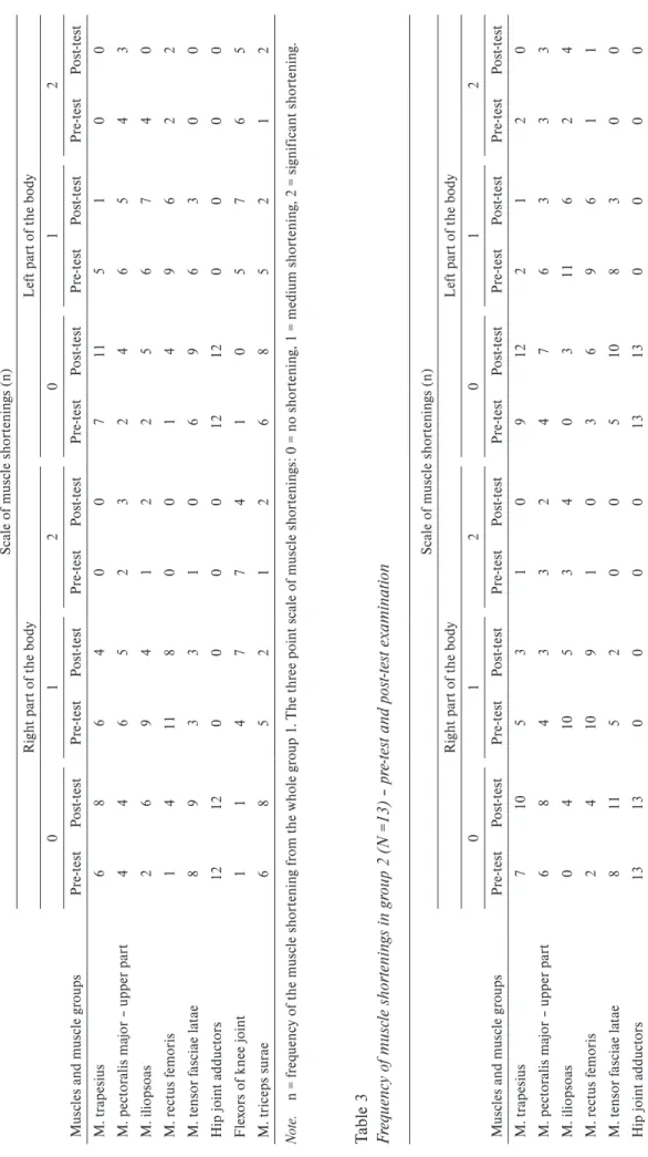
Pre-test measurements of muscle shortening in the group 2 proved that all examined muscles, apart from hip joint adductors, were shortened in different stages. For details see Table 3.
Similarly to the group 1, post-test examination after the combination of dynamic and static stretching pro-gram in the group 2 showed improvements in muscles shortenings in the following muscles – m. trapesius on both sides, m. pectoralis major on both sides, m. rectus femoris on both sides, m. tensor fasciae latae on both sides, knee flexors on both sides and m. triceps surae on both sides, too. Only in m. iliopsoas on both sides was found even more shortening after the dynamic stretching program.
Spine mobility – Thomayer’s test
Limited range of motion in spinal mobility was found during pre-test measurements in 75% of the partici-pants in the group 1 and in 54% of the participartici-pants in the group 2 (M ± SD: group 1: –3.0 ± 3.4 cm; group 2: –4.0 ± 6.4 cm).
Post-test measurements showed improved spine mobility in both groups. In the group 1 the percentage occurrence of individuals with limited range of motion was reduced to 17% and in the group 2 to 38% (mean ± SD: group 1: –2.0 ± 3.4 cm; group 2: –1.0 ± 6.0 cm).
Joint mobility – range of motion in selected joints
Changes of joint flexibility between pre-test and post-test in interaction with stretching program were not statistically significant (Table 4). The participants in the group 1 performed greater ROM in ankle during dorsal flexion then participants in the group 2. We found significant changes in pre-test and post-test in range of motion for ankle plantar flexion (p = .04,
ηp2= .18) after the combination of dynamic and static
stretching. Further, in group 2 we found that the range of motion of left hip in flexion increased significantly (p = .03, ηp2 = .20).
Range of motion of measured joints of both groups influenced by stretching is presented in Table 4.
ROM Results for group 1 provided evidence that the average range of motion in ankle in dorsal flexion reach population standards (Janda & Pavlů, 1998; Rychlíková, 2002) in both pre-test and post-test measurements. Above standard values in both measurements of plantar flexion were observed in all
participants from group 1. After the stretching pro-gram, range of joint mobility in ankle joint remained within standard or above standard values, but at the same time further decline in the range of motion in both tests was observed in group 1. In the same group were observed values below average in both pre-test and post-test measurements in hip joint flexion on both sides. After the stretching program, 5 participants (38%) achieved improved range of motion of the hip joint on the right and 9 participants (75%) on the left side. However reached values were still below average.
In the majority of cases from the group 1, range of motion during extension in horizontal abduction of shoulder joints was average and above average on both sides in both pre-test as well as post-test measurements. After the stretching program, improvement of range of motion appeared in 8 participants (67%) on the right side and in 9 participants (75%) on the left side.
Results of ranges of motion of group 2 provided evidence that the average range of motion in ankle joint dorsal flexion reached population standards in both pre-test and post-test measurements. Above aver-age values were observed in plantar flexion for all par-ticipants in both measurements. After the stretching program, ranges of motion in ankle remained within population standards or above average. But at the same time we noticed worsened range of motion in both – dorsal and plantar flexions.
Below average values were observed in the range of motion in flexion of the hip joint again on both pre-test and post-pre-test measurements. After the stretching program, 7 participants (54%) improved their range of motion on the right side and 9 participants (70%) on the left side from, however, values were still below average.
Similarly, extension in horizontal abduction of shoulder joint reached average values of range of motion on both sides in all participants in both mea-surements. After the stretching program, 8 participants (67%) improved their range of motion on the right side and 9 participants (75%) on the left side.
Discussion
Table 2 Freq
uenc
y of musc
le shor
tenin
gs in g
roup 1 (
N = 12
) – pr
e-te
st and pos
t-t es t e xamina tion Musc le
s and musc
le g
roups
Sc
ale of musc
le shor tenin gs ( n) Ri ght par
t of t
he bod
y
L
ef
t par
t of t
he bod y 0 1 2 0 1 2 Pr e-te st Po st -te st Pr e-te st Po st -te st Pr e-te st Po st -te st Pr e-te st Po st -te st Pr e-te st Po st -te st Pr e-te st Po st -te st M . t rape sius 6 8 6 4 0 0 7 11 5 1 0 0 M . pect or alis ma
jor – upper par
t 4 4 6 5 2 3 2 4 6 5 4 3 M . iliopsoa s 2 6 9 4 1 2 2 5 6 7 4 0 M . r ect us f emor is 1 4 11 8 0 0 1 4 9 6 2 2 M . t ensor f asciae la tae 8 9 3 3 1 0 6 9 6 3 0 0
Hip joint adduct
or s 12 12 0 0 0 0 12 12 0 0 0 0 F le xor
s of k
nee joint 1 1 4 7 7 4 1 0 5 7 6 5 M . t riceps sur ae 6 8 5 2 1 2 6 8 5 2 1 2 No te .
n = fr
eq
uenc
y of t
he musc
le shor
tenin
g fr
om t
he whole g
roup 1
. T
he t
hr
ee point sc
ale of musc
le shor
tenin
gs
: 0 = no shor
tenin
g, 1 = medium shor
tenin g, 2 = si gnif ic ant shor tenin g.
Table 3 Freq
uenc
y of musc
le shor
tenin
gs in g
roup 2 (
N =13
) – pr
e-te
st and pos
t-t es t e xamina tion Musc le
s and musc
le g
roups
Sc
ale of musc
le shor tenin gs ( n) Ri ght par
t of t
he bod
y
L
ef
t par
t of t
he bod y 0 1 2 0 1 2 Pr e-te st Po st -te st Pr e-te st Po st -te st Pr e-te st Po st -te st Pr e-te st Po st -te st Pr e-te st Po st -te st Pr e-te st Po st -te st M . t rape sius 7 10 5 3 1 0 9 12 2 1 2 0 M . pect or alis ma
jor – upper par
t 6 8 4 3 3 2 4 7 6 3 3 3 M . iliopsoa s 0 4 10 5 3 4 0 3 11 6 2 4 M . r ect us f emor is 2 4 10 9 1 0 3 6 9 6 1 1 M . t ensor f asciae la tae 8 11 5 2 0 0 5 10 8 3 0 0
Hip joint adduct
or s 13 13 0 0 0 0 13 13 0 0 0 0 F le xor
s of k
nee joint 4 4 3 5 6 4 3 3 5 6 5 4 M . t riceps sur ae 7 8 1 2 5 3 7 11 1 0 5 2 No te .
n = fr
eq
uenc
y of t
he musc
le shor
tenin
g fr
om t
he whole g
roup 2
. T
he t
hr
ee point sc
ale of musc
le shor
tenin
gs
: 0 = no shor
tenin
g, 1 = medium shor
Machado, Scaramussa, & Gomes, 2013; Kutáč & Gajda, 2006; Theodorou, Galazoulas, Zakas, Vergou, & Vamvakoudis, 2005).
In general a positive effect of stretching techniques on (1) compensation of overloaded or monotonously loaded muscles, (2) reduction of muscle shortenings, and (3) elimination of muscle imbalances (in combi-nation with strength developing techniques) is well established. Thus, inclusion of stretching exercises in training process of athletes of every age category would be expected. In our study it was hypothesized, that the status of joint and muscle system of youth soccer play-ers would be comparable with population standards, or it would match the norms after the stretching program. Our hypothesis was supported by Šrámková and Votík (2010) who studied issues of muscular imbalances and means of imbalance prevention as well as correction within the same age group. The research team discov-ered that application of specialized gymnastic training which requires good spine flexibility in all directions can correct developed muscle imbalances. Both con-stant corrections and increased performance of trunk flexion during training unit and at home training can positively affect functional status of movement system. Both examined groups showed muscle shorten-ings of different stages, apart from hip joint adductors without side asymmetries, in both measurements.
Changes of joint flexibility between pre-test and post-test in interaction with the stretching program were not statistically significant. Participants in the group 1 performed better in ankle during dorsal flexion then participants in the group 2. We can discuss the changes influenced by this age. It could be caused by the small sample and by growing periods.
In the current scientific literature we did not find any study dealing with a similar problem in youth soccer players so our results cannot be compared to existing data. However, a few similar studies may be mentioned. Kutáč and Gajda (2006) and Adamčák (2000) dealt with the stated problem in adolescent soc-cer players. Kutáč and Gajda (2006) studied muscle shortenings in loaded muscles of lower limbs and the trunk (knee flexors, m. iliopsoas, m. pectoralis major and m. rectus abdominis) in a group of soccer play-ers between 16–18 years of age from different perfor-mance categories. The most shortened muscles were knee flexors on both sides (more on the left side) in 22 out of 24 players of the lowest performance category. Interestingly the highest occurrence of muscle shorten-ings was found in players of the lowest performance category which includes city championship and city competition. They achieved worse results in tests of muscle imbalance despite the fact that these players train less than league players or players competing in Table 4
Comparison of ranges of motion (degrees) in selected joints between different intervention
Joint – movement Intervention
Descriptive statistics of ROM (degrees)
Statistical analyses
Pre-test Post-test
M SD M SD F p ηp2
Ankle DF S 27.0 4.9 23.6 9.8 0.02 .89 < .001
S+D 23.7 8.1 20.8 3.9
Ankle PF S 70.7 4.7 65.1 9.0 3.95 .06 .15
S+D 63.6 5.4 63.3 5.5
Hip FL right S 68.7 6.7 67.9 10.9 0.02 .88 < .001
S+D 71.6 7.5 71.3 6.7
Hip FL left S 67.3 6.5 70.5 5.1 0.01 .91 < .001
S+D 67.6 9.1 71.1 9.8
Shoulder EXTHA right S 33.0 16.3 37.4 10.3 1.23 .28 .05
S+D 39.9 15.8 38.2 16.4
Shoulder EXTHA left S 32.9 12.7 39.8 10.4 3.22 .09 .12
S+D 39.9 15.3 38.2 17.7
Note. ROM = range of motion. DF = dorsal flexion, PF = plantar flexion, FL = flexion, EXTHA = extension in horizontal abduction, S = static stretching (data of the group 1), S+D = static and dynamic stretching (data of the group 2). F = Fisher’s F ratio, ηp2 = partial
regional tournaments. A logical explanation of these results was offered at the end of the study, specifically in a summary of overall time players spend training and performing compensation activities in the train-ing session. The lowest performance group stated zero time for compensation activities in training. Adamčák (2000) focused mainly on muscle shortenings in lower limbs in 16 year old soccer players (N = 22), where the highest stage of muscle shortenings was measured in knee flexors – 90.9% on the right side and 81.8% on the left side. The next most shortened muscles or muscle groups were m. tensor fasciae latae, m. rectus femoris, hip joint adductors, m. iliopsoas and m. soleus.
The study of Malátová and Matějková (2011) was focused on muscle imbalance among soccer players between 21–33 years of age. The greatest improve-ment was achieved by compensatory exercise in the muscle group responsible for hip flexion. Shortening of lumbar muscles, hamstrings and muscles in the back of the neck was also successfully affected. Abdominal muscles were strengthened.
Spine mobility in participants of both groups was limited in the baseline measurements. However improvements were observed after the intervention for both groups. Adamčák (2000) found that the range of motion of all examined joints and spine was similar to the values of healthy population. In some case even slight hyper-mobility was present.
Limited range of motion in hip flexion on both sides is closely associated with muscle shortenings found in knee flexors. Changes in other study variables were not statistically or clinically significant. Also no statisti-cally or clinistatisti-cally significant changes were observed in interaction time (pre-test, post-test) and the stretching programs. We did not find any effect of the stretching programs.
The presented shortenings, mainly of lower limbs (hip joint flexors and knee flexors), and limited range of motion, especially in hip joints, suggest that inclu-sion of stretching techniques into training sesinclu-sions of youth soccer players is needed. In order to achieve the most benefits, the stretching program should be accompanied by strengthening exercises for weakened muscles.
The stretching method was chosen as the technique for compensation of sport load in the present study because its benefits for all kind of sports are well docu-mented (Aguilar et al., 2012; Amiri-Khorasani, 2013; Behm & Chaouachi, 2011; Magnusson & Renström, 2006; Nédélec et al., 2012; Rogan, Wüst, Schwitter, & Schmidtbleicher, 2013; Yuktasir & Kaya, 2009; Zakas, Doganis, Galazoulas, & Vamvakoudis, 2006; Zakas, Grammatikopoulou, Zakas, Zahariadis, & Vamvak-oudis, 2006). The benefits include: (1) improvements
in the range of joint motion, (2) prevention of muscle injuries and (3) improvements of physical performance. This technique does not require demanding space and material. The choice of the stretching method depends on one’s fitness capacity and abilities, on the situation where the stretching is performed, and on goals that an athlete wants to achieve.
The technique of static stretching is according to Alter (1999) one of the safest in terms of muscle injury, because all the movements, which are performed during muscle elongation, are slow and controlled. Alter (1999) recommends that stretching should be performed after the main part of the training session. Kovacs (2006) warns that the static form of stretching is often used within warm-up before sport activities, in spite of the fact that nowadays there are still no avail-able studies which would confirm, that it improves fit-ness or supports prevention from injury. This is the rea-son why the static stretching program was performed during the final part of the training session. According to Little and Williams (2006) static stretching does not appear to be detrimental to high-speed performance when included in a warm-up for professional soccer players. However, dynamic stretching during the warm-up was most effective as preparation for subsequent high-speed performance.
Amiri-Khorasani, Abu Osman, and Yusof (2011) recommended professional soccer players to incorpo-rate into warm-ups dynamic stretching exercises of the hip joint, hence increasing the chances of scoring and injury prevention during soccer games.
From a practical point of view, the aim of dynamic stretching is to stimulate muscles before sport activi-ties by short repeated stretches and, at the same time, to offer a relaxation for the joints of the most loaded body parts. The goal of static stretching after physical activity is to relax muscles by impulses from the Golgi tendon organs and to restore original length in loaded muscles.
The present study support previous findings and available knowledge. The results provided evidence that group 2 which performed both static and dynamic stretching at the beginning of the training session gained more significant improvements (both percent-age and symmetric decrease in muscle shortenings). Similar results were found in spine and joint mobility in the group 2. Group 1 improved after the stretch-ing program as well but the improvements were less significant.
results showed more significant improvements in the group 2 with combination of dynamic and static stretching programs. Analysis of training sessions of both groups showed some differences in a training organization. Time might be one of the factors respon-sible for the study results. The stretching program in group 1 may not have had enough time. Group 1 was influenced by the bus transport to the place of training session. It was not possibility to stay there a longer time. The second group commuted to training sessions individually. Another factor might be a coach (covariate human factor). There was a requirement that in both groups there should be two coaches during stretching phases of training sessions. It was always the case for group 2, but not for the group 1.
We concluded that different results in one group would be influenced by the personality of the coach or the number of coaches. In our case, one coach did not manage to correct performance of particular exer-cises enough and therefore incorrect muscle stretch-ing could have been performed. A similar situation is described by Kutáč and Gajda (2006), who emphasize the importance of the quality and professionalism of training process management and, of course, the importance of the time needed for stretching exercises so the stretching program is the most beneficial for desired compensation of muscle imbalances. In terms of the time necessary for compensation exercises, the authors suggest that 15 min from a 90 min training session is not sufficient. This fact was also confirmed by Fajfer (1990). Fajfer (1990) suggests at least 20 min of stretching for successful compensation within one training session.
For the future studies we would recommend to include a control group and only one intervention group as opposed to two. Also the duration of the stretching program could be extended. In addition it would be desirable to include one extra control assess-ment of the moveassess-ment system status after the end of the study, perhaps few weeks later, to make sure that coaches are still including a stretching program into training sessions.
Conclusions
The results detected some muscle imbalance in our team of youth soccer players. Muscle imbalance and related slight range of motion of the spine and joints was observed particularly in hip and knee joints flex-ors. The stretching program that included dynamic components at the beginning of training sessions (performed by group 2) seems to lead to a decrease of muscle shortenings. It could be use for prevention
device to decrease the injury possibilities in older years of soccer player history.
One of the reasons is a revealed presence of sig-nificant muscle shortenings and limited joint mobility already in youth soccer players.
Acknowledgment
The study was supported by research project 0021620864 “Active lifestyle in biosocial context” funded by the Ministry of Education, Youth and Sports and by the grant project of the Czech Science Founda-tion number P407-12-0166.
References
Adamčák, Š. (2000). Funkčné svalové poruchy u mladých futbalistov a možnosti ich odstraňovania [Functional muscle disorders in young soccer players and their removal options]. Telesná výchova a šport, 10(4), 27–30.
Adamčák, Š. (2005). Využitie strečingu v pohybovom režime žiakov základnej školy [Use of the stretching mode in the motion of elementary school pupils]. Telesná výchova a šport, 15(1), 13–15.
Aguilar, A. J., Distefano, L. J., Brown, C. N., Herman, D. C., Guskiewicz, K. M., & Padua, D. A. (2012). A dynamic warm-up model increases quadriceps strength and ham-string flexibility. Journal of Strength and Conditioning Research, 26, 1130–1141.
Alter, M. J. (1999). Strečink: 311 protahovacích cviků pro 41 sportů [Sport Stretch: 311 stretches for 41 sports] (V. Janda & T. Alfödi, Trans.). Praha: Grada.
Amiri-Khorasani, M. (2013). Kinematics analysis: The acute effect of different stretching methods on dynamic range of motion of lower extremity joints during soccer instep kick-ing. International Journal of Performance Analysis in Sport, 13, 190–199.
Amiri-Khorasani, M., Abu Osman, N. A., & Yusof, A. (2011). Acute effect of static and dynamic stretching on hip dynamic range of motion during instep kicking in professional soccer player. Journal of Strength and Conditioning Research, 25, 1647–1652.
Arnason, A., Andersen, T. E., Holme, I., Engebretsen, L., & Bahr, R. (2008). Prevention of hamstring strains in elite soc-cer: an intervention study. Scandinavian Journal of Medicine & Science in Sports, 18, 40–48.
Behm, D., & Chaouachi, A. (2011). A review of the acute effects of static and dynamic stretching on performance.
European Journal of Applied Physiology, 111, 2633–2651. Brügger, A. (1993). Kinesiologické aspekty omezení funkce
při pohybu a držení těla [Kinesiological aspects of func-tional limitations during movement and posture]. Rehabili-tácia, 26, 136–144.
Bunc, V. (2006). Zvláštnosti kondiční přípravy žen [Special fitness training for women]. In I. Čechovská, V. Bunc, & V. Novotná (Eds.), Fit programy pro ženy (pp. 9–23). Praha: Grada Publishing.
Bursová, M., Votík, J., & Zálabák, J. (2003). Kompenzační cvičení pro fotbalisty [Compensation exercise for soccer play-ers]. Praha: Olympia.
Cingálková, M., Bunc, V., & Cingálek, R. (2001). Kineziolo-gický rozbor jako prostředek kodhalení svalových dysbalancí a prevence zranění u fotbalistů[Use of kinesiologic analysis in injury prevention of football players]. In H. Válková & Z. Hanelová (Eds.), 2nd International conference Movement and Health (p. 532). Olomouc: Univerzita Palackého. Data Input. (2004). Manual NUTRI 4 – Multifrequency
soft-ware for the determination of body water, body composition and nutritional status. Pöcking: Author.
De Castro, J. V., Machado, K. C., Scaramussa, K., & Gomes, J. L. E. (2013). Incidence of decreased hip range of motion in youth soccer players and response to a stretching program: A randomized clinical trial. Journal of Sport Rehabilitation, 22, 100–107.
Dittmar, M. (2003). Reliability and variability of bioimped-ance measures in normal adults: Effects of age, gender, and body mass. American Journal of Physical Anthropology, 122, 361–370.
Dovalil, J., Choutka, M., Svoboda, B., Hošek, V., Perič, T., Potměšil, J., … Bunc, V. (2002). Výkon a trénink ve sportu
[Performance and training in sport]. Praha: Olympia. Fajfer, Z. (1990). Koordinační (obratnostní) schopnosti,
pohyb-livost (strečink) v systému hráče fotbalu [Coordination (dex-terity) capability, flexibility (stretching) in the system of soccer players]. Brno: ČFS.
Ferguson, C. J. (2009). An effect size primer: A guide for clini-cians and researchers. Professional psychology: Research and Practice,40, 532–538.
Friery, K. (2008). Incidence of injury and disease among for-mer athletes: A review. Journal of Exercise Physiology Online, 11(2), 26–45.
Gúth, A. (1994). Propedeutika v rehabilitácii [Propedeutics in rehabilitation]. Bratislava: Liečreh.
Houglum, P. A. (2001). Therapeutic exercises for athletic inju-ries. Champaign, IL: Human Kinetics.
Janda, V. (1981). Vyšetřování hybnosti [Investigation of the movement]. Praha: Avicenum.
Janda, V. (1996). Funkční svalový test [Functional muscle’s test]. Praha: Grada.
Janda, V., & Pavlů, D. (1993). Goniometrie [Goniometry]. Brno: IPVPZ.
Janura, M., & Zahálka, F. (2004). Kinematická analýza pohybu člověka [Kinematic analysis of human movement]. Olo-mouc: Univerzita Palackého.
Kovacs, M. S. (2006). The argument against static stretching before sport and physical activity. Athletic Therapy Today, 11(3), 6–8.
Kutáč, P., & Gajda, V. (2006). Kvalita hybných funkcí u hráčů kopané ve vztahu k jejich výkonnosti [The quality of motional functions of football players in relation to their performance]. Česká antropologie, 56(1), 85–87.
Kyle, U. G., Bosaeus, I., De Lorenzo, A. D., Deurenberg, P., Elia, M., Manuel Gomez, J., … ESPEN. (2004). Bioelectri-cal impedance analysis – part II: Utilization in cliniBioelectri-cal prac-tice. Clinical Nutrition, 23, 1430–1453.
Lewit, K. (1996). Manipulační léčba [Manipulation the-rapy]. Leipzig: Česká lékařská společnost Jana Evangelisty Purkyně.
Little, T., & Williams, A. G. (2006). Effects of differential stretching protocols during warm-ups on high-speed motor capacities in professional soccer players. Journal of Strength and Conditioning Research, 20, 203–207.
Magnusson, P., & Renström, P. (2006). The European college of sports sciences position statement: The role of stretch-ing exercises in sports. European Journal of Sport Science, 6, 87–91.
Malátová, R., & Matějková, P. (2011). Svalové dysbalance u hráčů fotbalu ve věku 21–33 let [The muscles imbalances of footballers and their compensation]. Studia Kinanthropo-logica, 12, 35–39.
Morse, D. T. (1999). Minisize2: A computer program for determining effect size and minimum sample for statistical significance for univariate, multivariate, and nonparamet-ric tests. Educational and Psychological Measurement, 59, 518–531.
Nédélec, M., McCall, A., Carling, C., Legall, F., Berthoin, S., & Dupont, G. (2012). Recovery in soccer: Part I – post-match fatigue and time course of recovery. Sports Medicine, 42, 997–1015.
Nováková, H. (1994). Význam flexibility u dětí a mládeže [The importance of flexibility in children and youth]. Tělesná výchova a sport mládeže, 60(5), 36–39.
Peterson, L., Renström, P., & Grana, W. A. (1986). Sports injuries: Their prevention and treatment. Sant Louis: Mosby Year Book.
Rogan, S., Wüst, D., Schwitter, T., & Schmidtbleicher, D. (2013). Static stretching of the hamstring muscle for injury prevention in football codes: A systematic review. Asian Journal of Sports Medicine, 4(1), 1–9.
Rychlíková, E. (2002). Funkční poruchy kloubů končetin: diag-nostika a léčba [Functional disorders of the joints of limbs: diagnostic and treatment]. Praha: Grada.
Šrámková, P., & Votík, J. (2010). Svalové dysbalance a mož-nosti jejich prevence a korekce u hráčů žákovské kategorie FC Viktoria Plzeň [Muscle imbalances and possibility of their prevention in case of players of student category of FC Viktoria Plzeň]. Studia Kinanthropologica, 11, 101–107. Theodorou, I., Galazoulas, C., Zakas, N., Vergou, A., &
Vam-vakoudis, E. (2005). The effect of stretching duration on the flexibility of lower extremities in junior soccer players.
Physical Training, 1(1), 1.
Trojan, S., Druga, R., Pfeiffer, J., & Votava, J. (1996). Fyziolo-gie a léčebná rehabilitace motoriky člověka [Physiology and rehabilitation of the human motorics]. Praha: Grada. Véle, F. (1997). Kineziologie pro klinickou praxi [Kinesiology
for clinical practice]. Praha: Grada.
Volpi, P., Melegati, G., Tornese, D., & Bandi, M. (2004). Mus-cle strains in soccer: A five year survey of an Italian major league team. Knee Surgery, Sports Traumatology, Arthroscopy, 12, 482–485.
Wong, P. L., & Hong, Y. (2005). Soccer injury in the lower extremities. British Journal of Sports Medicine, 39, 473–482. Yuktasir, B., & Kaya, F. (2009). Investigation into the long-term effects of static and PNF stretching exercises on range of motion and jump performance. Journal of Bodywork and Movement Therapies, 13, 11–21.
Zakas, A., Doganis, G., Galazoulas, C., & Vamvakoudis, E. (2006). Effect of acute static stretching duration on isoki-netic peak torque in pubescent soccer players. Pediatric Exercise Science, 18, 252–261.
Zakas, A., Grammatikopoulou, M. G., Zakas, N., Zahariadis, P., & Vamvakoudis, E. (2006). The effect of active warm-up and stretching on the flexibility of adolescent soccer players.