ORIGINAL ARTICLE
High-resolution Transmission Electron
Microscopy Characterization of the Structure
of Cu Precipitate in a Thermal-aged
Multicomponent Steel
Lizhan Han
1, Qingdong Liu
1,2*and Jianfeng Gu
1,2Abstract
High-dispersed nanoscale Cu precipitates often contribute to extremely high strength due to precipitation harden-ing, and whereas usually lead to degraded toughness for especially ferritic steels. Hence, it is important to understand the formation behaviors of the Cu precipitates. High-resolution transmission electron microscopy (TEM) is utilized to investigate the structure of Cu precipitates thermally formed in a high-strength low-alloy (HSLA) steel. The Cu precipi-tates were generally formed from solid solution and at the crystallographic defects such as martensite lath boundaries and dislocations. The Cu precipitates in the same aging condition have various structure of BCC, 9R and FCC, and the structural evolution does not greatly correlate with the actual sizes. The presence of different structures in an individual Cu precipitate is observed, which reflects the structural transformation occurring locally to relax the strain energy. The multiply additions in the steel possibly make the Cu precipitation more complex compared to the binary or the ternary Fe–Cu alloys with Ni or Mn additions. This research gives constructive suggestions on alloying design of Cu-bearing alloy steels.
Keywords: Cu precipitate, High-resolution transmission electron microscopy, Thermal aging, High-strength low-alloy steel
© The Author(s) 2019. This article is distributed under the terms of the Creative Commons Attribution 4.0 International License (http://creat iveco mmons .org/licen ses/by/4.0/), which permits unrestricted use, distribution, and reproduction in any medium, provided you give appropriate credit to the original author(s) and the source, provide a link to the Creative Commons license, and indicate if changes were made.
1 Introduction
The nanoscale Cu precipitates often endows alloyed steels with dramatically high strength but simultaneously lead to degraded impact toughness especially at cryo-genic temperature [1–3]. The nature of Cu precipitation in α-Fe matrix have been comprehensively investigated in aspect of mutual development of composition, struc-ture and morphology [4]. It has been established that the Cu precipitates contain significant concentration of Fe and are also rich in Ni and Mn at their initial nucleation stage [7]. With the precipitation reaction proceeding, Fe atoms are gradually expelled into the matrix and Ni and
Mn tend to segregate at the precipitate/α-Fe boundaries [8, 9]. Meanwhile, the structure of Cu precipitates evolves following a complicated BCC→9R→3R→FCC sequence [10, 11], and the BCC Cu precipitates are considered to be originate from the growth of B2 ordered domains, as reported by Wen et al. [12]. Accompanying the compo-sitional and structural evolution, the morphology of Cu precipitates generally changes from spherical to ellip-soidal to rod-like shape [4, 13]. The transformations in course of Cu precipitation collectively take place in order to reduce the related coherent strain energy, interfacial energy and inherent energy of Cu precipitates.
The structural evolution of Cu precipitates in α-Fe is extremely complex. It is known that the Cu atoms ini-tially experience a coherent precipitation to form a B2 or BCC structure in the BCC α-Fe [14], and then the meta-stable structures transform to an internally twinned mar-tensitic 9R structure (the stacking sequence of the closely
Open Access
*Correspondence: [email protected]
1 Institute of Materials Modification and Modelling, School of Materials Science and Engineering, Shanghai Jiao Tong University, Shanghai 200240, China
packed planes is ABC/BCA/CAB/A) by the repeated shear of {110}α-Fe planes with an orientation relationship
(011)α-Fe // (11-4)9R, [1–11]α-Fe // [-110]9R [10]. The
fol-lowing possible changes in the 9R structure, including the detwinning of typical twin variants by migration of intervariant (twins) boundary, rotation of closed-packed (009)9R basal planes to align more close to {110}α-Fe, and
elimination of regular stacking faults on the (009)9R basal
planes [15], can produce a more stable 3R structure with non-cubic heavily distorted FCC structure [11]. With subsequent diffusional growth of the 3R structure and the resultant lattice relaxation to minimize total free energy, the expected FCC structure is eventually formed following the Kurdjumov-Sachs orientation [15]. The 9R, 3R and FCC structures are all possibly be with twined or untwined structure, as the untwined 9R and twined 3R or FCC structures were all reported by Feng [16] and Heo et al. [17]. In addition, the critical size of Cu precipitates at which the structural transformation occurs also appear to be different. The complexity of the structural evolution of Cu precipitates is usually ascribed to the differences in the initial compositions and/or the actual heat treat-ments (tempering or ageing intensity) of the model or multicomponent steels investigated [18–25]. The defor-mation (stress or strain) and irradiation also prove to efficiently induce or accelerate BCC to 9R transformation [18, 22]. In addition, computational modeling also used to understand the structural and compositional evolution in Fe–Cu-X (X denotes Mn and/or Ni, etc.) systems [26–
32], and indicates that the structures of Cu precipitates are greatly influenced by alloying elements.
Hence, the structural development of Cu precipi-tates is extremely complex and need to be experimen-tally studied on more extended ferritic steels. The aim of the present work is to provide more information on the structural evolution of Cu precipitates in a thermal-aged multicomponent steel by using high-resolution transmis-sion electron microscopy (HRTEM).
2 Experimental Details
The experimental steel was received from IRIS, Shasteel China with compositions of 0.04 C, 0.24 Si, 0.84 Mn, 1.15 Cu, 1.94 Ni, 0.51 Mo, 0.50 Cr, 0.043 Nb, 0.009 Ti, 0.03 Al, 0.004 N, 0.007 P, and 0.004 S (wt. %) (More see reference [19]). The steel was solution treated at 900 °C for 30 min followed by water-quenching and thermally aged at 500 °C for 16 h when most of Cu precipitates were in overaged conditions. HRTEM observation were performed on a JEOL 2010F (Tokyo, Japan) at 200 kV. Although it has been found that the characteristic 9R “herring-bone” fringe pattern can only be well imaged with the beam direction closely parallel to a <111> α-Fe direction [11], most micrographs in this paper were
alternatively taken along a <001>α-Fe direction in order
to give different appearances of the Cu precipitates. Thin foil specimens were prepared by mechanically grinding sample slices to 80-μm thick and twin-jet electropolish-ing in a mixture of 90% alcohol and 10% perchloric acid at − 30 °C under 50 V.
3 Results and Discussion
Figure 1a shows a HRTEM micrograph taken along a [001]α-Fe direction of the sample aged at 500 °C for 16 h.
Several nanoscale Cu precipitates can be recognized by the strain field contrast (black domains marked by cir-cle) arose from lattice distortion and by the somewhat weak fringe contrast (marked by arrow) where the atomic columns are mostly ill-arranged. These precipitates are formed in association with linear dislocation as they gen-erally distributed in an arc curve (highlighted by dash line), which is consistent with the APT observation of Cu precipitates in a similar steel [19]. The Fast Fourier trans-form (FFT) conducted on two representative Cu precipi-tates (labeled A and B, respectively) embedded in α-Fe in the dashed square in Figure 1a shows that the diffraction spots from the precipitates exactly overlap with those from α-Fe matrix, as shown in Figure 1b, which indicates the identical BCC structure and the absolute coherency of the Cu precipitates and α-Fe matrix. Faint extra weak spots (marked by arrows) along g=[020]α-Fe are
respond-ing to the anomalous frrespond-inge spacrespond-ings in precipitate B, as discussed later. However, no evidence of the forma-tion of B2 ordered domains reported by Wen et al. [12] is found from the FFT pattern, although the image here is also taken along the [001] α-Fe direction and the steels
used are similarly alloyed with multiple elements. In a BCC Cu and B2-NiAl precipitation strengthened ferritic steel, Kapoor et al. [20] found that the B2 ordered dif-fraction become distinct only when the B2-NiAl precipi-tates have an average radius of 4.28 ± 1.47 nm (obtained
by APT analysis) after at least aging at 550 °C for 10 h. The B2 ordered diffraction is seemingly too weak to be detectable for the nanoscale features using the HRTEM, since the imaging mechanism and the working condi-tions (such as voltage employed) of HRTEM are both also different from that of the HAADF-STEM.
The corresponding inverse Fast Fourier transform (IFFT) of Figure 1b presents a more distinct atomic structure of the Cu precipitates, as shown in Figure 1c. In precipitate A, the ‘black’ strain contrast possibly is associated with the compositional fluctuation, since the nanoscale bcc Cu precipitates are thought to contain high concentrations of Fe, Ni and Mn [7]. Other studies [10–
readily to be imaged by using the phase contrast method of HRTEM. The precipitate/matrix interface is therefore roughly distinguished by the strain contrast and the pre-cipitate A is found to have a size approximately 6 nm in diameter (average of major and minor axis values). The (020)/(-200) interplanar spacings of the precipitate and α-Fe are carefully measured to be 2.116 Å/ 2.112 Å and 2.084 Å/2.067 Å, respectively, which gives rise to slight lattice deviation along [020] and [-200] directions. The small misfit inevitably induced coherent strain energy, which may serve as main driving force for bcc to 9R mar-tensitic transformation.
In precipitate B, the appearance of irregular striations (marked by arrows) along the [-200]α-Fe direction, which
correspond to the weak diffraction spots between g000
and g020 spots, manifests the distorted lattices in the
pre-cipitate and resultant ‘black’ strain contrast, as shown Figure 1c. One possibility that the anisotropic atoms of Cu segregate at given lattice planes of α-Fe can account for the anomalous atomic structure of the precipitate, which is consistent with the observation by Xu at al. [21] in a thermal-aged reactor pressure vessel (RPV) model steel with enhanced Cu content. The Cu segregation undoubtedly induces significant lattice misfit of α-Fe, which in turn impede the segregation occurring on con-tinual lattice planes and, the Cu atoms hence tend to peri-odically replace the Fe atoms. The Cu segregation seems to be rather irregular here and its periodicity generally possesses 2‒5 column spacings. However, as observed by Xu et al., the segregated Cu atoms can be regularly arranged every three sets of {110}α-Fe planes and the
peri-odical spacing is measured to be 0.6 nm, which is right consistent with the spacing of the herring-bone fringes of 9R structure [10]. The anomalous atom arrangement in the present nanoscale Cu precipitate seems to repre-sent an earlier nucleation stage. Moreover, note that the [001]α-Fe incidence direction may also cause the different
appearance of Cu precipitate in HRTEM. The Cu segre-gation at certain lattice planes of α-Fe preceding to the Cu precipitation is similar to the formation of G.P. zone in some Al alloys, which are both energetically favora-ble. Overall, regular segregation of Cu atom perceivably occurs periodically with the Cu precipitation proceed-ing, leading to the development of the BCC structured (such as precipitate A) and further the 9R structured Cu precipitates.
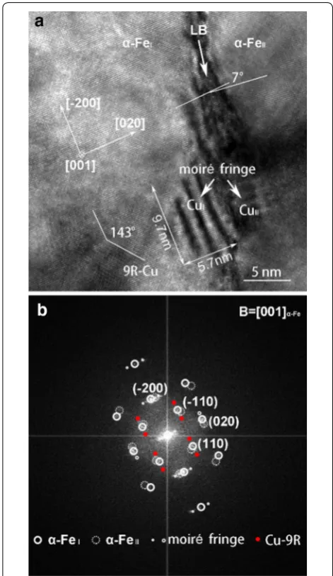
Figure 2a shows a HRTEM micrograph and Figure 2b the corresponding Fast Fourier Transform pattern with index illustration taken along a [001]α-Fe direction
of the sample aged at 500 °C for 16 h. A straight zone with distinct “black” strain contrast where the atoms are seriously ill arranged and the lattice is significant distorted indicates the appearance of a martensite lath
boundary (LB), which divides the whole ferritic matrix into α-FeI and α-FeII crystals. The angular deviation of
the two α-Fe crystals is measured to be approximately 7o, which is also consistent with the low angle
mar-tensite LBs. Two adjacent Cu precipitates (labeled CuI
and CuII detected in α-FeI and α-FeII, respectively) at
the LB exhibit evident Moiré fringes arising from the lattice overlap, which also brings about extra diffraction spots close to the g<200>α-Fe spots, as shown in Figure 2b.
The fringes are almost parallel to the [-200]α-FeI and
[020]α-FeII, respectively. The Moiré fringes, the intensity
of which is related to the in incidence direction as dis-cussed followed, make the fine structure of the Cu pre-cipitates difficult directly imaged for further analysis.
Compared to that formed on the dislocation (Figure 1), the Cu precipitates formed in association with LBs gen-erally have relatively larger sizes (approximately 7.8 nm for CuII), which is possibly attributed to faster diffusion
of related solutes along LBs than dislocations. The Cu precipitates are thought to have a possible 9R struc-ture as they are listed in the size range of 9R strucstruc-ture but which cannot be well established. A Cu precipitate with 9R structure, however, is easily recognized in α-FeI
containing only two twin-related variant segments. The characteristic herring-bone fringe patterns are not observed, that is, there is no stacking fault every three closely packed planes in the adjacent twin segments, but in which the striations become evident and their angles are approximately 143o. The (-1-14)
9R mirror
boundary lies parallel to <110>α-FeI, as shown by extra
diffraction spots in Figure 2b. It seems that it is not necessary to form SF in 9R structure, because the strain energy between 9R Cu and the a-matrix can be suffi-ciently reduced by formation of (114)9R twins. In
addi-tion, it is suggested the typical 9R structure is indeed not easily detected with [001]α-Fe direction [10, 11].
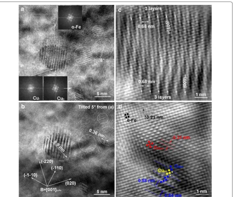
Figure 3 gives HRTEM analysis of several Cu precipi-tates in the same sample as that in Figures 1 and 2. The micrograph in Figure 3a taken along a [001] α-Fe
direc-tion shows the presence of Cu precipitates (labeled CuI
and CuII) recognized from the evident moiré fringes.
The general FFT patterns from the two Cu precipitates in the selected zones (dash square) are somewhat differ-ent, corresponding to their observed different configura-tion as discussed below. However, the FFT patterns from CuI and CuII are generally identical to that from α-Fe
because the main diffraction spots from the precipitates may overlap with the fundamental diffraction from α-Fe, except for the presence of extra weak diffraction spots corresponding to the complex structure of the two Cu precipitates after careful examination. In addition, three selected regions marked A, B and C (dash circle) show less distinct moiré fringes. In these regions, the fringes generally lie parallel to [020] α-Fe (regions A and B) or
just slightly deviate from [-110] α-Fe direction with an
angle of approximately 8o (Region C). It is interesting to
see that the moiré fringes changes in appearance as the bean orientation varies approximately 5o, as shown in
Figure 3b. At the same time, the corresponding diffrac-tion spots arising from these faint moiré fringes slightly move around after carefully conducting the FFT on these regions. The Cu precipitates possibly have a BCC struc-ture like those shown in Figure 1. However, the moiré fringe and their diffraction spots are not from the Cu pre-cipitates themselves but thought to be formed by over-lap of the (020) α-Fe with (020)BCC-Cu (region A and B) and
(-110)BCC-Cu (region C). The deviation of moiré fringes
from [-110]α-Fe direction in region C indicates a slightly
angular rotation of the BCC Cu precipitate to α-Fe, which is considered to facilitate the stress relaxation during BCC to 9R transformation.
Figure 3c and d give the atomic structure of the CuI
and CuII, respectively, by conducing IFFT on the
cor-responding FFT patterns in Figure 3a. The atomic col-umns are not readily distinguished with the interference from strain field and moiré fringe contrast. The CuI has
a heavily distorted untwined 9R structure. The perfect untwined 9R structure was previously found during aging of a Fe-3Si-2Cu alloy [17], but such irregular pre-cipitate of CuI with significantly distorted lattice with
SF were not reported before. The distorted structure of
the precipitates may be ascribed to the complex com-positional fluctuation with Ni and Mn segregation and partitioning between α-Fe and precipitates during their growth and coarsening. In addition, the distortion seems to be necessary in the formation of 9R precipitates. Ran-dom SF is inevitably generated to meet the invariant plane condition during the BCC-to-9R transformation. The CuII is a non-spherical 9R precipitate contains
sin-gle variants without the twin (intervariant) mirror plane. The 9R stacking sequence is randomly destroyed and the fringes appear to be not uniformly distributed, equal to 2‒5 times the (009)9R close-packed plane spacing. There
are several regions with different atom arrangement that indicates the coexisting of different structures of α-Fe,
Figure 3 a HRTEM image of Cu precipitates formed in α-Fe matrix; b HRTEM image with 5o tilted from a; c, d IFFT images of selected CuI and CuII in
BCC Cu, 9R Cu and FCC Cu by measuring the given lattice spacing. However, no direct evidence for these coexisting structure could be obtained from the diffrac-tion patterns in Figure 3a taken from this region, as the precipitate reflections arising from different structures are extremely weak and their proximity to matrix reflec-tion. It is suggested that the multiply structural transition of the Cu precipitate can occur via atomic relaxation in an individual precipitate, leading to the complex struc-ture of the Cu precipitates. In addition, the transforma-tion occurs at atomic scale and seems to be independent of the precipitate size. It is generally recognized that the structure of Cu precipitates roughly depends on their actual size. The exceptional case herein is possibly attrib-uted to the systematic effect of multiple solute additions especially Ni and Mn, etc. which greatly influence the nucleation and growth of the Cu precipitates.
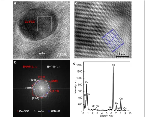
Figure 4 shows HRTEM analysis of a selected Cu pre-cipitate with a relatively large size. The prepre-cipitate in Figure 4a taken along a [111] α-Fe direction have a FCC
structure, as shown by the FFT image of Figure 4a in Figure 4b. Close observation of the diffraction patterns reveals the presence of faint extra spots marked by the blue squares, which is considered to correspond to the faults in the FCC structure. IFFT image in Figure 4c of the region in the white square in Figure 4a shows the fine structure of the precipitate adjacent to the matrix. Dis-tinct faults are observed with irregular crystal lattices, as marked by blue lines. EDS analysis in Figure 4d shows the presence of additional Ni and Mn as well as the carbide-forming elements Mo and Nb in the precipitate, which is possibly responded for the formation of faults even at the later coarsening stage. It seems that active diffusion of these heteroatoms facilities the relaxation of strain
energy by forming faults during the formation of FCC structure. Therefore, the structural evolution of Cu pre-cipitates seems to be more complex in multicomponent steels than that in binary or the ternary Fe–Cu alloys with merely Ni or Mn additions.
4 Conclusions
The Cu precipitates in a high-strength low-alloy steel isothermally aged at 500 °C for different time are char-acterized by using high-resolution transmission electron microscopy. The following conclusions are drawn from this study.
(1) The Cu precipitates preferentially nucleate at crys-tallographic defects such as dislocations and mar-tensite LBs.
(2) Although imaged in the same aging condition, the Cu precipitates have multiple structure of BCC, 9R and FCC and vary in a wide size region. However, the structural evolution does not greatly correlate with the actual sizes.
(3) The presence of different structures can exist in an individual Cu precipitate, reflecting the local occur-rence structural transformation to relax the strain energy.
(4) The complex Cu precipitation compared to that in the binary or the ternary Fe–Cu alloys with merely Ni or Mn additions is attributed to the multiply additions in the multicomponent steel.
Authors’ Contributions
QL and JG as in charge of the whole trial; LH wrote the manuscript; QL and LH assisted with sampling and laboratory analyses. All authors read and approved the final manuscript.
Authors’ Information
Lizhan Han, born in 1970, is currently an assistant researcher at School of Mate-rials Science and Engineering, Shanghai Jiao Tong University, China. He received his doctoral degree from Shanghai Jiao Tong University, China, in 2010. His research interests include heat treatment of heavy steel forges.
Qingdong Liu, born in 1982, is currently an assistant researcher at School of Materials Science and Engineering, Shanghai Jiao Tong University, China. He received his doctoral degree from Shanghai University, China, in 2013.
Jianfeng Gu, born in 1970, is currently a professor at School of Materials Science and Engineering, Shanghai Jiao Tong University, China.
Acknowledgements
The authors sincerely thanks to Dr. Jianchao Peng of Shanghai University for his critical discussion on TEM analysis and reading during manuscript preparation.
Competing Interests
The authors declare that they have no competing interests.
Funding
Supported by Startup Fund for Youngman Research at SJTU (SFYR at SJTU), National Basic Research Program of China (Grant No. 2011CB012904), and China Postdoctoral Science Foundation (Grant No. 2013M541517).
Author Details
1 Institute of Materials Modification and Modelling, School of Materials Science and Engineering, Shanghai Jiao Tong University, Shanghai 200240, China. 2 Collaborative Innovation Center for Advanced Ship and Deep-Sea Explora-tion, Shanghai Jiao Tong University, Shanghai 200240, China.
Received: 1 April 2019 Revised: 24 June 2019 Accepted: 25 September 2019
References
[1] A Saha, J Jung, G B Olson. Prototype evaluation of transformation tough-ened blast resistant naval hull steels: Part II. J. Computer-Aided Mater. Des., 2007, 14: 201-233.
[2] M D Mulholland, D N Seidman. Nanoscale co-precipitation and mechani-cal properties of a high-strength low-carbon steel. Acta Mater., 2011, 59: 1881-1897.
[3] Q D Liu, H M Wen, H Zhang, et al. Effect of multistage heat treatment on microstructure and mechanical properties of a high-strength low-alloy steel. Metall. Mater. Trans. A, 2016, 47A: 1960-1974.
[4] R P Kolli, D N Seidman. The temporal evolution of the decomposition of a concentrated multicomponent Fe–Cu-based steel. Acta Mater., 2008, 56: 2073-2088.
[5] J Takahashi, K Kawakami, Y Kobayashi. Consideration of particle- strength-ening mechanism of copper-precipitation-strengthened steels by atom probe tomography analysis. Mater. Sci. Eng. A, 2012, 535: 144-152. [6] Q D Liu, Y H Chen, C W Li, et al. Compositional variants of Cu-rich
precipi-tate in thermally aged ferritic steel. Acta Metall. Sin. (English Letters), 2018, 31: 465-470.
[7] M Schober, E Eidenberger, H Leitner, et al. A critical consideration of mag-netism and composition of (bcc) Cu precipitates in (bcc) Fe. Appl. Phys. A, 2010, 99: 697-704.
[8] D Isheim, M S Gagliano, M E Fine, et al. Interfacial segregation at Cu-rich precipitates in a high-strength low-carbon steel studied on a sub-nanometer scale. Acta Mater., 2006, 54: 841-849.
[9] R P Kolli, Z Mao, D N Seidman, et al. Identification of a Ni0.5(Al0.5-xMnx) B2 phase at the heterophase interfaces of Cu-rich precipitates in an α-Fe matrix. Appl. Phys. Lett., 2007, 91: 241903.
[10] P J Othen, M L Jenkins, G D W Smith, et al. Transmission electron micro-scope investigations of the structure of copper precipitates in thermally-aged Fe–Cu and Fe–Cu-Ni. Philos. Mag. Lett., 1991, 64: 383-391. [11] P J Othen, M L Jenkins, G D W Smith. High-resolution electron
micros-copy studies of the structure of Cu precipitates in α-Fe. Philos. Mag., 1994, 70A: 1-24.
[12] Y R Wen, A Hirata, Z W Zhang, et al. Microstructure characterization of Cu-rich nanoprecipitates in a Fe-2.5 Cu-1.5 Mn-40 Ni-1.0 Al multicomponent ferritic alloy. Acta Mater., 2013, 61: 2133-2147.
[13] R Monzen, K Takada, C Watanabe. Coarsening of spherical Cu particles in an α-Fe matrix. ISIJ Int., 2004, 44: 442-444.
[14] S Pizzini, K J Roberts, W J Phythian, et al. A fluorescence EXAFS study of the structure of copper-rich precipitates in Fe–Cu and Fe–Cu-Ni alloys. Philos. Mag. Lett., 1990, 61: 223-229.
[15] R Monzen, M Iguchi, M L Jenkins. Structural changes of 9R copper pre-cipitates in an aged Fe–Cu alloy. Philos. Mag. Lett., 2000, 80: 137-148. [16] L Feng. Crystal structure evolution and deformation characteristics of Cu-rich
nano-phase in RPV model steels. Shanghai University, China, 2014. [17] Y U Heo, Y K Kim, J S Kim, et al. Phase transformation of Cu precipitates
from bcc to fcc in Fe-3Si-2Cu alloy. Acta Mater., 2013, 61: 519-528. [18] S Lozano-perez, M L Jenkins, J M Titchmarsh. Evidence of
deformation-induced transformation of Cu-rich precipitates in and aged FeCu alloy. Philos. Mag. Lett., 2006, 86: 367-374.
[19] Q D Liu, S J Zhao. Cu precipitation on dislocations and interfaces in quench-aged steel. MRS Communi., 2012, 2: 127-132.
[20] M Kapoor, D Isheim, G Ghosh, et al. Aging characteristics and mechanical properties of 1600MPa body-centered cubic Cu and B2-NiAl precipita-tion-strengthened ferritic steel. Acta Mater., 2014, 73: 56-74.
[22] X M Bai, H Ke, Y Zhang, et al. Modeling copper precipitation hardening and embrittlement in a dilute Fe-0.3 at% Cu alloy under neutron irradia-tion. J. Nucl. Mater., 2017, 495: 442-454.
[23] Q D Liu, J F Gu. Hierarchical Cu precipitation in lamellated steel after multistage heat treatment. Philosophical Magazine, 2017, 97: 2361-2374 [24] Q D Liu, C W Li, J F Gu. Interactive formation of Cu-rich precipitate,
reverted austenite, and alloyed carbide during partial austenite reversion treatment for high-strength low-alloy steel. Journal of Materials Research, 2017, 32: 2325-2334.
[25] Q D Liu, J F Gu, C W Li. Regulation of Cu precipitation by intercritical tempering in a HSLA steel. J. Mater. Res., 2014, 29: 950-958.
[26] S Kajiwara. Theoretical analysis of the crystallography of the bcc to FCT martensitic transformation. The Japan Society of Applied Physics, 1976, 17(7): 435-446.
[27] L Qin, A Redermeier, C Dellago, et al. Rigid-lattice Monte Carlo study of nucleation kinetics in dilute bcc Fe–Cu alloys using statistical sampling techniques. Acta Mater., 2018, 159: 429-438
[28] J M Zhu, T L Zhang, Y Yang, et al. Phase field study of the copper precipi-tation in Fe–Cu alloy. Acta Mater., 2019, 166: 560-571.
[29] G Stechauner, E Kozeschnik. Thermo-kinetic modeling of Cu precipitation in α-Fe. Acta Mater., 2005, 100: 135-146
[30] S P Shu, P B Wells, N Almirall, et al. Thermodynamics and kinetics of core-shell versus appendage co-precipitation morphologies: An example in the Fe–Cu-Mn-Ni-Si system. Acta Mater., 2018, 157: 298-306.
[31] D Molnar, R Mukherjee, A Choudhury, et al. Multiscale simulations on the coarsening of Cu-rich precipitates in α-Fe using kinetic Monte Carlo, molecular dynamics, and phase-field simulations. Acta Mater., 2016, 60(20): 15-36.
![Figure 2the corresponding Fast Fourier Transform pattern with index illustration taken along a [001]a shows a HRTEM micrograph and Figure 2b direction](https://thumb-us.123doks.com/thumbv2/123dok_us/9583737.1941104/3.595.56.291.82.691/figure-corresponding-fourier-transform-illustration-micrograph-figure-direction.webp)