R E S E A R C H
Open Access
Increased production of hydrogen peroxide by
peripheral blood monocytes associated with
smoking exposure intensity in smokers
Suzana E Tanni
1,3*, Camila R Correa
2, Aparecida Y Angeleli
1, Simone A Vale
1, Liana S Coelho
1and Irma Godoy
1Abstract
Background:Smoking is known to be associated with oxidative stress; however, it has not been elucidated whether the oxidative response is influenced by the intensity of smoking exposure.
Objectives:Evaluate the effect of smoking exposure on the secretion of hydrogen peroxide (H2O2) by the peripheral blood monocytes of smokers.
Methods:A total of 25 smokers (50.3±8.8 years, 48% male) underwent the following evaluations: spirometry, pulse oximetry, body composition and total peripheral blood count. Peripheral blood monocyte (PBM) cultures were isolated and maintained, and IL-6 and TNF-αwere measured in the plasma and in the supernatants of spontaneous and stimulated cultures. H2O2was evaluated in the supernatants of the PBM cultures, and a subset of the PBM culture supernatants was stimulated with phorbol myristate acetate (PMA). We also evaluated 38 healthy controls (49.1±8.2 years, 42% male).
Results:The spontaneous and stimulated monocytes’secretion of H2O2were statistically higher in the smokers than in the healthy controls (p<0.001). The H2O2secretions were statistically significant higher after stimulation with PMA in both groups (p<0.001). In the multiple regression analysis, we identified a positive, statistically significant association between pack-years of smoking and the spontaneous secretion of H2O2by PBM culture, adjusted for potential confounding variables. The association between PBM culture secretion of H2O2and the production of TNF-αand IL-6 was not significant.
Conclusion:We identified a positive association between higher production of H2O2in smokers and higher smoking exposure during life. The influence of pack-years smoking may be a key modifiable factor in oxidative stress associated to smoking.
Keyword:Hydrogen peroxide, Smoking, Oxidative stress, Systemic inflammation, Cultured cells
Introduction
Smoking is a major agent of environmental pollution and is the leading preventable cause of death worldwide [1]. According to data from the World Health Organization, smoking is responsible for 85% of deaths due to chronic ob-structive pulmonary disease (COPD), 30% of cancer deaths and 25% of deaths from coronary disease [1]. The develop-ment of smoking-related diseases is associated with the
deleterious effects of cigarette smoke, including increased oxidative stress and inflammatory processes [2,3].
Oxidative stress is associated with higher concentra-tions of reactive oxygen specimens, including hydrogen peroxide (H2O2), and this phenomenon is a characteris-tic response of inflammatory cells to smoking [4-7]. Oxidative stress can further stimulate the inflammatory process in the airways, increasing the levels of cytokines and the accumulation of polymorphonuclear leukocytes (PMNs) [2,8-10]. Moreover, this stress may induce the oxidation of lipids, lipoproteins, DNA and other proteins and molecules in ways that impair normal cellular func-tion and enable the development of many diseases, such * Correspondence:[email protected]
1
Faculdade de Medicina de Botucatu, Disciplina de Pneumologia, Univ Estadual Paulista, UNESP, Botucatu, São Paulo, Brazil
3
Departamento de Clínica Médica da Faculdade de Medicina de Botucatu, UNESP, Distrito de Rubião Júnior, Botucatu, SP 18618-970, Brazil Full list of author information is available at the end of the article
as COPD and atherosclerosis. COPD patients present higher concentrations of H2O2 in exhaled breath con-densate compared with healthy controls [11], which might be associated with smoking exposure. Oxidative stress activates proteins that are associated with the de-struction of parenchymal tissue [11].
An association between active smoking and elevated oxidative stress in alveolar macrophages is well described in the literature [2,12,13]; however, whether the intensity of smoking exposure has an impact on oxidative stress has been less thoroughly investigated. In the study of van Beurden et al. [14], the production of superoxide by PMNs was not associated with the intensity of smok-ing exposure evaluated by smoksmok-ing history duration. Similarly, Yeh et al. [15] did not find an association with smoking history duration when protein carbonylation was used as a biomarker for oxidative stress. Therefore, the aim of this study was to assess the relationship be-tween systemic oxidative stress, represented by the se-cretion of H2O2 by peripheral monocytes, and pack-years of smoking.
Methods
We evaluated 25 active smokers (52% female) of at least 10 pack-years and 38 healthy controls self-identified as never smokers (57.9% female). All of the participants underwent routine clinical assessments, including spir-ometry and chest X-ray. The height and weight were measured, and the body mass index (BMI) was calcu-lated (kg/m2). The exclusion criteria included pharmaco-logical treatments in the last three months before enrollment in the study, a forced expiratory volume in the first second (FEV1)/Forced vital capacity (FVC) ratio < 0.70 or significant reversibility (increase >200 mL or >11% in the value of FEV1 expressed as percentage of the predicted value) 20 minutes after the inhalation of a β2-agonist (400 mcg fenoterol) in the spirometry test, diagnosis of any chronic disease, and inability to under-stand the study protocol.
The Research Ethics Committee of the Botucatu Medical School approved the study design, and all of the participants gave written informed consent.
Spirometry test and pulse oximetry
The FEV1and FVC were obtained from the flow-volume curve using a spirometer (Ferraris KOKO, Louisville, CO, USA) before and 20 minutes after the inhalation of a β2-agonist (400 mcg fenoterol) according to Brazilian guidelines [16]. The highest value of at least three mea-surements was selected and expressed as a percentage of reference values [16]. The pulse oximetry (SpO2%) was assessed by a portable oximeter (Nonin Medical, Plymouth, MN, USA).
Peripheral blood monocyte isolation and culture (PBM)
PBM cultures were isolated from heparinized blood drawn between 07:00 – 08:00 using a density gradient followed by plastic adherence, as described elsewhere [17]. In brief, the PBM cultures were isolated from heparinized blood using Histopaque 1077 (Sigma Chem-ical Co., St Louis, MO, USA) and washed three times with Hanks’solution (Sigma Chemical Co.). A cell sus-pension was prepared with PBMCs at 2×10⁶ cells·mL-1 in Roswell Park Memorial Institute (RPMI)-1640 medium (Sigma Chemical Co.) supplemented with 5% heat-inactivated autologous serum, 2 mM glutamine, 100 U·mL-1 penicillin and 10 mM HEPES (N-2-hydro-xyethylpiperazine-N-2-ethanesulfonic acid) buffer (that hereafter will be referred to as mRPMI) and incubated for 2 hours in a humidified atmosphere with 5% CO2at 37°C. Nonadherent cells were removed by three washes with plain RPMI and counted. The adherent cells were provided with fresh mRPMI at a volume of 1 mL·1×106 cells-1 and incubated for 24 hours. The supernatants were collected, centrifuged and stored at −80°C until cytokine analysis. After the supernatants were harvested, the adherent cells were stained with nonspecific esterase staining (Sigma Chemical Co.), and the cell viability was determined using a trypan blue exclusion test. The adherent cells contained > 95% monocytes; >98% were viable.
Interleukin (IL)-6 and tumor necrosis factor alpha (TNF-α) measurements
Fasting peripheral blood was collected in the early morning (07.00-08.00 hours), and the plasma was stored at −80°C until analysis. TNF-αand IL-6 in the spontan-eous PBM culture supernatant and plasma were assessed in duplicate by highly sensitive commercial kits using the enzyme-linked immunosorbent (ELISA) technique according to the manufacturer’s instructions (BioSource International Inc, CA, USA). The lower detection limits were 0.5 pg/mL for TNF-αand 0.16 pg/mL for IL-6.
H2O2production
was interrupted by the addition of 0.01 ml of 1 N NaOH. The samples were assayed in quadruplicate. The absorb-ance was determined using an ELISA reader at 620 nm against a blank containing phenol red and 1 N NaOH.
The results were expressed as nanomoles of H2O2/ 2×10⁵cells using the standard curve established in each assay and made up of known molar concentrations of H2O2in phenol red buffer solution. In these experimen-tal conditions, the curve was generated based on the fol-lowing concentrations: 0.5; 1.0; 1.5 and 2.0 nM of H2O2.
Statistical analysis
The results are presented as the means ± SD for nor-mally distributed variables and the median (range) when not normally distributed. The independent t test or Mann–Whitney was used to compare groups according to smoking status. The Chi-square test was used to com-pare proportions between groups. To explore the associ-ation of H2O2 with markers of systemic inflammation (TNF-α, IL-6 and neutrophils) and with pack-years of smoking, we performed multiple regression analysis with robust standard errors with and without adjustment for potential confounding variables. A p value of less than 0.05 denotes statistically significant difference (“STATA” 10.0 - Corp, College Station, TX, USA).
Results
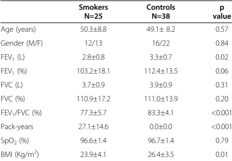
The demographic characteristics and the data related to the spirometry, pulse oximetry and BMI of the smokers and controls are shown in Table 1. The smokers exhib-ited lower values of FEV1 (L), FEV1/FVC (%) and BMI (kg/m2) compared with the healthy controls.
We identified systemic inflammation with higher num-bers of neutrophils (p=0.004) and higher serum levels of TNF-α(p<0.001) in active smokers when compared with
never-smoker healthy controls (Table 2). We did not find differences in the secretion of TNF-α and IL-6 be-tween the spontaneous PBM cultures of different groups.
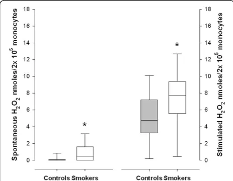
The spontaneous and stimulated PBM culture secre-tions of H2O2 were statistically higher in the smokers than in the healthy controls (p<0.001) (Figure 1). The H2O2 secretion increased significantly after stimulation with PMA in both groups (p<0.001).
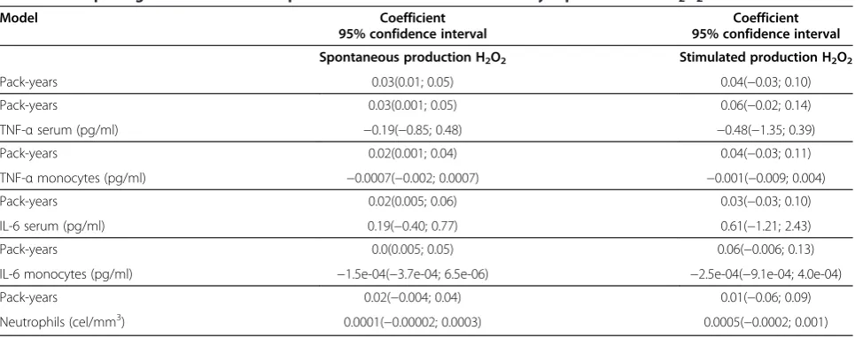
When the smokers and the controls were analyzed in the multiple regression analysis adjusted for age, gender, FEV1(L) and BMI, there was a significant positive asso-ciation between pack-years of smoking and the spontan-eous secretion of H2O2 by PBM culture (Table 3). However, the stimulated PBM culture secretion of H2O2 was not associated with smoking pack-years after adjust-ments for potential confounding variable. Pack-years of smoking also exhibited a positive correlation with the number of leukocytes in the peripheral blood (coeffi-cient=5.22, p<0.001) and the serum levels of TNF-α (coefficient=5.41, p=0.007).
We did not find associations between H2O2secretion with systemic inflammation evaluated by serum values and PBM culture secretion of TNF-α or IL-6 or the number of neutrophils in the peripheral blood (Table 3).
Discussion
This study aimed to evaluate the relationship between systemic oxidative stress and intensity of smoking expos-ure. The main finding was that the smokers exhibited significantly higher secretion of H2O2 than the healthy controls and that there was a significantly positive asso-ciation of oxidative stress with pack-years of smoking. These results confirm that smoking itself is an important determinant of oxidative stress and that a higher con-sumption of cigarettes is associated with higher levels of oxidative stress [12,14,20]. We also reinforce previous findings that smokers exhibit evidence of systemic in-flammation compared with healthy controls [7,21].
The relationship between intensity of smoking expos-ure, through the evaluation of smoking pack-years, and oxidative stress has previously been investigated in the literature. However, our study is the first to show a sig-nificant association between pack-years of smoking and the secretion of H2O2 by PBMs. Thomassen et al. [22] did not find a correlation between the number of pack-years of smoking and the secretion of H2O2 by alveolar macrophages when assessing smokers and non-smokers. Similarly, Puri et al. [23] found no correlation between exhaled ethane air levels and cumulative smoking status. It is difficult to compare our results with those of these studies. The study by Thomassen et al. [22] used differ-ent cultures of cells to evaluate the secretion of H2O2. Those authors assessed alveolar macrophages, whereas Table 1 Demographic characteristics
Smokers Controls p
value
N=25 N=38
Age (years) 50.3±8.8 49.1± 8.2 0.57
Gender (M/F) 12/13 16/22 0.84
FEV1(L) 2.8±0.8 3.3±0.7 0.02
FEV1(%) 103.2±18.1 112.4±13.5 0.06
FVC (L) 3.7±0.9 3.9±0.9 0.31
FVC (%) 110.9±17.2 111.0±13.9 0.20
FEV1/FVC (%) 77.3±5.7 83.3±4.1 <0.001
Pack-years 27.1±14.6 0.0±0.0 <0.001
SpO2(%) 96.6±1.4 96.7±1.4 0.79
BMI (Kg/m2) 23.9±4.1 26.4±3.5 0.01
M/F: male/female, FEV1: forced expiratory volume in the first second, FVC:
forced vital capacity, SpO2: pulse oximetry, BMI: body mass index. The values
we evaluated PBM cultures. Similarly, Puri et al. used a different outcome to quantify oxidative stress. However, the sample size of these studies was smaller than that in our study.
In this study, we demonstrated the higher secretion of H2O2in smokers by PBM cultures, either spontaneously or after stimulation. Nowak et al. [2] demonstrated increased content of H2O2in the expired breath conden-sate of cigarette smokers compared with controls. Our results are also in agreement with those of Ishida et al. [24], who identified higher H2O2production in alveolar macrophage cultures of rats exposed to cigarette smoke. In contrast to our results, van Beurden et al. [14] did not detect differences in the release of superoxide dis-mutase by PBM cultures, either spontaneously or after stimulation with PMA, between COPD current/ex-smoker patients and healthy controls, and the smoking history did not influence the results when compared by covariance analysis between groups. The authors
concluded that superoxide dismutase is only one of the oxidants that are produced by PBM culture; these cells also produce H2O2and hydroxyl radicals. Therefore, the fact that the secretion of H2O2by PBM culture is higher in smokers may indicate an imbalance in which the pro-duction of reactive oxygen species exceeds the capacity of the antioxidant defense systems in the systemic microenvironment. The oxidative stress may already be occurring to produce oxygen free radicals [25].
Our results showed higher secretion in H2O2 after stimulation with PMA in both groups, and the smokers exhibited higher production than the controls. This re-sult indicates that the smokers included in our study still have monocytes with efficient immune capacity [26]. We can speculate that monocyte phagocytosis in smokers is still functional, although we did not evaluate the im-mune capacity in our study. In contrast, Correa et al. showed lower fungicidal activity and secretion of H2O2 by the monocytes of patients with peripheral athero-sclerosis obliterans compared with those of control subjects.
Cigarette smoke induces the enhanced recruitment of mononuclear phagocytes and polymorphonuclear cells into the lower airways [2,12], and these cells modify oxy-gen metabolism and release additional H2O2 and other reactive oxygen species [25]. In our results, we identified a statistically higher number of neutrophil cells in the peripheral blood of smokers in comparison with that of healthy controls and a positive association between the numbers of leukocytes cells and pack-years of smoking. These observations are in accordance with a previous study that demonstrated an accelerated release of poly-morphonuclear leukocytes from the bone marrow after smoking exposure [6].
The systemic inflammation induced by smoking includes neutrophilia and cytokines [6,27,28]. In our study, we found higher levels of TNF-αin smokers com-pared with healthy controls. Similarly, Tanni et al. [28] and Pretescu et al. [9] showed higher TNF-α levels in smokers compared with healthy non-smokers. We did Table 2 Peripheral blood cells, serum values and spontaneous monocyte secretion of TNF-αand IL-6
Smokers Controls p
N=25 N=38
Leukocytes (cel/mm3) 7249.2±2071.7 6110.5±1111.8 0.09
Neutrophils (cel/mm3) 4406.7±1802.7 3392.2±796.6 0.004
Lymphocytes (cel/mm3) 1879.6±480.5 1885.8±512.9 0.96
TNF-αserum (pg/ml) 5.2 (4.3-5.8) 3.6 (3.4-3.9) <0.001
TNF-αmonocytes(pg/ml)* 34.6 (16.1-55.7) 57.4 (22.2-100.9) 0.09
IL-6 serum (pg/ml) 0.40 (0.20-0.80) 0.30 (0.2-0.4) 0.45
IL-6 monocytes (pg/ml) * 1392.6 (272.6-2277.7) 1006.3 (281.9-3048.9) 0.93
TNF-α: tumor necrosis factor alpha, IL-6: interleukin 6, TNF-αand IL-6 monocytes*: measurements of the supernatant of spontaneous peripheral monocyte cultures. The values are presented as the means and standard deviations or as medians and quartiles. The statistical analysis was performed by t test or Mann–Whitney.
not find differences in the cytokine secretion by the PBM culture in both groups, and the literature reveals controversial results that are dependent on the cell cul-ture methods [7,21]. In agreement with our results, Ryder et al. [21] utilized cell cultures without stimulus and did not find increased values of TNF-α production by PBM cultures in smokers compared with controls. In contrast, Zeidel et al. [7] identified increased production of the pro-inflammatory cytokines (IL-1β, IL-6 and TNF-α) in smokers compared with non-smoking sub-jects, however, these authors analyzed cell cultures sti-mulated with lipopolysaccharide.
The limitations of our study need to be addressed. First, this study is cross sectional, and we are not explaining the cause-and-effect relationships among events. Second, we did not evaluate whether different smoking pack-years or cigarettes-day can induce greater secretion of H2O2to confirm our results. Third, we did not assess the other biomarkers of oxidative stress and antioxidants to better understand the biological response to smoking pack-years.
In conclusion, we identified an association between higher secretions of H2O2 and smokers compared with healthy controls. The influence of pack-years smoking may be a key modifiable factor in oxidative stress asso-ciated to smoking.
Abbreviations
BMI: Body mass index; COPD: Chronic obstructive pulmonary disease; FEV1: Forced expiratory volume in the first second; FVC: Forced vital capacity; H2O2: Hydrogen peroxide; IL: Interleukin; PBM: Peripheral blood monocyte; PMA: Phorbol myristate acetate; PMN: Polymorphonuclear leukocytes; RPMI: Roswell park memorial institute; TNF-α: Tumor necrosis factor alpha.
Competing interests
None of the authors has any potential conflicts of interest.
Authors' contributions
The authors’responsibilities were as follow. SET: performed selection and the medical assessment of the individuals, statistical analysis and interpret the data and draft the final manuscript; CRC: laboratory analysis; AYOA: conducted the laboratory analysis; SAV: performed the medical assessment; LSC: performed the medical assessment, IG: had overall responsibility for the study, designed the research, analyzed and interpret the data, and wrote the final manuscript. All the authors contributed to the revision of the manuscript.
Funding
Research Grant from FAPESP (Fundação de Amparo à Pesquisa do Estado de São Paulo, São Paulo, Brazil) N º 03/05285-1.
Author details
1
Faculdade de Medicina de Botucatu, Disciplina de Pneumologia, Univ Estadual Paulista, UNESP, Botucatu, São Paulo, Brazil.2Faculdade de Medicina de Botucatu, Univ Estadual Paulista, UNESP, Departamento de Patologia, Botucatu, São Paulo, Brazil.3Departamento de Clínica Médica da Faculdade de Medicina de Botucatu, UNESP, Distrito de Rubião Júnior, Botucatu, SP 18618-970, Brazil.
Received: 18 May 2012 Accepted: 13 November 2012 Published: 21 November 2012
References
1. WHO. World Health Organization:Chronic respiratory diseases. www.hoint/en. 2. Nowak D, Antczak A, Krol M, Pietras T, Shariati B, Bialasiewicz P,et al:
Increased content of hydrogen peroxide in the expired breath of cigarette smokers.Eur Respir J1996,9:652–657.
3. Corrêa CR, Dias-Melicio LA, Calvi SA, Lastória S, Soares AM:Activation of monocytes and cytokine production in patients with peripheral atherosclerosis obliterans.J Inflamm2011,29:23.
4. Lerner L, Weiner D, Katz R, Reznick AZ, Pollack S:Increased pro-inflammatory activity and impairment of human monocyte differentiation induced by in vitro exposure to cigarette smoke.
J Physiol Pharmacol2009,60(Suppl 5):81–86.
5. Wirtz PH, von Känel R, Kunz-Ebrecht S, Ehlert U, Fischer JE:Enhanced glucocorticoid sensitivity of cytokine release from circulating leukocytes stimulated with lipopolysaccharide in healthy male smokers.
Brain Behav Immun2004,18:536–543.
6. van Eeden SF, Hogg JC:The response of human bone marrow to chronic cigarette smoking.Eur Respir J2000,15:915–921.
Table 3 Multiple regression models for spontaneous and stimulated monocyte production of H2O2
Model Coefficient Coefficient
95% confidence interval 95% confidence interval
Spontaneous production H2O2 Stimulated production H2O2
Pack-years 0.03(0.01; 0.05) 0.04(−0.03; 0.10)
Pack-years 0.03(0.001; 0.05) 0.06(−0.02; 0.14)
TNF-αserum (pg/ml) −0.19(−0.85; 0.48) −0.48(−1.35; 0.39)
Pack-years 0.02(0.001; 0.04) 0.04(−0.03; 0.11)
TNF-αmonocytes (pg/ml) −0.0007(−0.002; 0.0007) −0.001(−0.009; 0.004)
Pack-years 0.02(0.005; 0.06) 0.03(−0.03; 0.10)
IL-6 serum (pg/ml) 0.19(−0.40; 0.77) 0.61(−1.21; 2.43)
Pack-years 0.0(0.005; 0.05) 0.06(−0.006; 0.13)
IL-6 monocytes (pg/ml) −1.5e-04(−3.7e-04; 6.5e-06) −2.5e-04(−9.1e-04; 4.0e-04)
Pack-years 0.02(−0.004; 0.04) 0.01(−0.06; 0.09)
Neutrophils (cel/mm3) 0.0001(−0.00002; 0.0003) 0.0005(−0.0002; 0.001)
H2O2: peroxide hydrogen; TNF-α: tumor necrosis factor alpha; IL-6: interleukin 6. Monocyte production of H2O2was stimulated by phorbol myristate acetate.
7. Zeidel A, Beilin B, Yardeni I, Mayburd E, Smirnov G, Bessler H:Immune response in asymptomatic smokers.Acta Anaesthesiol Scand2002,
46:959–964.
8. Barbieri SS, Amadio P, Gianellini S, Zacchi E, Weksler BB, Tremoli E:Tobacco smoke regulates the expression and activity of microsomal
prostaglandin E synthase-1: role of prostacyclin and NADPH-oxidase.
FASEB J2011,25:3731–3740.
9. Petrescu F, Voican SC, Silosi I:Tumor necrosis factor-alpha serum levels in healthy smokers and nonsmokers.Int J Chron Obstruct Pulmon Dis2010,
5:217–222.
10. Lima DF, Coleta KD, Tanni SE, Godoy I, Silveira LVA, Godoy I:Potentially modifiable predictors of mortality in patients treated with long-term oxigen therapy.Respir Med2010,105:470–476.
11. Dekhuijzen PN, Aben KK, Dekker I, Aarts LP, Wielders PL, van Herwaarden CL,et al:Increased exhalation of hydrogen peroxide in patients with stable and unstable chronic obstructive pulmonary disease.
Am J Respir Crit Care Med1996,154:813–816.
12. Guatura SB, Martinez JA, Santos Bueno PC, Santos ML:Increased exhalation of hydrogen peroxide in healthy subjects following cigarette
consumption.Sao Paulo Med J2000,118:93–98.
13. Bloomer RJ, Solis AD, Fisher-Wellman KH, Smith WA:Postprandial oxidative stress is exacerbated in cigarette smokers.Br J Nutr2008,99:1055–1060. 14. van Beurden WJ, Wielders PL, Scheepers PJ, van Herwaarden CL, Dekhuijzen
PN:Superoxide production by peripheral polymorphonuclear leukocytes in patients with COPD.Respir Med2003,97:401–406.
15. Yeh CC, Graham Barr R, Powell CA, Mesia-Vela S, Wang Y, Hamade NK,et al:
No effect of cigarette smoking dose on oxidized plasma proteins.
Environ Res2008,6:219–225.
16. Pereira CAC, Neder JA:Diretrizes para testes de função pulmonar.
J Bras Pneumol2002,28:S2–S238.
17. deGodoy I, Donahoe M, Calhoun WJ, Mancino J, Rogers RM:Elevated TNF-alpha production by peripheral blood monocytes of weight-losing COPD patients.Am J Respir Crit Care Med1996,153:633–637.
18. Pick E, Keisari Y:A simple colorimetric method for the measurement of hydrogen peroxide produced by cells in culture.J Immunol Methods1980,
8:161–170.
19. Pick E, Mizel D:Rapid microassays for the measurement of superoxide and hydrogen peroxide production by macrophages in culture using an automatic enzyme immunoassay reader.J Immunol Methods1981,
46:211–226.
20. Ludwig PW, Hoidal JR:Alterations in leukocyte oxidative metabolism in cigarette smokers.Am Rev Respir Dis1982,126:977–980.
21. Ryder MI, Saghizadeh M, Ding Y, Nguyen N, Soskolne A:Effects of tobacco smoke on the secretion of interleukin-1 beta, tumor necrosis factor-alpha, and transforming growth factor-beta from peripheral blood mononuclear cells.Oral Microbiol Immunol2002,17:331–336. 22. Thomassen MJ, Barna BP, Wiedemann HP, Farmer M, Ahmad M:Human
alveolar macrophage function: differences between smokers and nonsmokers.J Leukoc Biol1988,44:313–318.
23. Puri BK, Treasaden IH, Cocchi M, Tsaluchidu S, Tonello L, Ross BM:A comparison of oxidative stress in smokers and non-smokers: an in vivo human quantitative study of n-3 lipid peroxidation.BMC Psychiatry2008,
8(Suppl 1):S4.
24. Ishida T, Pinkerton KE, Takeuchi M:Alveolar macrophage from cigarette smoke-exposed mice inhibits B lymphocyte proliferation stimulated with LPS.Respiration2008,77:91–95.
25. Opara EC:Oxidative stress.Dis Mon2006,52:83–98.
26. DeChatelet LR, Shirley PS, Johnston RBJ:Effect of phorbol myristate acetate on the oxidative metabolism of human polymorphonuclear leukocytes.Blood1976,47:545–554.
27. Tappia PS, Troughton KL, Langleyevans SC, Grimble RF:Cigarette-smoking influences cytokine production and antioxidant defenses.
Clin Sci1995,88:485–489.
28. Tanni SE, Pelegrino NR, Angeleli AY, Correa C, Godoy I:Smoking status and tumor necrosis factor-alpha mediated systemic inflammation in COPD patients.J Inflamm2010,7:29.
doi:10.1186/1476-9255-9-45
Cite this article as:Tanniet al.:Increased production of hydrogen peroxide by peripheral blood monocytes associated with smoking exposure intensity in smokers.Journal of Inflammation20129:45.
Submit your next manuscript to BioMed Central and take full advantage of:
• Convenient online submission
• Thorough peer review
• No space constraints or color figure charges
• Immediate publication on acceptance
• Inclusion in PubMed, CAS, Scopus and Google Scholar
• Research which is freely available for redistribution