Spring 2019
CAR T Cell Therapy
CAR T Cell Therapy
Megan Bonnett
Iowa State University, [email protected]
Follow this and additional works at: https://lib.dr.iastate.edu/creativecomponents
Part of the Analytical, Diagnostic and Therapeutic Techniques and Equipment Commons
Recommended Citation Recommended Citation
Bonnett, Megan, "CAR T Cell Therapy" (2019). Creative Components. 141.
https://lib.dr.iastate.edu/creativecomponents/141
CAR T Cell Therapy
by
Megan Bonnett
A paper submitted to the graduate faculty in partial fulfillment of the requirements for the degree
of Master of Science in Biomedical Sciences
Program of Study Committee: Jon Mochel
Abstract
With chemotherapy and radiation being the most common forms of cancer treatment,
some cancer cells are able to develop resistance and persist in the body. Because of this
resistance, there is a need for a new cancer therapy that can attack these persistent cells. CAR T
cell therapy utilizes the patient’s own immune system to attack and potentially eliminate these
cancer cells. These cells have successfully been used against B cell malignancies, however, not
much success has been seen when used against solid tumors. Solid tumors pose many challenges
and threats these T cells must overcome in order to produce any anti-tumor effect. Challenges
researchers run into include finding a suitable antigen to target, successfully trafficking to and
infiltrating the tumor, and overcoming the microenvironment of the tumor. Researchers are
continuously trying to figure out ways to overcome these challenges and increase the success of
Introduction
In the United States, cancer is the second most common cause of death, estimating
610,000 deaths in 2018 (Cancer Statistics, 2018). There is a necessity for a new and improved
cancer treatment option. The primary forms of treatment for most types of nonsurgical cancers
are chemotherapy and radiation. One of the disadvantages of these therapies is that cancers have
been known to develop resistance over time. This development of resistance has created a strong
need for a new, effective form of treatment that decreases relapse rate. However, there has been
little advancement in the development of a form of treatment that is capable of completely
eliminatinglingering malignant
cells. A promising form of treatment
utilizes the patient’s own immune
system, notably lymphocytes, to
control and potentially eliminating
malignant cells and the cancer all
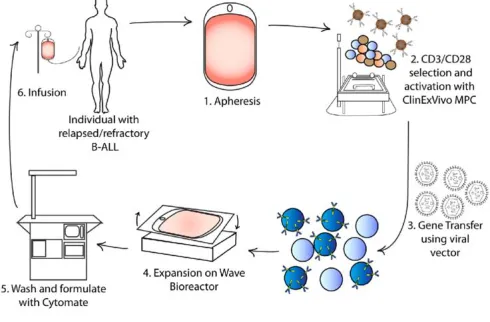
together (Figure 1) (Almåsbak,
Aarvak, & Vemuri, 2016). Years
ago, Gross and colleagues
developed and demonstrated the idea of redirecting cytotoxic T lymphocytes to the tumor cells.
They concluded their work with the statement, “Chimeric T cell receptors with antitumor
specificity will enable testing feasibility of this approach in combating human tumors” (Gross,
Waks, & Eshhar, 2006). Gross and colleagues laid the groundwork for a series of generations
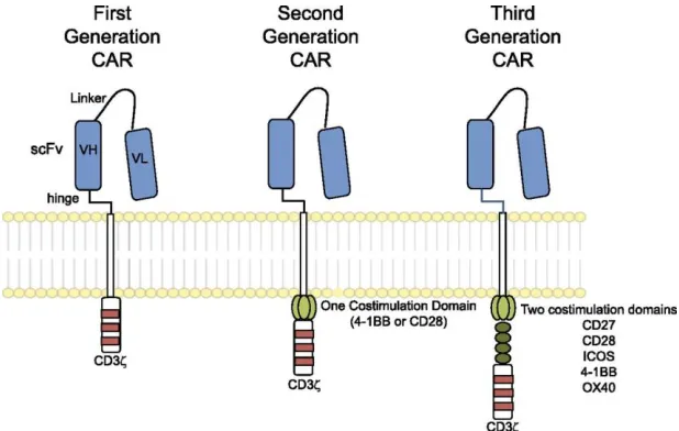
utilizing the immune system and cytotoxic T lymphocytes as a way to target tumor cells. First
[image:4.612.276.521.318.479.2]generation chimeric antigen receptors (CARs) were created by directly fusing a tumor targeting
antibody single chain variable fragment (scFv) to the signaling domain of the T cell receptor
(TCR) signaling complex
member CD3ζ (zeta). The first
generation showed high
target-cell specific killing in vitro and
showed preclinical efficacies in
mouse tumor models, however,
when these T cells were used
clinically in ovarian cancer, the
results were not nearly as
promising (Kershaw et al., 2006).
There was no reduction of tumor
burden in 14 patients due to a lack of specificity trafficking of the T cells to the tumor and short
persistence of the T cells. Further research determined that first generation T cells were
susceptible to activation induced cell death (AICD) in the absence of exogenous costimulation
(Park & Brentjens, 2015). After years of continuous research, second generation CAR expressing
T cells were developed.
According to Essand and Loskog, second generation CARs were constructed to provide
signaling through CD3ζ chain and CD28 costimulatory molecule by placing the signaling
domains in series as a single gene multidomain product (Essand & Loskog, 2012) (Finney,
Lawson, Bebbington, & Weir, 1998). This design was able to mediate 20 times more IL-2
production upon stimulation with solid-phase antigen when compared with first generation
[image:5.612.245.554.107.303.2]CARs. It was discovered that second generation CARs with the costimulatory signals, from B7
and tumor necrosis factor receptor (TNFR), in series with CD3ζ, promote self-sufficient clonal
expansion and enhanced effector function in resting human T cells. In order to create the most
effective second generation CAR T cells, studies were done to compare various signaling
domains. Various studies reported increased persistence, tumor localization, and antitumor
activity of CAR T cells with a 4-IBB signaling domain when compared with CARs with CD28
signaling domain (Essand & Loskog, 2012).
As a way to try and improve second generation CARs, third generation CARs were
constructed containing a CD3ζ chain, CD27, CD28, ICOS, and OX-40 or 4-IBB signaling
domain (Figure 2). Ideally, these receptors would provide a full complement of activation,
proliferation, and survival signals for enhanced antitumor activity; however, using third
generation CARs has been somewhat disadvantageous. Despite the promising preclinical results,
there were concerns of triggering lethal cytokine storms within patients treated. Another concern
of the third generation CARs, is the reduction of the signal threshold to a level at which the
activation of grafted T cells can occur without triggering antigens (Essand & Loskog, 2012).
With these advances in CAR T cell technology and the many studies that have been
performed using all three generations of CAR, these cells have been successful or on the road to
success when treating blood borne and solid tumors. For B-cell malignancies, CAR T cells are
expected to be a mainstream therapy, especially for refractory or relapsed B-cell malignancies
(Almåsbak et al., 2016). By changing the antigen T cells target, CARs can be used for many
different types of cancers, both hematologic and solid. Though treatment of hematologic cancers
with CAR T cells has been more successful, treating solid cancers has posed difficult challenges
Review
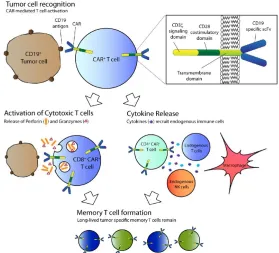
CAR T cells have changed the
way various cancers are treated. The
targeting of tumor cells begins when a
CAR on a T cell binds to its antigen on
the tumor, leading to signaling and
activation. Activation can lead to the
killing of tumor cells through the release
of granzyme and perforin or by the
activation of other immune system
components by CD4+ T cell cytokine
release. The use of T cells can lead to the formation of memory T cells, developing long-lived T
cells that are specific to that specific tumor type (Figure 3) (Davila, Bouhassira, et al., 2014).
Treatment of B cell malignancies with CAR T cells
Using the mechanism described above, CAR T cells have shown much promise and are
expected to be the mainstream treatment. B cell malignancies include B cell acute lymphoblastic
leukemia, B cell non-Hodgkin’s lymphoma, chronic lymphocytic leukemia, and Hodgkin’s
lymphoma. Many of the studies conducted (65%) that involved T cell therapy were directed
towards hematological malignancies. Of those studies, CD19 was the most common antigen
targeted in B cell cancers, used over 80% of the time. There are studies to investigate other
[image:7.612.282.561.81.334.2]potential target antigens, such as CD20, CD22, CD30, ROR1, κ light chain, CD123, CD33,
CD133, CD138, and B-cell maturation antigen (Berger et al., 2014) (Dotti, Gottschalk, Savoldo,
& Brenner, 2014) (Carpenter et al., 2013). The most successful outcomes have come by targeting
CD19, CD20, or CD30, however, the most advancement has been seen by using CD19-specific
CAR T cells for B cell acute lymphoblastic leukemia with complete remission rates of 70-94%
(Z. Wang, Wu, Liu, & Han, 2017). One of the first successful studies of the use of CD19 was
conducted at the National Cancer Institute in 2010. Follicular lymphoma patients were treated
with this form of CAR T cell therapy, developing partial remission after receiving chemotherapy
and an infusion of retrovirally transduced T cells that expressed a second generation CD19 CAR
with a CD28 costimulation domain (Kochenderfer et al., 2010). Shortly after this study showed
success, a group at the University of Pennsylvania showed antileukemia efficacy in T cells
transduced with a lentiviral vector carrying a CD19 CAR with a 4-IBB costimulation domain
(Kalos, Levine, et al., 2011) (Kalos, June, Levine, Bagg, & Porter, 2011). Of the three end-stage
advanced chronic lymphocytic leukemia patients treated, two developed complete remission and
the other developed a partial response. This study exhibited the expansion of CAR T cells in vivo
correlated with clinical responses (Almåsbak et al., 2016).
Treatment of solid tumors with CAR T cells
Much success has been shown with blood-borne tumors, but little has been seen with
solid tumors. There has been a steady rise in the number of clinical trails that focus on solid
tumors. These clinical trials target carcinoembryonic antigen, diganglioside GD2, mesothelin,
interleukin 13 receptor α (IL13Rα), human epidermal growth factor receptor 2 (HER2),
fibroblast activation protein, and L1 cell adhesion molecule (Gill, Maus, & Porter, 2016) (Fousek
trials have used GD2 CARs to target neuroblastoma, 3 of 11 patients develop complete
remission, and HER2 CARs for sarcoma, 4 of 17 patients showing stable disease (Louis et al.,
2011) (Ahmed et al., 2015). The reasons for such little success are yet to be discovered and pose
to have many factors. When compared to blood-borne malignancies, solid tumor landscapes
contain many barriers (Newick, Moon, & Albelda, 2016). In order for CAR T cells to impact
solid tumors, they must successfully travel to the site of the tumor regardless of potential T-cell
chemokine receptor-tumor-deprived chemokine mismatches. Also, CAR T cells must infiltrate
the stromal elements of the tumors in order to elicit tumor-associated antigens-specific
cytotoxicity, despite antigen loss of heterogeneity. Even if the CAR T cells make it to the tumor
and are able to infiltrate it, the T cells must overcome the environment, the presence of
suppressive soluble factors and cytokines, suppressive immune cells, and T-cell intrinsic
negative regulatory mechanisms and overexpression of inhibitory molecules. Within the
environment of the tumor, there is oxidative stress, the T cells would have to overcome
nutritional depletion, and the T cells would experience a low pH and hypoxia (Newick et al.,
2016) (Knochelmann et al., 2018).
Difficulties faced when treating solid tumors with CAR T cells
One of the obstacles that must be overcome when using CAR T cell therapy against solid
tumors is determining which tumor-associated antigen is optimal and would be the most
effective. In blood-borne malignancies, CD19 is consistently expressed, leading to effective
treatment with CD19 CAR T cells. However, with solid tumors, identifying specific antigens to
target has posed a challenge. Currently, 30 different tumor-associated antigens have been
CAR T cell therapy because they are only expressed on tumor cells (Restifo, Dudley, &
Rosenberg, 2012). With further research, neoantigens were discovered to be a product of
tumor-specific mutations and are highly individualized, not making them an ideal target of CAR T cell
therapy. Despite tumor-specific mutations of neoantigens, many generalized neoepitopes have
been identified (Newick et al., 2016).
Instead of using only tumor-specific antigens, antigens expressed only during
developmental growth have been potential targets of CAR T cell therapy. An example of an
antigen expressed during development and is restricted in normal adult tissues is CEA. This has
been a target for mouse studies, showing evidence of tumor eradication by CEA-CAR T cells
(Chmielewski et al., 2012). Targeting this antigen has caused serious side effects, specifically
transient colitis, in three metastatic colon cancer patients (Parkhurst et al., 2011). Other types of
antigens have been researched and are currently being tested in mouse studies and clinical trials
(Newick et al., 2016).
As soon as an antigen has been identified, generated, and infused into a cancer patient, a
major obstacle that is immediately encountered is the ability of the CAR T cells to get to the
tumor and successfully infiltrate it. This process is dependent on the expression of adhesion
receptors on T cells and the tumor endothelium, and a “pairing” between the chemokine
receptors on the CAR and the chemokines tumors secrete. However, there is often a mismatch of
chemokine/chemokine receptor, with tumors producing very small amounts of ligands that
enhance CD8+ recruitment (Harlin et al., 2009). Often times, tumors that express less
chemokines evade host surveillance by impairing the recruitment of effector T cells and their
ability to infiltrate the solid tumor. Some chemotherapy drugs are able to enhance the
(Hong et al., 2011). Studies have shown that two groups enhance the infiltration of T cells and
augment antitumor activity of the CAR T cells, these groups are GD2 in neuroblastoma and
CCR2 on CAR T cells (Craddock et al., 2010). One of the main struggles scientists face is the
individuality of each form of cancer (Knochelmann et al., 2018). In order to create effective
CAR T cells and traffic them to the correct areas, where the solid tumors are located, the unique
chemokine profiles have to be determined and understood.
Once the CAR T cells have made it to the solid tumors, another obstacle is run into, the
microenvironment of the tumor. Normally, the microenvironment of the tumor is hostile, both
physically and metabolically. A couple of physical barriers the T cells must overcome are stroma
and high tissue pressure that prevents extravasation. To overcome these barriers, some studies
used FAP-CAR T cells to reduce the number of tumor fibroblasts. Other studies used specific
CAR T cells that secrete an enzyme that degrades the matrix. These two methods, FAP-CAR T
cells and enzyme secreted CAR T cells, have only been tested in animal models, they have yet to
be tested on humans (L.-C. S. Wang et al., 2014) (Caruana et al., 2015).
Despite the hostility of the physical environment of the tumor, the metabolic landscape
poses a threat to the viability of CAR T cells. Tumor microenvironments are, often times, rich in
suppressor cytokines, such as TGF-β and IL-4, and inhibitory molecules, such as PD-L1. PD-L1
helps the tumor escape the host immune system. In order to overcome this, researchers have
added domains to the CAR T cells. These domains either limit suppressive signaling or convert
the suppressive signals into activating signals (Foster et al., 2008). Recently, studies have used a
chimeric cytokine receptor to bind IL-4. When IL-4 binds the receptor, the therapeutic IL-7
signal pathway phosphorylates STAT5 and polarizes the cell toward an inflammatory Th1
switch receptor. This switch receptor was designed to convert an exhaustive stimulus into a
costimulatory signal, enhancing cytokine production and efficacy (Liu et al., 2016)
(Knochelmann et al., 2018).
Though the suppressive cytokines are a major hurdle that must be overcome in order for
the CAR T cells to survive the tumor microenvironment, other microenvironment issues include
hypoxia and nutrient starvation. Because of hypoxia and nutrient starvation, the generation of
lactate is increased, leading to acidosis within the tumor (Fischer et al., 2007). The deficiency in
nutrients, specifically tryptophan, arginine, and lysine, can activate the integrated stress response
within the T cells. The activation of the integrated stress response can cause shutdown of
translation and stimulate an autophagy response in effector T cells as a way to generate a source
of nutrients (Howie, Waldmann, & Cobbold, 2014) (Newick et al., 2016).
Adverse effects of CAR T cells
CAR T cells have provided a way to attack refractory or relapsed malignancies, but there
are some adverse effects of using CARs. These adverse effects can be immediate or delayed,
mild or severe, or persist for a short or long period of time. One of most prevalent adverse effects
is the onset of immune activation, known as cytokine release syndrome (Lee et al., 2014). This
results in the increase in inflammatory cytokines, specifically IL-10 and IL-6. Clinical features
include high fever, malaise, fatigue, myalgia, nausea, anorexia, tachycardia, capillary leak, and
many more. The severity of CRS is determined by the burden of the disease at the time of
infusion; patients with high tumor burden experience more severe CRS. Researchers have been
studying the use of IL-6R blockade as a treatment for CRS while looking at its effect on
Another adverse effect reported with the use of CAR T cell therapy is on-target/off-tumor
recognition. This often occurs because the epitope targeted by the CAR T cells is often shared
with normal cells in other tissues (Curran, Pegram, & Brentjens, 2012). The severity of this
on-target/off-tumor ranges from manageable lineage depletion to severe toxicity possibly leading to
death. This is often seen in various organ systems: gastrointestinal, pulmonary, and hematologic.
On-target/off-tumor recognition was seen in one trial utilizing a carboxyanhydrase-IX-specific
CAR T cell for renal cell carcinoma. This utilization of these specific CARs led to the
development of cholestasis because of carboxyanhydrase-IX expression on bile duct epithelium
(Lamers et al., 2006). An example of manageable lineage depletion was seen when
CD19-specific CAR T cells were used. The CD19-CD19-specific CARs targeted normal B cells resulting in
B-cell aplasia, which required an infusion of pooled immunoglobulin as prophylaxis from
infectious complications (Kochenderfer et al., 2010). On the other hand, on-target/off-tumor
recognition can be lethal. An example of this was seen in a patient treated with CAR T cells
specific for the cancer-associated antigen HER-2/neu. This patient quickly developed respiratory
failure and multi-organ dysfunction, ultimately leading to death. The death of the patient was
attributed to reactivity against pulmonary tissue expression of HER-2/neu and the high dose of
infused CAR T cells (Morgan et al., 2010).
Recent technological advances in T cell engineering with retroviral and plasmid vectors
allow the generation of high numbers of tumor targeting T cells by introducing tumor specific T
cell receptors (TCR) or CARs. CARs exhibit high-affinity major histocompatibility complex
(MHC) independent recognition of any surface antigen (Almåsbak et al., 2016). The use of
retroviral vectors has produced adverse effects within patients, from the retroviral vector
leading to induced malignant transformation has been a concern of researchers. However, there
have been no reported cases of transformation after the infusion of genetically modified T cells.
This can be due to the fact that the LMO-2 oncogene is silent in T cells, therefore, making it an
undesirable location of retroviral integration. Overall, the risk of insertional oncogenesis
following gene transfer into T cells is low, but there is still a risk it may occur (Scholler et al.,
2012).
Managing toxicity of CAR T cell therapy
Of the adverse effects cited above, there are a few more worth mentioning. Following
CAR T cell therapy, there is a risk of neurological toxicity, anaphylaxis, and off-target antigen
recognition (Bonifant, Jackson, Brentjens, & Curran, 2016). Though there are many risks to be
concerned about, there are many measures being taken to manage toxicity and these adverse
effects, especially in hematologic malignancies. The most common toxicity developed by
patients after the administration of CD19-specific T-cells is the uncontrolled immune activation
in the form of cytokine release syndrome. Tocilizumab had demonstrated IL-6R blockade,
resulting in the reversal of cytokine release syndrome symptoms. Corticosteroids have also been
used and have demonstrated immunosuppressive qualities. However, the prolonged use of
corticosteroids has resulted in the diminished persistence and efficacy of CAR T cells (Davila,
Riviere, et al., 2014).
As this technology has progressed and developed through the years, the integration of
suicide genes has allowed for the selective depletion of CAR T cells, helping with the wide range
of toxicities that can develop (Marin et al., 2012). Another way to selectively deplete CAR T
triggering of cell death via the infusion of the associated monoclonal antibody. The expression of
these cell-surface antigens allows for the selection and tracking of the genetically modified T
cells (Philip et al., 2014).
CAR T cell therapy in dogs
The development and use of CAR T cells in humans has sparked interest in using this
therapy in canines. Canine cancers closely parallel those in humans through the biology,
behavior, and genetic aspects. In dogs, Non-Hodgkin’s Lymphoma is the most common cancer
and has posed many difficulties when treating. Combinations of chemotherapeutic agents have
shown success by leading to remission in about 75% of dogs, however, most dogs relapse within
six to nine months. Dr. Nicola Mason, a professor at eh University of Pennsylvania’s School of
Veterinary Medicine, and Dr. Avery Posey, an instructor at the University of Pennsylvania’s
School of Medicine, have developed CAR T cell therapy for canine use. In 2018, a first-in-dog
clinical trial took place at the University of Pennsylvania School of Veterinary medicine. Patients
had relapsed, refractory B cell lymphoma or leukemia. Much success was seen in the preliminary
results, but further research must be done to improve the persistence and function of these CAR
T cells. With the use of these canine models, veterinary oncologists and researchers hope to
establish better therapeutic platforms that will improve the effectiveness in both blood-borne
cancers and solid tumors and reduce side effects in both humans and canine patients (Lee &
Conclusion
CAR T cell therapy is a cancer treatment option that will someday become mainstream
for many types of cancers. In order for this to be achieved, the safety and efficacy of this
treatment has to be improved, and with further research, it will be. Thus far, CAR T cells have
demonstrated efficacy and success in various hematological cancers. Clinical data seems
promising in solid tumors, including neuroblastoma and tumors overexpressing mesothelin,
HER2 and EGFR. However, utilizing CAR T cells for solid tumors has posed quite a challenge
and has not shown much success. As research continues, more types of cancers will be targeted
and the safety and efficacy will be increased. In order for this treatment to be effective, it must
work at the cellular level and it has to be affordable. As this treatment becomes more mainstream
and generates success, the industrialization of the development of these cells will become more
Bibliography
Ahmed, N., Brawley, V. S., Hegde, M., Robertson, C., Ghazi, A., Gerken, C., … Wels, W. S.
(2015). Human Epidermal Growth Factor Receptor 2 (HER2) –Specific Chimeric Antigen
Receptor–Modified T Cells for the Immunotherapy of HER2-Positive Sarcoma. Journal of
Clinical Oncology, 33(15), 1688–1696. https://doi.org/10.1200/jco.2014.58.0225
Almåsbak, H., Aarvak, T., & Vemuri, M. C. (2016). CAR T Cell Therapy: A Game Changer in
Cancer Treatment. Journal of Immunology Research, 2016(signal 1).
https://doi.org/10.1155/2016/5474602
Berger, C., Balakrishnan, A., Hudecek, M., Kosasih, P. L., Sommermeyer, D., Berger, M., …
Riddell, S. R. (2014). Safety of Targeting ROR1 in Primates with Chimeric Antigen
Receptor-Modified T Cells. Cancer Immunology Research.
https://doi.org/10.1158/2326-6066.cir-14-0163
Bonifant, C. L., Jackson, H. J., Brentjens, R. J., & Curran, K. J. (2016). Toxicity and
management in CAR T-cell therapy. Molecular Therapy - Oncolytics.
https://doi.org/10.1038/mto.2016.11
Cancer Statistics. (2018, April 27). Retrieved March 29, 2019, from
https://www.cancer.gov/about-cancer/understanding/statistics
Caruana, I., Savoldo, B., Hoyos, V., Weber, G., Liu, H., Kim, E. S., … Dotti, G. (2015).
Heparanase promotes tumor infiltration and antitumor activity of CAR-redirected T
lymphocytes. Nature Medicine. https://doi.org/10.1038/nm.3833
Chmielewski, M., Hahn, O., Rappl, G., Nowak, M., Schmidt-Wolf, I. H., Hombach, A. A., &
pancreatic carcinomas without inducing autoimmune colitis in mice. Gastroenterology.
https://doi.org/10.1053/j.gastro.2012.06.037
Craddock, J. A., Lu, A., Bear, A., Pule, M., Brenner, M. K., Rooney, C. M., & Foster, A. E.
(2010). Enhanced tumor trafficking of GD2 chimeric antigen receptor T cells by expression
of the chemokine receptor CCR2b. Journal of Immunotherapy.
https://doi.org/10.1097/CJI.0b013e3181ee6675
Curran, K. J., Pegram, H. J., & Brentjens, R. J. (2012). Chimeric antigen receptors for T cell
immunotherapy: Current understanding and future directions. Journal of Gene Medicine.
https://doi.org/10.1002/jgm.2604
Davila, M. L., Bouhassira, D. C. G., Park, J. H., Curran, K. J., Smith, E. L., Pegram, H. J., &
Brentjens, R. (2014). Chimeric antigen receptors for the adoptive T cell therapy of
hematologic malignancies. International Journal of Hematology.
https://doi.org/10.1007/s12185-013-1479-5
Davila, M. L., Riviere, I., Wang, X., Bartido, S., Park, J., Curran, K., … Brentjens, R. (2014).
Efficacy and Toxicity Management of 19-28z CAR T Cell Therapy in B Cell Acute
Lymphoblastic Leukemia. Sci Transl Med., 6(224).
https://doi.org/10.1126/scitranslmed.3008226.Efficacy
Dotti, G., Gottschalk, S., Savoldo, B., & Brenner, M. K. (2014). Design and development of
therapies using chimeric antigen receptor-expressing T cells. Immunological Reviews.
https://doi.org/10.1111/imr.12131
Finney, H. M., Lawson, A. D., Bebbington, C. R., & Weir, A. N. (1998). Chimeric receptors
Journal of Immunology (Baltimore, Md. : 1950).
https://doi.org/10.4049/jimmunol.172.1.104
Fischer, K., Hoffmann, P., Voelkl, S., Meidenbauer, N., Ammer, J., Edinger, M., … Kreutz, M.
(2007). Inhibitory effect of tumor cell-derived lactic acid on human T cells. Blood.
https://doi.org/10.1182/blood-2006-07-035972
Foster, A. E., Dotti, G., Lu, A., Khalil, M., Brenner, M. K., Heslop, H. E., … Bollard, C. M.
(2008). Antitumor activity of EBV-specific T lymphocytes transduced with a dominant
negative TGF-β receptor. Journal of Immunotherapy.
https://doi.org/10.1097/CJI.0b013e318177092b
Fousek, K., & Ahmed, N. (2015). The evolution of T-cell therapies for solid malignancies.
Clinical Cancer Research. https://doi.org/10.1158/1078-0432.CCR-14-2675
Gill, S., Maus, M. V., & Porter, D. L. (2016). Chimeric antigen receptor T cell therapy: 25 years
in the making. Blood Reviews. https://doi.org/10.1016/j.blre.2015.10.003
Gross, G., Waks, T., & Eshhar, Z. (2006). Expression of immunoglobulin-T-cell receptor
chimeric molecules as functional receptors with antibody-type specificity. Proceedings of
the National Academy of Sciences. https://doi.org/10.1073/pnas.86.24.10024
Harlin, H., Meng, Y., Peterson, A. C., Zha, Y., Tretiakova, M., Slingluff, C., … Gajewski, T. F.
(2009). Chemokine expression in melanoma metastases associated with CD8 + T-CeII
recruitment. Cancer Research. https://doi.org/10.1158/0008-5472.CAN-08-2281
Hong, M., Puaux, A. L., Huang, C., Loumagne, L., Tow, C., Mackay, C., … Abastado, J. P.
(2011). Chemotherapy induces intratumoral expression of chemokines in cutaneous
melanoma, favoring T-cell infiltration and tumor control. Cancer Research.
Howie, D., Waldmann, H., & Cobbold, S. P. (2014). Nutrient sensing via mTOR in T cells
maintains a tolerogenic microenvironment. Frontiers in Immunology.
https://doi.org/10.3389/fimmu.2014.00409
Kalos, M., June, C. H., Levine, B. L., Bagg, A., & Porter, D. L. (2011). Chimeric Antigen
Receptor–Modified T Cells in Chronic Lymphoid Leukemia. New England Journal of
Medicine. https://doi.org/10.1056/nejmoa1103849
Kalos, M., Levine, B. L., Porter, D. L., Katz, S., Grupp, S. A., Bagg, A., & June, C. H. (2011). T
cells with chimeric antigen receptors have potent antitumor effects and can establish
memory in patients with advanced leukemia. Science Translational Medicine.
https://doi.org/10.1126/scitranslmed.3002842
Kershaw, M. H., Westwood, J. A., Parker, L. L., Wang, G., Eshhar, Z., Mavroukakis, S. A., …
Hwu, P. (2006). A phase I study on adoptive immunotherapy using gene-modified T cells
for ovarian cancer. Clinical Cancer Research.
https://doi.org/10.1158/1078-0432.CCR-06-1183
Knochelmann, H. M., Smith, A. S., Dwyer, C. J., Wyatt, M. M., Mehrotra, S., & Paulos, C. M.
(2018). CAR T Cells in Solid Tumors: Blueprints for Building Effective Therapies.
Frontiers in Immunology, 9(July), 1–20. https://doi.org/10.3389/fimmu.2018.01740
Kochenderfer, J. N., Dudley, M. E., Kassim, S. H., Somerville, R. P. T., Carpenter, R. O.,
Maryalice, S. S., … Rosenberg, S. A. (2015). Chemotherapy-refractory diffuse large B-cell
lymphoma and indolent B-cell malignancies can be effectively treated with autologous T
cells expressing an anti-CD19 chimeric antigen receptor. Journal of Clinical Oncology.
Kochenderfer, J. N., Wilson, W. H., Janik, J. E., Dudley, M. E., Stetler-Stevenson, M., Feldman,
S. A., … Rosenberg, S. A. (2010). Eradication of B-lineage cells and regression of
lymphoma in a patient treated with autologous T cells genetically engineered to recognize
CD19. Blood. https://doi.org/10.1182/blood-2010-04-281931
Lamers, C. H. J., Sleijfer, S., Vulto, A. G., Kruit, W. H. J., Kliffen, M., Debets, R., …
Oosterwijk, E. (2006). Treatment of metastatic renal cell carcinoma with autologous
T-lymphocytes genetically retargeted against carbonic anhydrase IX: first clinical experience.
Journal of Clinical Oncology : Official Journal of the American Society of Clinical
Oncology. https://doi.org/10.1200/JCO.2006.05.9964
Lee, D. W., Gardner, R., Porter, D. L., Louis, C. U., Ahmed, N., Jensen, M., … Mackall, C. L.
(2014). Current concepts in the diagnosis and management of cytokine release syndrome.
Blood. https://doi.org/10.1182/blood-2014-05-552729
Lee, W., & Mason, N. (2018, April 27). The Hope of CAR-T Therapy for People and Pets.
Retrieved April 5, 2019, from
http://www.acfoundation.org/blog/the-hope-of-car-t-therapy-for-people-and-pets
Leen, A. M., Sukumaran, S., Watanabe, N., Mohammed, S., Keirnan, J., Yanagisawa, R., …
Vera, J. F. (2014). Reversal of Tumor Immune Inhibition Using a Chimeric Cytokine
Receptor. Molecular Therapy. https://doi.org/10.1038/mt.2014.47
Liu, X., Ranganathan, R., Jiang, S., Fang, C., Sun, J., Kim, S., … Moon, E. K. (2016). A
chimeric switch-receptor targeting PD1 augments the efficacy of second-generation CAR T
cells in advanced solid tumors. Cancer Research.
Louis, C. U., Savoldo, B., Dotti, G., Pule, M., Yvon, E., Myers, G. D., … Brenner, M. K. (2011).
Antitumor activity and long-term fate of chimeric antigen receptor-positive T cells in
patients with neuroblastoma. Blood. https://doi.org/10.1182/blood-2011-05-354449
Marin, V., Biagi, E., Philip, B., Tettamanti, S., Pule, M., Pizzitola, I., … Cribioli, E. (2012).
Comparison of Different Suicide-Gene Strategies for the Safety Improvement of Genetically
Manipulated T Cells. Human Gene Therapy Methods. https://doi.org/10.1089/hgtb.2012.050
Maude, S. L., Barrett, D., Teachey, D. T., & Grupp, S. A. (2014). Managing cytokine release
syndrome associated with novel T cell-engaging therapies. Cancer Journal (United States).
https://doi.org/10.1097/PPO.0000000000000035
Maus, M. V., Grupp, S. A., Porter, D. L., & June, C. H. (2014). Antibody-modified T cells:
CARs take the front seat for hematologic malignancies. Blood.
https://doi.org/10.1182/blood-2013-11-492231
Morgan, R. A., Yang, J. C., Kitano, M., Dudley, M. E., Laurencot, C. M., & Rosenberg, S. A.
(2010). Case report of a serious adverse event following the administration of t cells
transduced with a chimeric antigen receptor recognizing ERBB2. Molecular Therapy.
https://doi.org/10.1038/mt.2010.24
Newick, K., Moon, E., & Albelda, S. M. (2016). Chimeric antigen receptor T-cell therapy for
solid tumors. Molecular Therapy - Oncolytics, 3(January), 16006.
https://doi.org/10.1038/mto.2016.6
Park, J. H., & Brentjens, R. J. (2015). Are All Chimeric Antigen Receptors Created Equal?
Journal of Clinical Oncology. https://doi.org/10.1200/jco.2014.57.5472
Parkhurst, M. R., Yang, J. C., Langan, R. C., Dudley, M. E., Nathan, D. A. N., Feldman, S. A.,
regression of metastatic colorectal cancer but induce severe transient colitis. Molecular
Therapy. https://doi.org/10.1038/mt.2010.272
Philip, B., Kokalaki, E., Mekkaoui, L., Thomas, S., Straathof, K., Flutter, B., … Pule, M. (2014).
A highly compact epitope-based marker/suicide gene for easier and safer T-cell therapy.
Blood. https://doi.org/10.1182/blood-2014-01-545020
Restifo, N. P., Dudley, M. E., & Rosenberg, S. A. (2012). Adoptive immunotherapy for cancer:
Harnessing the T cell response. Nature Reviews Immunology.
https://doi.org/10.1038/nri3191
Scholler, J., Brady, T. L., Binder-Scholl, G., Hwang, W. T., Plesa, G., Hege, K. M., … June, C.
H. (2012). Decade-long safety and function of retroviral-modified chimeric antigen receptor
T cells. Science Translational Medicine. https://doi.org/10.1126/scitranslmed.3003761
Wang, L.-C. S., Lo, A., Scholler, J., Sun, J., Majumdar, R. S., Kapoor, V., … Albelda, S. M.
(2014). Targeting Fibroblast Activation Protein in Tumor Stroma with Chimeric Antigen
Receptor T Cells Can Inhibit Tumor Growth and Augment Host Immunity without Severe
Toxicity. Cancer Immunology Research. https://doi.org/10.1158/2326-6066.CIR-13-0027
Wang, Z., Wu, Z., Liu, Y., & Han, W. (2017). New development in CAR-T cell therapy. Journal