Detecting DNA Base Modifications Using
Single Molecule, Real-Time Sequencing
Introduction
Base modifications are important to the
understanding of biological processes such as gene expression, host-pathogen interactions, DNA damage, and DNA repair1. Single Molecule, Real-Time (SMRT®) sequencing has the potential to revolutionize the study of base modifications through direct detection of unamplified source material. Traditionally, it has been a challenge to study the wide variety of base modifications that are seen in nature. Most high-throughput techniques focus only on cytosine methylation and involve both bisulfite sequencing to convert unmethylated cytosine nucleotides to uracil nucleotides, and comparison of sequence reads from bisulfite-treated and untreated samples2. SMRT sequencing, in contrast, does not require base conversion within the source material in order to detect base modifications. Instead, the kinetics of base addition is measured during the normal course of sequencing. These kinetic measurements present characteristic patterns in response to a wide variety of base modifications – researchers at PacBio have been able to observe unique kinetic characteristics for over 25 base modifications3.
As a result of this breakthrough methodology to detect base modifications, it is now possible to sequence modifications other than 5-methylcytosine (5-mC). Bacterial modifications such as
6-methyladenine (6-mA), 4-methylcytosine (4-mC), or more recently identified eukaryotic modifications such as 5-hydroxymethylcytosine4 (5-hmC), are also accessible to study using a single sequencing method on the PacBio®RS system.
As our understanding of kinetic information grows, the analysis of base modifications using SMRT technology will continue to become easier and faster.
Types of Base Modification in Biology
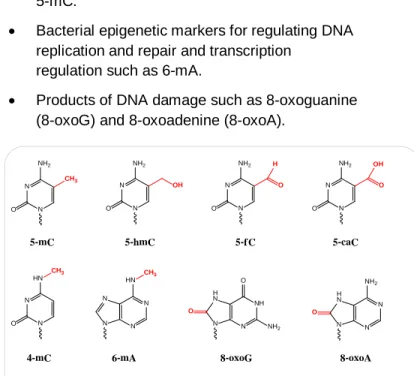
DNA base modifications have a variety of functional roles which include:
• Epigenetic markers for influencing gene expression such as mC, hmC,
5-formylcytosine fC), and 5-carboxylcytosine (5-caC).
• Bacterial identity markers for affecting host-pathogen interactions such as 6-mA, 4-mC, and 5-mC.
• Bacterial epigenetic markers for regulating DNA replication and repair and transcription
regulation such as 6-mA.
• Products of DNA damage such as 8-oxoguanine (8-oxoG) and 8-oxoadenine (8-oxoA).
N N NH2
O N
N NH2
O
CH3
OH
N N HN
O
CH3
N N N N
HN
CH3
NH N N H N
O
NH2
O
5-mC 5-hmC
4-mC 6-mA 8-oxoG
N N NH2
O
O
5-fC
N N NH2
O
O
H OH
N N H N
NH2
O 5-caC
N
8-oxoA
Today, the most commonly studied base modification is 5-mC, commonly referred to as “methylation” despite the fact that other bases are also naturally modified by methyl groups.
Figure 1. Molecular structures of base modifications including 5-mC, 5-hmC, 5-fC, 5-caC, 4-mC, 6-mA, 8-oxoG, and 8-oxoA.
Methylation has gathered interest from researchers in a variety of disciplines including early
developmental biology, cancer biology, and neurological disorders. Methylation and
de-methylation help regulate gene expression and have been linked to several human diseases through mechanisms such as deactivating tumor suppressors or activating oncogenes5.
Other modifications, such as 6-mA in bacteria, have been studied with lower resolution methods – such as chromatography or through methylation’s
protective effect against restriction endonucleases – because they are not easily accessible with standard sequencing techniques. This modification is
associated with basic functions such as DNA replication and repair6. It is also common in protists and plants, and some studies suggest that it may also be present in mammalian DNA7.
SMRT sequencing is capable of detecting 6-mA as well as other common bacterial base modifications. As a result, the technology is expected to increase our understanding of a broad array of biological processes.
The potential benefits of detecting base modification, using SMRT sequencing, include:
• Single-base resolution detection of a wide
variety of base modifications (including those in Figure 1 and more).
• Single-molecule resolution over long-read distances.
• Unamplified double-stranded input DNA, which means that strand-specific modifications, such as hemimethylation, are detectable.
• Hypothesis-free base detection which allows discovery of unknown or unexpected
modifications through the effects on sequencing kinetics (as described below).
Studying Polymerase Kinetics with
SMRT
®Sequencing
SMRT Sequencing allows the observation of single DNA polymerases reading individual molecules of DNA in real time. Therefore, the kinetic
characteristics of DNA polymerization are
observable on a single-molecule basis. The kinetic characteristics, such as the time duration between two successive base incorporations, are altered by the presence of a modified base in the DNA
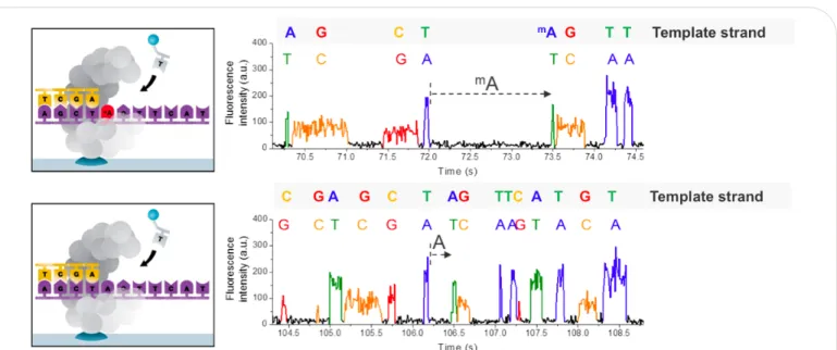
template3. This is observable as an increased space between fluorescence pulses, which is called the interpulse duration (IPD), as shown in Figure 2.
Figure 2. Principle of detecting modified DNA bases during SMRTsequencing. The presence of the modified base in the DNA template (top), shown here for 6-mA, results in a delayed incorporation of the corresponding T nucleotide, i.e. longer
These changes in the DNA polymerase speed, relative to an amplified control template lacking modified bases, can be measured for each template position to indicate the presence of modified bases in the DNA template. In order to quantify the change in IPD distributions between a control sample and a native sample, we define the IPD ratio as the ratio of the mean IPD at a site in the native sample to the mean IPD at the same site in the amplified control.
An alternative method calculates IPD ratios based on a computational model rather than using an unmodified control sample. This model is referred to as the “in silico control”. The model must be trained to recognize the kinetics of a specific sequencing chemistry. The in silico control has obvious benefits in reducing the time required to perform an initial experiment and avoids using an amplified control. The local sequence context of unmodified DNA influences polymerase dynamics. As such, the in
silico control uses a predicted value of the mean IPD
at each reference position that is based on the local sequence context.
Kinetic effects are not necessarily limited to just the nucleotide incorporation opposite the modified base position in the DNA template. This is because the DNA polymerase is in intimate contact with the DNA over an extended region of approximately twelve
bases, and the modified base can impart effects on the polymerase dynamics at several positions over this region. This results in kinetic “signatures” which can aid in identifying the type of base modification. For example, for the three common bacterial identity markers8 (see Figure 4):
• 5-mC has typical characteristic kinetic signals two and six bases downstream of the
methylated position
• 4-mC just at the position of the modification • 6-mA at the modified position and often five
bases downstream
Different signal magnitudes translate to different amounts of sequencing-fold coverage required to obtain similar confidence levels for modification detection.
The current in silico control algorithms have proven reliable for studying 6-mA in bacteria, for example in methylome motif analysis. However, when studying subtle kinetic effects such as those for native 5-mC, using an amplified control is more accurate than
Figure 3. Image from SMRT® View software using the in silico control showing detection of 6-mA at GATC motifs using IPD ratios. For each template position (x-axis), the ratio (y-axis, purple bar up for forward strand and orange bar down for reverse strand) of average
interpulse durations (IPDs) for each strand of the native DNA to the control is plotted. Excursions from the baseline indicate the presence of a modified base (6-mA, in this example, marked with indicators at the top along with a seven-base context in 5’ -> 3’ orientation), slowing down the polymerase at the position of the modification9.
Figure 4. Kinetic signatures for the three common bacterial identity markers 5-mC, 4-mC, and 6-mA. The methylated template positions are highlighted in red. Note that the kinetic signatures vary both in magnitude as well as in the length of the region over which the polymerase dynamics are affected.
using the in silico control. Future commercial products are expected to improve use of the sequence context to better identify and interpret modification signals.
Based on the measurement and analysis principle outlined above, a typical experiment for detecting DNA base modifications can be designed as follows:
• Obtain native DNA of interest and prepare a SMRTbell™ library for SMRT sequencing (see the Pacific Biosciences Sample Preparation and Sequencing Guide).
• If using an amplified control design, use a small aliquot of the original DNA sample and perform a whole-genome amplification (WGA) reaction to obtain the control DNA sample lacking any base modifications. Then prepare a SMRTbell library for this sample.
• Perform adequate SMRT sequencing (for both samples when using an amplified control) to obtain the necessary coverage needed for the modifications under study, and sufficient overall coverage to characterize the genome (or portion of the genome) of interest.
• Use the bioinformatics tools to perform kinetic analysis for base modification9.
Examples of Current Applications
Direct DNA sequencing of base modifications requires that the following general considerations be fulfilled during the experimental design:
1. Unamplified DNA is necessary. Amplification of DNA through PCR, WGA, or other techniques, will result in the loss of base modifications. Therefore, enough native DNA must be available to make a sequencing library. With version 2 sequencing reagents (“C2 chemistry”), the sample requirement for preparing a ~500 bp SMRT sequencing library is 250 ng of input DNA.
2. As DNA input requirements are reduced over time, a smaller or targeted region of a genome is expected to be sufficient for studies without amplification requirements.
3. The different magnitudes of kinetic signal, for the different base modifications, translate to different amounts of sequencing fold coverage required to obtain high confidence levels of detection. We recommend the minimum sequencing fold coverage per strand as follows9:
4-methylcytosine 25x 5-methylcytosine 250x 5-hydroxymethylcytosine 250x glucosylated 5-hydroxymethylcytosine1 25x hydroxymethylcytosine enriched with
the Hydroxymethyl Collector™ Kit 2
5x
6-methyladenine 25x
8-oxoguanine 25x
4. The genome, or portion of a genome, to be interrogated also has to be compatible with the current throughput performance of the PacBio
RS. With C2 Chemistry, the base throughput of a two hour run on the PacBio RS is
approximately 100 Mb. Studying methyladenine in E. coli requires 4 to 6 SMRT Cells per
sample,which can be run in one afternoon. Over time, we expect SMRT technology throughput to increase, making larger genome sizes more practical to sequence for base modifications.
Applications particularly suitable for SMRT sequencing based on the criteria above include:
• Sequencing of E. coli to find the sites of 5-mC, 4-mC, and 6-mA modification when studying virulence, gene expression, or pathogen-host interactions. See the Pacific Biosciences Technical Note - Detecting DNA Base
Modifications for more detailed information on
strategies for experimental design and motif analysis in bacteria.
• Isolating and purifying mitochondrial, chloroplast or other small genomes, containing known or novel DNA base modifications.
• Enriching portions of a larger genome (e.g., isolation of DNA regions modified with 5-hmC,
1
Generated by T4 Phage β-glucosyltransferase (Josse, J. and Kornberg, A. (1962) J. Biol.Chem., 237, 1968-1976)
2
Available from Active Motif
(http://www.activemotif.com/catalog/775/hydroxymethyl-collector-trade)
and enriched by specific biotinylation chemistries of 5-hmC and subsequent streptavidin pulldown). Normalizing IPDs with the in silico control is particularly useful for this type of experiment, where generating a completely overlapping set of amplified control sequence data can be a challenge.
Conclusion
SMRT sequencing is the only commercially available technology capable of measuring the kinetics of base incorporation during a sequencing run. This kinetics information can be used to identify sites in the target DNA that have been chemically modified in a variety of ways (including methylation,
formylation, carboxylation, and more). These base modifications are associated with several biological processes including gene expression and DNA oxidative damage.
Most of these modifications have not been extensively studied due to difficulties in adapting experimental techniques to a higher throughput sequencing method – we expect that these modifications will be possible to study with SMRT sequencing. Examples of novel studies that may be performed on the PacBio RS today include full-genome bacterial modification studies and hydroxymethylcytosine studies in mammalian genome regions enriched for that modification. We anticipate that over time the range of target genomes that will be addressable by SMRT sequencing will improve and ultimately address regions as small as single genes as well as larger whole genomes. We further expect that the advances afforded by SMRT sequencing will enhance genetic studies into gene regulation, DNA damage and repair, bacterial virulence, and other important biological pathways. Novel DNA modifications could potentially be discovered, expanding the utility of sequencing to even broader areas of study.
References
1.Trygve Tollefsbol (2010) Handbook of Epigenetics: The New Molecular and Medical Genetics. Academic Press.
2. Clark S.J., Statham A., Stirzaker C., Molloy P.L. & Frommer, M. DNA methylation: bisulphite modification and analysis. Nat. Protocols 1, 2353–2364 (2006).
3. Flusberg BA, Webster DR, Lee JH, Travers KJ, Olivares EC, Clark TA, Korlach J, Turner SW (2010) Direct detection of DNA methylation during single-molecule, real-time sequencing. Nature Methods 7:461-465.
4. Kriaucionis S. & Heintz N. The nuclear DNA base 5-hydroxymethylcytosine is present in purkinje neurons and the brain. Science 324, 929–930 (2009); Tahiliani, M. et al. Conversion of 5-methylcytosine to 5-hydroxymethylcytosine in mammalian DNA by MLL partner TET1. Science 324, 930–935 (2009).
5. Esteller M. Epigenetics in cancer. New England Journal of Medicine 358, 1148-1159 (2008).
6. Marinus M.G. & Casadesus, J. Roles of DNA adenine methylation in host-pathogen interactions: mismatch repair, transcriptional regulation, and more. FEMS Microbiol. Rev. 33, 488–503 (2009).
7.Ratel D, Ravanat JL, Berger F, Wion D, N6-methyladenine: the other methylated base of DNA. BioEssays 28,309-315 (2006). 8. Roberts R.J., Vincze T., Posfai J. and Macelis D. REBASE--a database for DNA restriction and modification: enzymes, genes and genomes. Nucleic Acids Res, 38, D234-236 (2010). 9. SMRT Analysis version 1.3.1 or higher, and/or “DNA Modification Detection with SMRT Sequencing using R” found at https://github.com/PacificBiosciences/R-kinetics.
Research Use Only. Not for use in diagnostic procedures. © Copyright 2012, Pacific Biosciences of California, Inc. All rights reserved. Information in this document is subject to change without notice. Pacific Biosciences assumes no responsibility for any errors or omissions in this document. Certain notices, terms, conditions and/or use restrictions may pertain to your use of Pacific Biosciences products and/or third party products. Please refer to the applicable Pacific Biosciences Terms and Conditions of Sale and to the applicable license terms at http://www.pacificbiosciences.com/licenses.html.
Pacific Biosciences, the Pacific Biosciences logo, PacBio, SMRT and SMRTbell are trademarks of Pacific Biosciences in the United States and/or certain other countries. All other trademarks are the sole property of their respective owners. PN 001-640-287-02