Study of UV-B Mutation Effect on pH
Resistance and Lipid Production of Microalgae
Botryococcus braunii
Thea PrastiwiSoedarmodjo, Fanina Aulia Rachma, Hakun Wirawasista Aparamarta, and Arief Widjaja
AbstractMicroalgae Botryococcus braunii is a potential biodiesel producer as an alternative for fossil fuels due to its high
lipid content. UV-B mutations were carried out to see the effect in microalgae growth at various pHs (3-8). Reduction of nitrogen levels was carried out to see the effect on the growth and lipid production of microalgae. UV-B mutation increased the ability of growth and resistance of B. braunii against low pH. Under low nitrogen conditions, the growth of B. braunii cells would not continue for a longer time. B. braunii which grow in nitrogen depletion medium produced lipid content greater than normal nitrogen. UV-B light mutation also increased the lipid content of B. braunii. At 7 days of incubation, the mutation not only increased lipid content, but also significantly increased the triacylglycerols (TAG) content of B.
braunii lipids.
KeywordsMicroalgae, Botryococcus braunii, Nitrogen Depletion, pH, mutation, UV-B.
I.INTRODUCTION1
nergy is now an absolute necessity and must be fulfilled. Almost all facilities and infrastructure supporting human life are driven by energy. Until now, energy as driving force of the human economy is still supplied by fossil fuel. Fossil energy is limited and less environmentally friendly. The combustion process produces unfavorable effects on the environment and health as greenhouse effects, due to the content of carbon dioxide (CO2), Sulphur dioxide (SO2), and nitrogen oxides (NOx) [1].
Indonesia has been widely known as a maritime country. Two-thirds of its territory is ocean with a 108,000 km coastline [2]. One living thing that grows and thrives in the ocean is algae. Microalgae are one of the producers of oil or lipid as a potential biodiesel feedstock. Microalgae use solar energy and carbon dioxide to produce biomass. Microalgae cells grow in water media that is why microalgae have a high level of efficiency in terms of the use of water, carbon dioxide, and other nutrients [3]. Microalgae have advantages over other oil-producing plants such as jatropha, sunflowers, or palm oil for land growth is not competing with conventional agricultural land, has a very high growth rate and has a better lipid composition as biodiesel feedstock [4]. Some of the common problems encountered in microalgae cultivation as a producer of lipid relate to its resistance to changes in pH. CO2 is a carbon source for microalgae while the addition of CO2 will reduce its pH value due to the reaction with H2O produce HCO3- [5].
Algae are diverse group of photosynthetic organisms ranging from unicellular (microalgae) to multicellular (macro algae) forms. They have chlorophyll as primary photosynthetic pigment and do not have a common ancestor. Commonly, algae population falls under two broad categories, (1) microalgae: microscopic algae that grows in fresh water and marine environment and (2)
Thea Prastiwi Soedarmodjo, Fanina Aulia Rachma, Hakun Wirawasista Aparamarta, and Arief Widjaja are Departement of Chemical Engineering, Institut Teknologi Sepuluh Nopember, Surabaya, 60111, Indonesia. E-mail: [email protected].
macro algae: comparatively large, multicellular organisms that grows in marine environment. Microalgae are microscopic, unicellular and phototropic organisms that fall under three categories, namely Diatoms (Bacillariophyceae), Green algae (Chlorophyceae) and Golden algae (Chrysophyceae). Diatoms are the dominant life forms in phytoplankton and probably represent the largest group of biomass producers on earth. Green algae are abundantly found in fresh water than in marine water. Golden algae are similar to diatoms and produce oils and carbohydrates. Microalgae are efficient producers of lipids and other great metabolites that work by utilizing nutrients in the presence of solar energy [6].
One of the microalgae that can be processed into biodiesel is Botryococcus braunii. Microalgae B. braunii
are a single-cell green microalga from the
Chlorophyceae class that live in freshwater to marine water [1]. B. braunii is one of microalgae with the largest lipid contain, 25-75% dry weight [4]. It contains 41.1% free fatty acids, 2.1% triacylglycerols, 1.8% diacylglycerols, and 1.7% monoacylglycerols [7]. Types of fatty acids found in B. braunii lipids dominated by oleic and palmitic acid [8].
Biodiesel is a mixture of alkali ether and fatty acids obtained from the transesterification process of vegetable or animal oils. Diesel raw materials are hydrocarbons which contain 8-10 carbon atoms per molecule while hydrocarbons contained in vegetable oils on average are 16-20 carbon atoms per molecule so that vegetable oils have a higher viscosity and low combustion as fuel [1]. Through esterification and transesterification process, microalgae lipid can be converted into biodiesel [8].
Mutation in microorganisms is carried out to improve its nature. Research using UV-B mutagen show that irradiated microbes with UV-B ray at the right dose will produce higher activity than native ones. UV radiation will cause changes in gene composition as mutant genes that can cause certain enhancement in microorganisms [9]. Microalgae as one of microorganisms are often investigated for its influence on UV-B radiation. Skerratt et. al. (1998), examined the effect of UV-B ray mutations on the lipid content of Antarctic marine phytoplankton microorganisms. It was found that lipid levels per cell
increased when exposed to high levels of UV-B caused by increased concentrations of free fatty acids and may show degradation of complex lipids during high levels of UV-B irradiation [10]. In the study of Xue et. al., it was reported that exposure to UV-B ray continuously decreased the chlorophyll content, but increased UV-B absorbent compounds in algae [11]. The decrease in photosynthesis in algae, especially at high UV-B radiation doses, was influenced by direct effect (effects on photosystem) and indirect effect (pigment decreased). The decrease in chlorophyll pigments and photosynthesis resulted lower biomass. However, algae have evolved and formed a defence mechanism to protect itself from the damaging effects of UV-B radiation [11].
Microalgae during photosynthesis utilize solar energy and some essential nutrients to synthesize biomass compounds and multiply cells [12]. Limitation of nutrients such as nitrogen can affect the metabolism of microalgae such as the amount of lipids and photosynthesis. Nitrogen is an essential component of many macromolecules such as DNA, RNA, chlorophyll and protein, known as one of the most important nutrients for microalgae. Nitrogen deficiency causes a decrease in photosynthesis and protein synthesis but increases the synthesis of lipids and carbohydrates [13]. Nitrogen deficiency leads to stress conditions for all organisms because nitrogen is the main component of proteins and nucleic acids in living cells [1]. Decrease levels of nitrogen in the substrate of microalgae
Chlorella vulgaris showed that the lipid production of microalgae has increased [5].
The research aimed to study the effect of UV-B rays on pH resistance and lipid production of microalgae B. braunii. The effect of nitrogen concentration and harvesting time on growth and lipid content on native microalgae B. braunii and those that are mutated in UV-B ray were investigated to obtain best condition under which high-lipid content could be achieved.
II.METHOD A. Materials
A microalgae strain of B. braunii was obtained from Balai Budidaya Air Payau Jepara, Central Java, Indonesia. The microalgae were diluted in marine water from Balai Budidaya Air Payau Situbondo, East Java, Indonesia with salinity levels of 21 ppm.
B. Medium and Cultivation Condition
The normal nutrition medium for cultivation of B. braunii was Walne nutrient, which had a composition per 1 Liter of solvent referred to Isnansetyo & Kurniastuty (1995): 100 mg/L NaNO3; 45 mg/L Na2EDTA; 33.6 mg/L H3BO3; 20 mg/L NaH2PO4.2H2O; 1.3 mg/L FeCl3.6H2O; 0.36 mg/L MnCl2.4H2O; 0.1 mg/L Vitamin B1; 0.005 mg/L Vitamin B12 [14]. This normal nutrition medium resulted in a nitrogen content of 100 mg/L medium. The nitrogen depletion medium was provided by eliminating the addition of NaNO3 to result in a medium with a nitrogen content of 0.03 mg/L medium.
C. Algae pre-culture
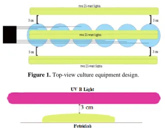
This stage was conducted to get 100 million cells/mL microalgae. Microalgae were measured using counting chamber method. The experiment was carried out in Erlenmeyer 500 mL. All media were sterilized at 121°C for 15-20 minutes at 1 atmosphere pressure. 250 mL of microalgae strain was poured into Erlenmeyer slowly then mixed with 250 mL of marine water with microalgae strain to reach a concentration of 1:1. Diffuser was inserted into Erlenmeyer to provide air supply and air circulation that occurred continuously with a flow rate of 2.5 L/min. Two 21-watt lights were installed on the front, rear and Erlenmeyer sides with a distance of 3 cm from the Erlenmeyer. The lights were set to 24 hours bright and 0 hours dark, with a light intensity of 6000 lux or approximately 77.08 µE/m2s. Equipment design is shown in Figure 1.
The cultivation was done for 7 days. Microalgae commonly double their biomass within 24 hours [6]. The number of microalgae cells was analyzed every 24 hours.
D. Algae mutation
Referred to Zul et. al., mutation was carried out with germicidal UV-B light treated in darkened space [9]. During radiation, the petri lid was opened so UV-B ray transmissions were not blocked. Before being mutated by Figure 1. Top-view culture equipment design.
Figure 2. Installation of UV-B mutation equipment.
TABLE 1.
NUMBER OF INITIAL B. BRAUNIICELLS AND AFTER MUTATIONS
USING UV-BRAY
Run Number of B.braunii cells (cell/mL) Average death cells Initial cells Mutated cells
Run 1 110,333,333 74,666,666
30.84 % Run 2 101,666,666 69,666,666
Run 3 105,000,000 74,833,333
Figure 3.B.braunii growth curves for 7 days of pre-culture during normal nutrition medium.
20 40 60 80 100 120
0 24 48 72 96 120 144 168
N
u
m
b
er
o
f
ce
ll
s
(m
il
li
o
n
c
ell
s/m
L
)
UV-B ray, microalgae were sampled to analyze the initial cell count. Microalgae were exposed to UV-B ray straight above the target cell with a distance of 3 cm for 3 minutes shown in Figure 2. To find out the number of cells that survive from the UV-B mutation process, microalgae were sampled again.
E. Algae culture in various pHs
B. braunii was cultured at pH 3-8 to determine the growth of native and mutated B. braunii in various pHs. Native and mutated microalgae, each of these were divided into 6 samples for culture in pH 3-8. The cultivation was carried out in normal nutrition medium. The number of cells was measured before conditioning different pHs by adding citric acid 1 M and checking with pH indicator paper. Number of microalgae cells was analyzed every 12 hours for 7 days. Other conditions of incubation such as light intensity, gas flow rate and temperature were all the same as the corresponding pre-culture condition.
F. Algae Culture in Various Nitrogen Concentrations B. braunii was cultured during various nitrogen concentrations that was nitrogen normal and nitrogen depletion to determine the growth of mutated and native
B. braunii. Native and mutated microalgae, each of these were divided into 2 samples for normal nutrition medium (nitrogen normal) and nitrogen depletion medium. The cultivation was carried out for 7 and 20 days at which time the cells were harvested, and the lipid content were measured. Number of microalgae cells was analyzed every 24 hours. Other conditions of incubation such as light intensity, gas flow rate and temperature were all the same as the corresponding pre-culture condition.
G. Lipid Extraction
Microalgae cells were harvested by centrifugation at 8500 rpm for 10 minutes at 20°C. The solid phase was separated carefully using Whatman filter paper in which two pieces of filter papers were applied twice to provide complete separation [5]. After separation, wet algae were dried using a cooling method by storing it in the refrigerator (4°C) for 12 hours. Drying was continued using an oven at 60°C for 2 hours. Dry algae were extracted by n-hexane using Soxhlet extraction. In each
extraction used 150 mL of n-hexane. The extraction process is carried out for 6 hours until the solvent becomes clear. A mixture of n-hexane and lipids were separated by distillation. Distillation was carried out for approximately 2 hours using a distillation flask and Liebig condenser. The temperature of the distillation operation was maintained at 70°C.
H. Gas Chromatography Analysis
Glycerides and FFA in crude lipid of microalgae was analyzed using Gas Chromatography (GC). Quantitative analysis was carried out with Shimadzu GC-2010 gas chromatography (Kyoto, Japan) which was equipped with a flame ionization detector. The separation was carried out with DB-5HT (5%-phenyl)-methylpoly-siloxane non-polar column (15 m x 0.32 mm ID; with a film thickness of 0.1 μm). Injector and detector temperature were set at 370ºC. Initial column temperature was 80ºC, increased to 305ºC at a rate of 15ºC/minute and then increased to 335ºC at a rate of 5ºC/minute. Column temperature was maintained at 335ºC for 5 minutes. Furthermore, the column temperature was increased to 365ºC at a rate of 15ºC/minute. Split ratio 1:50, used nitrogen as a gas carrier with a linear rate of 30cm/s at temperature of 80ºC. 20 mg of sample was dissolved in 1 mL of ethyl acetate, and 1 μL of it was injected into the GC.
III.RESULTS AND DISCUSSION
A. Microalgae Mutation
Microalgae are the most primitive form of plants. While the mechanism of photosynthesis in microalgae is similar to that of higher plants, they are generally more efficient converters of solar energy because of their simple cellular structure [3].
Figure 3 shows B. braunii cells growth for 7 days of pre-culture. It is seen that the cells growth of B. braunii
each day increased up to the day of 7 to reach 100 million cells/mL microalgae. Calculation of cell numbers was done by counting chamber method, only living cells were counted. Differences in living and dead cells could be seen in the color of microalgae. In optimal growth conditions, microalgae were green to deep dark green. Meanwhile in the opposite condition, microalgae were Figure 4. Native B. braunii growth curves in various pHs during
normal nutrion.
10 20 30 40 50 60 70 80 90 100
0 24 48 72 96 120 144 168
N
u
m
b
er
o
f
ce
ll
s
(m
il
li
o
n
c
ell
s/m
L
)
Time (hour) pH 8
pH 7 pH 6 pH 5 pH 4 pH 3
Figure 5. Mutated B. braunii growth curves in various pHs during normal nutrition
10 20 30 40 50 60 70
0 24 48 72 96 120 144 168
N
u
m
b
er
o
f
ce
ll
s
(m
il
li
o
n
c
ell
s/m
L
)
Time (hour) pH 8
yellowish green. In a dead state, microalgae tend to be yellow to clear. Microalgae pre-culture results were then used as a seed for a mutation treatment using UV-B ray.
Mutation treatment was carried out to produce mutation of B. braunii with 27-31% of death cells. The number of initial cells, living cells after UV-B exposure and the death cells percentage of B. braunii were presented in Table 1. Mutation was carried out with 3 minutes exposure time with average death cells of 30.84% from 3 repetitions. Living cells after exposure to UV-B ray were referred to as mutated cells.
B. Mutation Effect on Microalgae Growth in Various pHs
Each microalgae species has a tolerant value or a minimum pH value that allows for its survival. Most microalgae grow in normal pH conditions, pH 6-8 [15]. Using higher concentration of CO2 may result in decreasing the pH since unutilized CO2 will be converted to H2CO3. On the other hand, if there is not enough CO2
gas supply, algae will utilize carbonate to maintain its growth. Since algae use CO2 (aq) from bicarbonate as a compensation of lacking enough CO2 from gas supply, this will result in increase of pH [5].
UV-B ray was exposure to determine its effect on cell growth in media with pH 3-8. Acid conditioning was carried out by adding 1 M citric acid to the medium and maintained for 7 days of incubation.
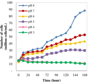
The cell growth of native B. braunii at pH 3-8 is shown on figure 4. It can be seen that algae grow well at pH 6-8. It has the best condition to grow at pH 6-8. According to Dayananda et. al., microalgae B. braunii can grow at a pH of 6-8.5 [16].
Figure 4 shows that B. braunii cells growth slow further in culture medium with lower pH. In culture medium pH 5, algae cells growth increased until the 6th day but decreased on the day of 7. In culture medium pH 3 and 4, algae cells growth decreased from the first day to the 7th day. In an experiment conducted by Hifney and Basset (2014), it was concluded that microalgae B. Figure 6. Native and mutated B. braunii cells growth curve for 7
days incubation during normal nutrition and nitrogen starvation.
10 20 30 40 50 60 70 80
0 24 48 72 96 120 144 168
Nu
m
b
er
o
f
ce
ll
s
(m
il
li
o
n
c
el
ls
/m
L
)
Time (hour) Mutation, N Normal
Mutation, N Depletion Native, N Normal
Figure 7. Native and mutated B. braunii cells growth curve for 20 days incubation during normal nutrition and nitrogen starvation.
10 20 30 40 50 60 70 80
0 48 96 144 192 240 288 336 384 432 480
N
u
m
b
er
o
f
ce
ll
s
(m
il
li
o
n
c
ell
s/m
L
)
Time (hour) Mutation, N Normal Mutation, N Depletion Native, N Normal Native, N Depletion
TABLE 2.
COMPARISON OF NATIVE AND MUTATED B. BRAUNIICELL GROWTH FOR 7DAYS INCUBATION DURING NORMAL NUTRITION t
(hour)
Mutation Native
Cx (cell/mL) dCx/dt Μ (hour-1) Cx (cell/mL) dCx/dt Μ (hour-1)
0 35,166,666 40,666,666
24 36,833,333 69444.4 0.00189 45,000,000 180555.6 0.00401 48 37,000,000 6944.4 0.00019 48,500,000 145833.3 0.00301 72 39,666,666 111111.1 0.00280 49,166,666 27777.8 0.00056 96 49,833,333 423611.1 0.00850 50,833,333 69444.4 0.00137 120 52,500,000 111111.1 0.00212 53,000,000 90277.8 0.00170 144 56,500,000 166666.7 0.00295 60,000,000 291666.7 0.00486 168 60,500,000 166666.7 0.00275 71,833,333 493055.6 0.00686
μ rata-rata 0.00303 0.00320
TABLE 2.
COMPOSITION OF MAJOR LIPID COMPONENTS UNDER DIFFERENT NUTRITION CONDITION
Microalgae culture conditions FFA MAG DAG TAG (%)
Native, N Normal 7d 17.622 22.097 4.453 0.824
Native, N Depletion 7d 17.283 22.791 4.908 0.885
Mutation, N Normal 7d 7.089 10.774 9.956 12.908
Mutation, N Depletion 7d 12.055 16.907 5.752 11.071
Native, N Normal 20d 10.998 12.261 12.383 5.976
Native, N Depletion 20d 13.607 18.966 6.311 4.511
Mutation, N Normal 20d 15.002 19.299 6.650 1.414
braunii could not grow optimally at acidic pH conditions (6> pH), because the amount of chlorophyll in B. braunii
decreased in proportion to the acidic pH. Decreasing chlorophyll content produces lower biomass [17].
Figure 5 shows the cells growth of mutated B. braunii
at pH 3-8. As well as native B. braunii, mutated B. braunii cells growth slow further in culture medium with lower pH. In culture medium pH 3-8, mutated algae could grow well. In culture media pH 8, the growth of native algae was better than mutated algae. However, in lower culture medium, mutated algae could grow better than native B. braunii. Compared to native algae, mutated algae still could grow well in culture medium pH 3 and 4. It can be seen that mutation by UV-B ray gave an effect to microalgae B. braunii. Warmadewi explained that mutation aim to deal with native changes which at times will arise with genetic changes that can be inherited to their offspring. In the changes that occur, mutated character is more adaptable than the original, so that the original character allows it to disappear from circulation and create new traits [18]. This new characteristic allows B. braunii to be more resistant to the pH of a low medium.
On the microalgae B. braunii growth curve shows different growth rates. It was highly influenced by the pH conditions of the microalgae medium culture. Ammar explained that in the growth of microalgae culture there were about four phases of growth [19]. The first phase is the induction phase or lag phase which increases slightly in the growth rate along with cell absorption. In the second phase, the growth rate increased exponentially. This depends on many factors such as species or types of algae, light intensity, and media temperature. The third phase is a constant phase where the cell density becomes relatively constant. Eventually the growth rate decreases when cell division decreases due to several factors that influence the growth rate such as nutrient concentration, pH, CO2 dissolved, risk of light and contamination [19].
The growth medium of microalgae B. braunii generally has a pH of 8. In this situation, UV-B mutation is not necessary because B. braunii can grow very well at pH 8. However, under conditions of growth medium with a lower pH such as due to the addition of CO2, UV-B mutation in microalgae B. braunii need to be done. This is based on the medium of cultivation with more acidic
conditions or below pH 8, B. barunii with mutation by UV-B ray could grow better than nativeB. braunii. As far as we know, this is the first report concerning the changes in the reproductive capacity of microalgae cells in low pH conditions upon exposing to UV-B ray.
C. Mutation Effect on Microalgae Growth in Various Nitrogen Concentrations
Nitrogen affects the growth of microalgae cells such as lipid and protein content. In conditions where nitrogen levels are limited, the production of lipids in cells will increase; this condition is commonly called Nitrogen Starvation [1].
During nitrogen starvation conditions, microalgae B. braunii need to find out their changes after exposure UV-B ray. Figure 6 and Figure 7 show the comparison of native and mutated B. braunii cells growth for 7 and 20 days of incubation during normal nutrition and nitrogen starvation conditions. Nitrogen starvation conditions caused native and mutated B. braunii get into the fourth phase of cells growth as a decrease of the cells number faster than the normal nutrition. Since nitrogen is the main nutrient that limits the growth of microalgae, a very high concentration of nitrogen in the growth medium of microalgae can cause deactivation production of the pigments for photosynthesis. Therefore, high nutrient concentrations take longer to reach the growth stability phase [19].
Combination treatment between UV-B radiation exposure and nitrogen limitation on B. braunii
cultivation increased the inhibition of photosynthesis intensively. Under the more severely nitrogen limited conditions, the damage rate increased. Unexpectedly, repair rates were also stimulated under nitrogen limited conditions, although this was insufficient to counteract the increase in damage, so the overall effect of nitrogen limitation was an enhancement of UV-B radiation-induced inhibition of photosynthesis. This result coincides with the study by Shelly et. al. using microalgae Dunaliella tertiolecta (Chlorophyceae) [20].
Comparison of native and mutated B. braunii cells growth calculated by specific growth cells (μ) and the result is shown in Table 2. NativeB. braunii had higher specific cells growth than mutated B. braunii. NativeB. braunii had specific growth cells of 0.00320/hour, while mutated B. braunii had specific growth cells of 0.00303/hour. It shows that nativeB. braunii had better cells growth compared to mutated B. braunii. Mutation by UV-B ray on microalgae has an influence on growth, survival, pigmentation, metabolism and photosynthesis from microalgae. Increased UV-B radiation will reduce the chlorophyll content and decrease photosynthesis (effects on photosystem). This reduction in chlorophyll content and photosynthesis results in lower biomass [9]. However, comparison of nativeand mutated B. braunii
cells growth needs further discussion due to the difference was not significant.
Figure 7 shows native and mutated B. braunii cells growth in the normal nutrition medium for 20 days of incubation decreased the number of cells on the 13th day. This was due to insufficient amounts of nutrients for culture up to 20 days, so it was necessary to add nutrients again.
Figure 8. Comparison of lipid content of native and mutated
B.braunii at 7 and 20 days incubation during normal nutrition and nitrogen starvation
0 10 20 30 40 50
Native, N Normal
Native, N Depletion
Mutation, N Normal
Mutation, N Depletion
L
ip
id
C
o
n
te
n
t
(%
)
Algae Conditions 7 d
D. Effect of Nitrogen Concentration on Lipid Production of Nativeand Mutated B. braunii
Nitrogen affects lipid and protein content in microalgae cells. Microalgae can utilize sources of nitrate, ammonia, or nitrogen. Nitrogen limitations can be considered as an efficient environmental pressure to increase lipid accumulation in microalgae [1].
Comparison of lipid content of nativeand mutated B. braunii is shown in Figure 8. Lipid contents at 7 days of incubation for nativeand mutated B. braunii were higher than 20 days of incubation. It could be influenced by the number of B. braunii cells at 7 days incubation higher than 20 days of incubation. Since the amount of nutrients was insufficient for culture up to 20 days, so it was necessary to add nutrients. Microalgae growth decreases when cell division decreases due to several factors that influence the growth rate such as nutrient concentration factors [19].
Figure 8 shows nitrogen starvation conditions produced greater lipid content compared to the normal nutrition, both in nativeand mutated B. braunii for 7 and 20 days of incubation. When incubated into a low-nitrogen containing medium, B. braunii accumulate more total lipid in their cells. Widjaja et. al reported the similar behaviour of C. vulgaris in which under conditions of nitrogen starvation there will be more lipid content than normal nitrogen levels. Microalgae need to be made under stress conditions to produce high lipids [5]. Sheehan et. al hypothesized that the reason for the increase in lipid content was that nitrogen depletion leads to the inhibition of cell division without a gradual decrease in lipid productions which result in accumulation of fat in cells [3].
Native B. braunii, both at 7 days and 20 days of incubation, showed lower lipid content compared to mutated B. braunii. Under nitrogen starvation conditions, mutated B. braunii also produce higher lipid content compared to nativeB. braunii. According to Skerratt et. al., mutations with irradiated UV-B make the lipid content of Antarctic marine phytoplankton microorganism increase. This is due to the increased concentration of free fatty acids and shows the degradation of complex lipids during high levels of UV-B irradiation [10]. Combination treatment of mutation by UV-B ray with culture under nitrogen starvation condition could produce higher lipid content of microalgae.
Lipid composition during normal nutrition and nitrogen starvation were analyzed using gas chromatography and the results are shown in Table 3. The biggest TAG content among all the results of the analysis was mutated
B. braunii during normal nutrition at 7 days of incubation. Table 3 shows that mutations gave different affect to B. braunii lipid composition. It can be seen at 7 days of incubation, the mutation with UV-B ray on microalgae B. braunii could increase the TAG content compared to nativeB. braunii significantly. At 20 days of incubation, mutation gave different effect to B. braunii, mutated algae result lower TAG content than native algae. Mutated B. braunii in 20 days of incubation showed lower TAG content than mutated B. braunii in 7 days of incubation, but it was contrary to nativeB. braunii.
Nitrogen starvation conditions also gave different affect to B. braunii lipid composition. At 7 days of incubation, native B. braunii in nitrogen starvation condition showed higher TAG content than normal nutrition condition and mutated B. braunii in nitrogen starvation condition showed lower TAG content than normal nutrition condition. But it was contrary to 20 days of incubation, native B. braunii in nitrogen starvation condition showed lower TAG content than normal nutrition condition and mutated B. braunii in nitrogen starvation condition showed higher TAG content than normal nutrition condition. However, TAG content comparison between normal nutrition and nitrogen starvation conditions exhibited no significant difference in each another conditions. Nitrogen starvation conditions and the length of time of culture could cause B. braunii cells to die, resulting in lower TAG content. According to Emmauel et. al, an increase in the amount of FFA in oil or fat samples shows the hydrolysis of triglycerides. This reaction occurs by the action of the lipase enzyme and is an indicator of inadequate processing and storage conditions, such as high temperatures, relative humidity and tissue damage. The source of the enzyme can come from the tissue where oil or fat is extracted or comes from contaminants from other cells including microorganisms [21]. The lipid composition produced by microalgae B. braunii
from this study is similar to that reported by Hidalgo et. al, in which FFA contents of B. braunii lipid were higher than TAG contents [7].
IV.CONCLUSION
Microalgae B. braunii was chosen as the subject of investigating the lipid production due to its easy growth and its significant lipid content. Factors responsible for the increase of lipid production such as pH, nitrogen concentration and harvesting time were investigated.
This study examined the changes in growth and lipid content of nativeand UV-B mutated B. braunii. UV-B mutated B. braunii could grow better and be more resistant to low pH than native B. braunii. Nitrogen concentrations in nutrients greatly influence the growth and lipid content of B. braunii. Under nitrogen starvation, the growth of B. braunii cells would not continue for a longer time. B. braunii which grow in nitrogen depletion medium produced lipid content greater than normal nitrogen. UV-B-ray mutation also increased B. braunii lipid content. At 7 days of incubation, the mutation not only increased lipid content, but also significantly increased the TAG content of B. braunii lipids.
ACKNOWLEDGEMENT
All of the experimental works of this research was funded by the Laboratory of Biochemical Technology, Department of Chemical Engineering, Institut Teknologi Sepuluh Nopember. The authors expressed sincere thanks for its support.
REFERENCES
[1] S. Amini and R. Susilowati, “Produksi Biodiesel dari Mikroalga Botryococcus braunii,” Squalen, vol. 5, pp. 23–32, 2010.
Diluncurkan,” 2019. [Online]. Available:
http://www.pushidrosal.id/berita/5256/%0ADATA- KELAUTAN-YANG-MENJADI-RUJUKAN-NASIONAL%0A--DILUNCURKAN/%0A.
[3] J. Sheehan, T. Dunahay, J. Benemann, and P. Roessler, “A Look Back at the U.S. Department of Energy’s Aquatic Species Program-Biodiesel from Algae,” Prepared for U.S. Department of Energy’s Office of Fuels Development, 1998. [4] Y. Chisti, “Biodiesel from Microalgae,” Biotechnol. Adv.,
vol. 25, pp. 294–306, 2007.
[5] A. Widjaja, C. C. Chien, and Y. H. Ju, “Study of Increasing Lipid Production from Fresh Water Microalgae Chlorella vulgaris,” J. Taiwan Inst. Chem. Eng., vol. 40, pp. 13–20, 2009.
[6] B. Bharathiraja et al., “Aquatic Biomass (Algae) as A Future
Feedstock for Bio-Refineries: A Review on Cultivation, Processing and Products,” Renew. Sustain. Energy Rev., vol. 47, pp. 634–653, 2015.
[7] P. Hidalgo, G. Ciudad, M. Mittelbach, and R. Navia, “Biodiesel production by direct conversion of Botryococcus braunii lipids: Reaction kinetics modelling and optimization,”
Fuel, vol. 153, pp. 544–551, 2015.
[8] M. Faried, M. Samer, E. Abdelsalam, R. S. Yousef, Y. A. Attia, and A.S. Ali, “Biodiesel Production from Microalgae: Processes, Technologies and Recent Advancements,” Renew. Sustain. Energy Rev., vol. 79, pp. 893–913, 2017.
[9] D. Zul, A. Chainulfiffah, and I. Febrianis, “Mutagenesis pada Kluyveromyces Marxianus T-2 Penghasil Inulinase Ekstraselular dengan Sinar Ultraviolet,” J. Natur Indones., vol. 6, pp. 24–28, 2003.
[10] J. H. Skerratt, A. D. Davidson, P. D. Nichols, and T. A. Mcmeekin, “Effect of UV-B on Lipid Content of Three Antartic Marine Phytoplankton,” Phytochemistry, vol. 49, pp. 999–1007, 1998.
[11] L. Xue, Y. Zhang, T. Zhang, L. An, and X. Wang, “Effects of Enhanced Ultraviolet-B Radiation on Algae and Cyanobacteria,” Crit. Rev. Microbiol., vol. 31, pp. 79–89,
2005.
[12] G. Markou, D. Vandamme, and K. Muylaert, “Microalgal and Cyanobacterial Cultivation: The Supply of Nutrients,”
Water Reasearch, vol. 65, pp. 186–202, 2014.
[13] N. Agirman and A. K. Cetin, “Effect of Nitrogen Limitation on Growth, Total Lipid Accumulation and Protein Amount in Scenedesmus acutusas Biofuel Reactor Candidate,” Nat. Sci. Discov., vol. 3, pp. 33–38, 2017.
[14] A. Isnansetyo and Kurniastuty, Teknik Kultur Phytoplankton Zooplankton, Pakan Alami untuk Pembenihan Organism Laut. Yogyakarta: Kanisius, 1995.
[15] Hadiyanto and M. Azim, Mikroalga, Sumber Pangan dan Energi Masa Depan. Semarang: UPT UNDIP Press, 2012. [16] C. Dayananda, R. Sarada, T. R. Shamala, and G. A.
Ravishankar, “Influence of Nitrogen Sources on Growth, Hydrocarbon and Fatty Acid Production by Botryococcus braunii,” Asian J. Plant Sci., vol. 5, pp. 799–804, 2006. [17] F. Hifney and R. A. Basset, “Photosynthesis, Respiration and
Carotenoid Contents in the Green Alga Botryococcus braunii at Elevated Nutrient Levels,” J. Biol. Earth Sci., vol. 4, pp. B191–B198, 2014.
[18] A. Warmadewi, “Mutasi Genetik,” Universitas Udayana, 2017.
[19] H. Ammar, “Cultivation of Microalgae Chlorella vulgaris in Airlift Photobioreactor for Biomass Production Using Commercial NPK Nutrients,” Al-Khwarizmi Eng. J., vol. 12, pp. 90–99, 2016.
[20] K. Shelly, P. Heraud, and J. Beardall, “Nitrogen Limitation in Dunaliella Tertiolecta (Chlorophyceae) Leads to Increased Susceptibility to Damage by Ultraviolet-B Radiation but also Increased Repair Capacity,” J. Phycol., vol. 38, pp. 713–720, 2002.