The handle
http://hdl.handle.net/1887/20899
holds various files of this Leiden University
dissertation.
Cannabinoids and zebrafish
Cannabinoids and zebrafish
ISBN: 978-94-6203-345-0
Printed by: Wöhrmann Print Service
Cover art and designed by
Cannabinoids and zebrafish
PROEFSCHRIFT
ter verkrijging van
de graad van Doctor aan de Universiteit Leiden,
op gezag van Rector Magnificus prof.mr. C.J.J.M. Stolker,
volgens besluit van het College voor Promoties
te verdedigen op woensdag 22 mei 2013
klokke 10:00 uur
door
Muhammad Tayyab Akhtar
geboren te Rahim Yar Khan (Pakistan)
Promotiecommissie
Promotor: Prof. Dr. R. Verpoorte
Co-promotores: Dr. F. van der Kooy
Dr. Y.H. Choi
Overige leden: Prof. Dr. S. Gibbons (The School of Pharmacy,
London)
CONTENTS
Chapter 1
General introduction 9
Chapter 2
Biotransformation of cannabinoids 21
Chapter 3
Hydroxylation and further oxidation of Δ9-tetrahydrocannabinol by alkane-degrading bacteria. 53
Chapter 4
Hydroxylation and glycosylation of Δ9-THC by Catharanthus roseus cell suspension culture analyzed by HPLC-PDA and mass
spectrometry 73
Chapter 5
Developmental effects of cannabinoids on zebrafish larvae 89
Chapter 6
Metabolic effects of cannabinoids in zebrafish (Danio rerio)
embryo determined by 1H NMR metabolomics. 117
Chapter 7
Metabolic effects of carrier solvents and culture buffers in zebrafish embryos determined by 1H NMR metabolomics. 141
Summary 163
Samenvatting 171
9
CHAPTER 1
GENERAL INTRODUCTION
Muhammad Tayyab Akhtar
10
Cannabis is an annual, dioecious plant, more rarely monoecious, native to
central Asia. Cannabis most likely originates from Neolithic China and its
preparations were known to the ancient Assyrians, Scythians, Chinese, Indians,
and Persians. However the complete history of domestication is not known but
the first known evidence of the use of cannabis as a medicine was found in
China 500 years ago (Hanuš and Mechoulam 2005). Preparations of cannabis
have been used by man for their euphoric effects for over 4000 years (Uhl
2006). More than 700 varieties of Cannabis have been reported, however, the
species and chemotaxonomic classification of this plant is quite controversial.
Schultes et al. (1974) described three putative varieties of cannabis, Cannabis
sativa L. (tall branched plants for fiber, seed and psychotropic use), Cannabis
indica Lam. (short, broad leaflets plants utilized to produce hashish), Cannabis
ruderalis Jan. (short, unbranched road side plants). More recently, de Meijer
developed a classification system of cannabis based on the cannabinoid contents
(de Meijer et al. 2003). For forensic and legislative purposes, the cannabis types
are classified into the drug types and the fiber types (Hazekamp and Fischedick
2012). This classification is based on the contents of the psychoactive
cannabinoid Tetrahydrocannabinol (THC). The ratio (THC + CBN)/CBD is
determined for identifying the phenotypes of cannabis plants (Fetterman et al.
1971). If the ratio found is greater than 1, the cannabis variety is classified as
drug type; if it is less than 1, it is a fiber type. More simply, cannabis strains
cultivated for fiber and/or seed purposes (with low content of THC) are referred
as fiber type, while the strains cultivated for medicinal or recreational use (with
high content of THC) are recognized as drug type (Hillig and Mahlberg 2004,
Hossein and Isaac 2007).
The basic material of all the cannabis products is the plant Cannabis sativa L.
(Hazekamp 2007). Therefore, in literature more often, the variety C. sativa L.
11 cannabis and vice versa. Cannabis sativa encompass both drug and nondrug
varieties. Nondrug varieties are called hemp and drug varieties are referred to as
marijuana (Hossein and Isaac 2007). The enormous number of products derived
from C. sativa have greatly increased the attention for the chemistry and
pharmacology of the plant. The demand for hemp made products is increasing
in the global market, including fibers hemp leaves, hemp seed derivatives, oil,
flour, beverages (beer, lemonade and liqueur) and cosmetic products
(Lachenmeier et al. 2004). Hemp is becoming the centre of attention for
sustainable economic development in USA (Tun 2005). The medicinal
applications of marijuana are also intensively investigated. A large number of
clinical reports favor the use of marijuana as an effective remedy against e.g.
central thalamic pain, dystonia, familial Mediterranean fever, multiple sclerosis,
chronic pain, depression, anxiety, migraine and sleeping difficulties (Ware et al.
2005). Cannabis sativa is a rich source of a variety of compounds; more than
500 chemical compounds have been identified in this plant, including
cannabinoids, terpenoids and flavonoids. The synergism between these
compounds might play a role in the therapeutic potential of cannabis (Stott and
Guy 2004).
The term “cannabinoids” represents a group of C1, C3 and C5 side chains
terpenophenolic compounds only found until now in C. sativa (Cannabaceae).
The highest concentration of cannabinoids is present in resinous form in the
buds and flowering tops of the female plants (Mechoulam and Goani 1967).
After the purification and structure elucidation of ∆9-Tetrahydrocannabinol (∆9
-THC) (Gaoni and Mechoulam 1964), Cannabidiol (CBD), Cannabiniol (CBN),
and ∆8
-Tetrahydrocannabinol (∆8-THC) (El-Sohly 2006) were extensively
studied and found to possess a variety of potentially useful pharmacological
activities in addition to the psychotropic effects for which cannabis is well
12
sativa have many distinct therapeutic properties (Sirikantaramas et al. 2007).
The analgesic, emetic, oxidative, neuroprotective and
anti-inflammatory properties could be helpful in the treatment of many neurological
disorders (Carter and Weydt 2002). Physiological actions as anticonvulsant,
antidepressant, hypotensive, bronchodilation and lowering of intraocular
pressure have led to a number of investigations on the possible development of
useful medicines from the naturally occurring and synthetic cannabinoids. The
∆9
-THC is the psychoactive component of the cannabis plant (Scotter et al.
2010), while the major non psychoactive constituents include CBD, CBN,
cannabigerol (CBG) and cannabichromene (CBC) (Gaoni and Mechoulam
1966). In the plant these compounds occur in their respective acidic form. i.e.
having a carboxylic acid group. The acidic cannabinoids, which are non–
psychotropic, undergo decarboxylation upon heat treatment (e.g. smoking) to
yield the physchotropic cannabinoids.
The ∆9
-THC is known to be responsible for the main psychotropic effects of the
cannabis made preparations (Ashton 2001). It was first identified in 1964 by
Mechoulam and Gaoni (Gaoni and Mechoulam 1964). Sometimes, ∆9-THC is
used as a marker in evaluating the drug intensity of various cannabis based
preparations (hashish, marijuana, hash oil) (Gambaro et al. 2002). The ∆9-THC
acts on the central nervous system (CNS) and caused euphoria, relaxation,
tachycardia and alteration in blood pressure; hallucinations also appear at high
doses (Pellegrini et al. 2005). It has been used in a number of disease states (e.g.
pain, anxiety, asthma, glaucoma, and hypertension) (Abbott et al. 1977). ∆9
-THC mediates many of these effects by acting on the cannabinoid receptor type
1 (CB1) (Tseng and Craft 2004, Varvel et al. 2005). The synthetic ∆ 9
-THC is
being prescribed with the brand name “Marinol”. In 1986, the U.S food and
drug administration (FDA) approved Marinol for the treatment of nausea and
13 drug to treat loss of appetite and weight loss in patients infected with HIV (the
virus causes AIDS). A U.K based company (GW Pharmaceuticals) sells the
drug named Sativex, which contains the two best known cannabinoids, ∆9-THC
and CBD. Sativex has been approved in Canada, New Zealand and eight
European countries. This drug is effective against muscle spasms associated
with multiple sclerosis (MS), cancer and neuropathic pain.
After 5 decades of research, a lot is known about the chemistry and
pharmacology of ∆9
-THC. Surely, ∆9-THC has been approved by FDA and is
being marketed to the pharmacies of U.S.A and Canada. But still the poor water
solubility of ∆9
-THC is an issue which needs to be addressed. ∆9-THC is a
lipophilic compound and only slightly soluble in water. The human body
contains high lipid contents, which are not only the body fats but also present in
brain and cell membranes. Lipid-soluble drugs leave the blood rapidly and tend
to accumulate in the fatty tissues (Hollister 1998). Likewise, ∆9-THC binds
strongly to plasma protein and other fatty tissues, which prolongs its release
from the body (Paton 1975). The intravenously administered ∆9-THC to a
human can persists longer than three days in the plasma and its metabolites can
be detected in the urine or feces even for 8 days (Kanter and Hollister 1977,
Lemberger et al. 1970). Although, there is no report of continuing effects of the
drug, the long persistence of ∆9
-THC in the body and the slow clearance of its
metabolites might cause side effects. So, there is a need to structurally transform
the compound to increase its polarity and its rapid release from the body.
Biotransformations are the chemical reactions carried out by cells, organs and
enzymes. These reactions are used to structurally modify the compounds by
exploring the unique properties of biocatalysts (Giri et al. 2001). ∆9-THC was
transformed into a number of new derivatives by using mammalian enzyme
14
pharmacokinetics. Mammalian transformations were useful to reveal the various
metabolic pathways of ∆9
-THC in different mammalian species, but for the
industrial scale production of the potential compounds by means of mammalian
cell culture or tissue culture is too expensive to develop cannabinoid based
drugs. Microorganisms may perform similar biotransformations as mammalians
and could be suitable source for large scale production of new cannabinoids.
The biotransformation potential of microorganisms and their enzymes for the
production of new modified compounds is well acknowledged. The attractive
characteristics of microorganisms make them favorable for biotransformation
studies because of reduction in the number of animals used, ease of setup and
manipulation (can easily be scaled up), and maintenance of stock cultures is
simple and cost effective. Plants are able to produce a number of different
chemicals which cannot easily be produced by synthetic means. Plant cell
cultures are used to produce a variety of secondary metabolites including
phenolics, steroids, alkaloids, coumarins and terpenoids. Plant enzymes act as
unique biocatalysts and can successfully transform exogenous substrate to novel
substances. Microorganisms and plant cell cultures have successfully produced
new cannabinoids, which are structurally similar to the compounds obtained
from mammalian transformations (This thesis). The only need is to scale up the
production of these metabolites for further bioassays, clinical studies and
potential commercial uses.
The zebrafish embryo is emerging as a prominent model of developmental
biology, for disease studies and for drug discovery. A number of unique
characteristics of the zebrafish embryo makes it an attractive model: small size,
ease of maintenance, rapid development, large number of offspring, small
amount of compound required for testing, and its optical transparency (Dahm
and Geisler 2006). The embryos hatch approximately 2-3 dpf (Days post
15 (Rubinstein 2003). The transparency of embryos and young larvae provide
insight in the organ formation and their functions in developmental processes
(Schwerte and Fritsche 2003). This optical transparency and access to all the
developmental stages give an opportunity to study developing pathologies at
different stages (Lieschke and Currie 2007). Genome sequencing has revealed a
great deal of homology between the zebrafish and other vertebrates (including
humans) (Schwerte and Fritsche 2003). A number of sophisticated mutagenesis
and screening approaches have been developed in zebrafish embryos and made
it a model of choice for the study of a wide variety of human diseases. Different
protocols and automated imaging systems have been established for the
behavioral analyses of zebrafish larvae. These systems produce a large set of
data and facilitate high-throughput genetic, pharmacological and environmental
screening (Creton 2009). A range of simple sensorimotor responses appear in
the early developmental stages of the zebrafish larvae. These features represent
the brain development and are useful for behavioral investigations (Souza and
Tropepe 2011).
The zebrafish larvae are not only useful for high throughput analyses or acute
toxicity but also helpful to understand the mechanisms of toxicity and possible
adverse and long term effects of hazardous chemicals. The effect of added
chemical entities finally leads to an alteration in gene transcription and protein
expression, which ultimately affects the metabolic profile of the organism.
Metabolites are the final product of gene expression and metabolomic profiling
leads to the understanding of possible important events taking place in a cell,
tissue or organism (Hayashi et al. 2011). A qualitative and quantitative
measurement of all the metabolites present in a system at a particular time is
called Metabolomics. Metabolomics, together with transcriptomics and
proteomics, reflects the condition of the system and may show e.g. the effect of
16
fingerprinting has been successfully introduced in zebrafish larvae to predict
different embryonic stages (Hayashi et al. 2011, Hayashi et al. 2009). A
multi-analytical approach (including 1H NMR, GC/MS and LC/MS) was also used to
study the biochemical profile of livers of male and female zebrafish (Ong et al.
2009).
The CB1 (Lam et al. 2006) and CB2 (Rodriguez-Martin et al. 2007) receptors
have been identified in zebrafish. A high level of sequence conservation of the
CB1 receptor has been shown for zebrafish and mammals (Lam et al. 2006).
Involvement of the CB1 receptor is also reported in the hatching process of
zebrafish embryo (Migliarini and Carnevali 2009). The cannabinoid receptors in
zebrafish embryos can help in studies to gain further insight in the
pharmacology of cannabinoids and it might also be helpful to resolve some
unclear features of the cannabinoids mode of action, like the phenomenon of
tolerance and dependence caused by cannabinoid based drugs
(Rodriguez-Martin et al. 2007).
Aim of the thesis
The aim of the present study was:
- To investigate the potential of bacterial strains and plant cell cultures to
produce polar derivatives of ∆9
-THC.
- To examine the possibilities of using the zebrafish embryo to screen for
cannabis receptor affecting compounds.
For the biotransformation, a library of microorganisms able to survive on
hydrocarbons was available for screening. Methods for the analysis of
cannabinoids and their metabolites and for their isolation and structure
17 For the zebrafish screening the developmental, behavioral and metabolic effects
of known cannabinoids in zebrafish embryos by using whole mount staining,
visual motor response test needed to be explored. Furthermore a novel systems
biology approach, using metabolomics was developed to study the effect of the
cannabinoids.
References
Abbott BJ, Fukuda DS, Archer RA. 1977. Microbiological transformation of cannabinoids. Experientia 33: 718-720.
Ashton CH. 2001. Pharmacology and effects of cannabis: a brief review. Br J Psychiatry 178: 101-106.
Carter GT, Weydt P. 2002. Cannabis: Old medicine with new promise for neurological disorders. Curr Opin Investig Drugs 3: 437-440.
Creton R. 2009. Automated analysis of behavior in zebrafish larvae. Behav Brain Res 203: 127-136.
Dahm R, Geisler R. 2006. Learning from small fry: the zebrafish as a genetic model organism for aquaculture fish species. Mar Biotechnol (NY) 8: 329-345.
de Meijer EP, Bagatta M, Carboni A, Crucitti P, Moliterni VM, Ranalli P, Mandolino G. 2003. The inheritance of chemical phenotype in Cannabis sativa L. Genetics 163: 335-346.
El-Sohly M, ed. 2006. Marijuana and the Cannabinoids New york: Humana Pr Inc.
Fetterman PS, Keith ES, Waller CW, Guerrero O, Doorenbos NJ, Quimby MW. 1971. Mississippi-grown cannabis sativa L.: Preliminary observation on chemical definition of phenotype and variations in tetrahydrocannabinol content versus age, sex, and plant part. J Pharm Sci 60: 1246-1249.
Gambaro V, Dell'Acqua L, Fare F, Froldi R, Saligari E, Tassoni G. 2002. Determination of primary active constituents in Cannabis preparations by high-resolution gas chromatography/flame ionization detection and high-performance liquid chromatography/UV detection. Anal Chim Acta 468: 245-254.
Gaoni Y, Mechoulam R. 1964. Isolation strucure and partial synthesis of an active constituent of hashish. J Amer Chem Soc 86: 1646.
Gaoni Y, Mechoulam R. 1966. Cannabichromene, a new active principle in hashish. Chem Commun 20-21.
18
Hanuš L, Mechoulam R. 2005. Cannabinoid chemistry: an overview Cannabinoids as Therapeutics. Mechoulam R, Eds, Birkhäuser, Basel p. 23-46.
Hayashi S, Yoshida M, Fujiwara T, Maegawa S, Fukusaki E. 2011. Single-embryo metabolomics and systematic prediction of developmental stage in zebrafish. Z Naturforsch C 66: 191-198.
Hayashi S, Akiyama S, Tamaru Y, Takeda Y, Fujiwara T, Inoue K, Kobayashi A, Maegawa S, Fukusaki E. 2009. A novel application of metabolomics in vertebrate development. Biochem Biophys Res Commun 386: 268-272.
Hazekamp A. 2007. Cannabis: extracting the medicine. Leiden university , Leiden.
Hazekamp A, Fischedick JT. 2012. Cannabis - from cultivar to chemovar. Drug Test Anal 4: 660-667.
Hillig KW, Mahlberg PG. 2004. A chemotaxonomic analysis of cannabinoid variation in Cannabis (Cannabaceae). Am J Bot 91: 966-975.
Hollister LE. 1998. Health aspects of cannabis: revisited. Int J Neuropsychopharmacol 1: 71-80.
Hossein H, Isaac K. 2007. Hypercholesterolemic Effect of Drug-Type Cannabis sativa
L. Seed (Marijuana Seed) in Guinea Pig. Pak j nutr 6: 59-62.
Kanter SL, Hollister LE. 1977. Marihuana metabolites in urine of man. VII. Excretion patterns of acidic metabolites detected by sequential thin layer chromatography. Res Commun Chem Pathol Pharmacol 17: 421-431.
Lachenmeier DW, Kroener L, Musshoff F, Madea B. 2004. Determination of cannabinoids in hemp food products by use of headspace solid-phase microextraction and gas chromatography-mass spectrometry. Anal Bioanal Chem 378: 183-189.
Lemberger L, Silberstein SD, Axelrod J, Kopin IJ. 1970. Marihuana: studies on the disposition and metabolism of Delta-9-tetrahydrocannabinol in man. Science 170: 1320-1322.
Lieschke GJ, Currie PD. 2007. Animal models of human disease: zebrafish swim into view. Nat Rev Genet 8: 353-367.
Mechoulam R, Goani Y. 1967. Recent advances in the chemistry of hashish. Fortschr Chem Org Naturst 25: 175-213
Migliarini B, Carnevali O. 2009. A novel role for the endocannabinoid system during zebrafish development. Mol Cell Endocrinol 299: 172-177.
Monique CB, Stephen S, Eds. 1976. Pharmacology of Marihuana: Philadelphia, PA, U.S.A. : Lippincott Williams & Wilkins.
Ong ES, Chor CF, Zou L, Ong CN. 2009. A multi-analytical approach for metabolomic profiling of zebrafish (Danio rerio) livers. Mol Biosyst 5: 288-298.
19 Pellegrini M, Marchei E, Pacifici R, Pichini S. 2005. A rapid and simple procedure for the determination of cannabinoids in hemp food products by gas chromatography-mass spectrometry. J Pharm Biomed 36: 939-946.
Rodriguez-Martin I, de Velasco EM, Rodriguez RE. 2007. Characterization of cannabinoid-binding sites in zebrafish brain. Neurosci Lett 413: 249-254.
Rubinstein AL. 2003. Zebrafish: from disease modeling to drug discovery. Curr Opin Drug Discov Devel 6: 218-223.
Scholz S, Fischer S, Gundel U, Kuster E, Luckenbach T, Voelker D. 2008. The zebrafish embryo model in environmental risk assessment--applications beyond acute toxicity testing. Environ Sci Pollut Res Int 15: 394-404.
Schultes RE, Klein WM, Plowman T, Lockwood TB. 1974. Cannabis: An example of taxonomic neglect. Bot museum leaf 23: 337-367.
Schwerte T, Fritsche R. 2003. Understanding cardiovascular physiology in zebrafish and Xenopus larvae: the use of microtechniques. Comp Biochem Phys A 135: 131-145.
Scotter EL, Abood ME, Glass M. 2010. The endocannabinoid system as a target for the treatment of neurodegenerative disease. Br J Pharmacol 160: 480-498.
Sirikantaramas S, Taura F, Morimoto S, Shoyama Y. 2007. Recent Advances in
Cannabis sativa Research: Biosynthetic Studies and Its Potential in Biotechnology. Curr Pharm Biotechnol 8: 237-243
Souza BR, Tropepe V. 2011. The role of dopaminergic signalling during larval zebrafish brain development: a tool for investigating the developmental basis of neuropsychiatric disorders. Rev Neurosci 22: 107-119.
Stott CG, Guy GW. 2004. Cannabinoids for the pharmaceutical industry. Euphytica 140: 83-93.
Tseng AH, Craft RM. 2004. CB(1) receptor mediation of cannabinoid behavioral effects in male and female rats. Psychopharmacology (Berl) 172: 25-30.
Tun L. 2005. Sustainable development: Building a case for hemp. Journal of textile and apparel, technology and management 4.
Uhl M. 2006. Determination of Cannabinoids in Human Hair. Analytical and Practical Aspects of Drug Testing in Hair, CRC Press P. 127-141.
Varvel SA, Bridgen DT, Tao Q, Thomas BF, Martin BR, Lichtman AH. 2005. Delta-9-tetrahydrocannbinol accounts for the antinociceptive, hypothermic, and cataleptic effects of marijuana in mice. J Pharmacol Exp Ther 314: 329-337.
21
CHAPTER 2
Biotransformation of Cannabinoids
Muhammad Tayyab Akhtar, Frank van der Kooy, and Robert Verpoorte
22
Abstract
Cannabinoids are terpenophenolic compounds consisting of an aromatic
polyketide and derived from the geranyl diphosphate C10 terpenoid unit. They
are the active constituents in Cannabis sativa and have been utilized in a
number of cannabis-based medicines. Biotransformation of cannabinoids is an
important field of xenobiochemistry and toxicology and the study of the
metabolism of these compounds can lead to the discovery of new compounds,
unknown metabolites with unique structures and new therapeutic entities.
Different fungi, bacteria, plants and animal cells have been used for the regio-
and stereoselective transformation of cannabinoids. All of the above mentioned
organisms have distinct enzymes which catalyze the conversion of a specific
cannabinoid at different positions and thus provide a variety of derivatives. All
organisms are able to transform the alkyl side chain where as mammalians are
unique in the formation of the carboxy derivatives. This review article assesses
the current knowledge on the biotransformation of ∆8-THC, CBN, CBD with
particular focus on ∆9
-THC.
Introduction
Pharmacological properties of cannabis are well known and documented in
literature (Carter and Weydt 2002; Russo 2007). The term “cannabinoids”
represents a group of C1, C3 and C5 side chains terpenophenolic compounds
found until now only in Cannabis sativa L (Cannabaceae) (Mechoulam and
Goani 1967). Cannabinoids, the active compounds in C. sativa (Sirikantaramas
et al. 2007) have distinct therapeutic properties. After the purification and
structure elucidation, the best known cannabinoids, ∆9-Tetrahydrocannabinol
(∆9
-THC) (Gaoni and Mechoulam 1964), Cannabidiol (CBD), Cannabiniol
(CBN), and ∆8
-Tetrahydrocannabinol (∆8-THC) (Fig. 1) were extensively
23 activities in addition to the psychotropic effects for which cannabis is well
known (Monique and Stephen 1976). Such physiological actions as
anticonvulsant, anti-emetic, anti-oxidative, neuroprotective, anti-inflammatory,
antidepressant, hypotensive, bronchodilation and lowering of intraocular
pressure have led to a number of investigations on the possible development of
useful medicines from the naturally occurring and synthetic cannabinoids. THC
is the psychoactive component of the cannabis plant (Scotter et al. 2010), while
the major non psychoactive constituents include CBD, CBN, cannabigerol
(CBG) and cannabichromene (CBC) (Fig. 1) (Gaoni and Mechoulam 1966). In
the plant these compounds occur in their respective acidic form. i.e. having a
carboxylic acid group. The acidic cannabinoids, which are non psychotropic,
undergo decarboxylation upon heat treatment (e.g. smoking) to yield the
physcotropic cannabinoids. Δ9-THC has been used to treat number of disease
states (e.g. pain, anxiety, asthma, glaucoma, hypertension) but also possesses
some undesirable pharmacological side effects (e.g. euphoria, tachycardia, etc.)
(Abbott et al. 1977). To minimize these undesirable side effects new
cannabinoids have been prepared by de novo synthesis (Pars and Howes 1977)
and by chemical and microbiological conversions (Abbott et al. 1977; Binder
and Meisenberg 1978; Christie et al. 1978; Robertson et al. 1978b) of both
naturally occurring and synthetic cannabinoids. This review assesses the
available information regarding the production of various compounds obtained
from the biotransformation of cannabinoids by using different microorganisms,
plant cells and mammalian cells. In this review the benzoypyran numbering
system is used instead of the monoterpene numbering system (under the
monoterpene system 9-THC is known as 1-THC) (Fig. 2).
Biotransformation
24
5 6
OH
O
OH
O
OH
O
OH
HO
1 2
3 4
O
HO OH
HO
Figure. 1: Chemical structures of the major cannabinoids. 1: ∆9-THC, 2: ∆8-THC, 3: CBD, 4: CBN, 5: CBG, 6: CBC.
25 synthetic compounds into substances having specifically modified structures
(Venisetty and Ciddi 2003). Biotransformation is a vital process for survival, in
that it transforms absorbed nutrients into substances required for normal bodily
functions and to detoxify xenobiotics. These reactions convert non polar
compounds into more polar compounds, making them water soluble and more
easily excretable for the organism (Asha and Vidyavathi 2009). In this way
biotransformation serves as an important defense mechanism in that, toxic
xenobiotics and body wastes are converted into less harmful substances which
can be easily excreted from the body. The pathways of xenobiotic metabolism
can be divided into phase І and phase II reactions. Phase I includes oxidative,
reductive and hydrolytic reactions. In these reactions, a polar group is
introduced, so the molecule becomes more polar and thus improves excretion
from the cell/or body. In phase II, polar endogenous molecules like glucoronic
acid, sulfate, glycine, glutathione etc. are attached to form highly water soluble
substances. These reactions are known as conjugation reactions (Smith and
Rosazza 1983).
Active metabolites obtained from biotransformation
Plant, animal, microbial cells and purified enzymes are being used to carry out
specific conversions of complex substances. This approach has great potential
to generate novel products (Giri et al. 2001). Biotransformation reactions yield
chemically stable metabolites which can be pharmacologically active (Baillie et
al. 2002, Fura et al. 2004, Garattini 1985, Guengerich 2001, Kumar and
Surapaneni 2001) and might thus significantly contribute to the therapeutic
effect of a drug. Some of these have even been developed as drugs in their own
right. Important examples are atorvastatin (Lennernas 2003; Williams and Feely
26
fluoxetine (Cheer and Goa 2001), cetirizine (Golightly and Greos 2005) and
fexofenadine (Golightly and Greos 2005, Meeves and Appajosyula 2003).
In a traditional prodrug-based approach to drug discovery, biotransformation
reactions can play a key role to convert pharmacologically inactive compounds
to active metabolites with strong therapeutic effects through phase I (oxidative
or reductive) and phase II (conjugative) metabolism. Some of the important
biotransformation reactions are aliphatic or aromatic carbon hydroxylation,
epoxidation, heteroatom oxidation (N, S, and P), reduction, glucuronidation,
sulfation and acetylation.
Microorganisms in biotransformation
The biotransformation potential of microorganisms and their enzymes for the
production of a large library of novel metabolites is well documented (Nikolova
and Ward 1993, Schulze and Wubbolts 1999, Ward and Singh 2000). The use
of microbial cell cultures for transformation of natural products is favorable
because they mimic mammalian biotransformation (Smith and Rosazza 1983).
Microbial models offer a number of advantages over the use of animals in
metabolism studies, such as a reduction in the number of animals used
(particularly in the early phases of drug development), ease of setup and
manipulation (microbial models can be scaled up easily for the preparation of
large quantities of metabolites) and cost effectiveness. In addition, methods for
genetic modification of microbes are well established (Rathbone and Bruce
2002). Microbial strains with specific properties for certain chemical
conversions may be selected or even engineered for stereoselective and
regiospecific conversions (Abourashed et al. 1999, Demain 2000, Rathbone and
Bruce 2002, Smith and Rosazza 1983). The maintenance of stock cultures of
microorganisms is also easier and cheaper than the maintenance of mammalian
27 utilized to perform reactions which are difficult to perform chemically
(Venisetty and Ciddi 2003).
Plants and plant cells in biotransformation
Plants are able to produce a number of diverse biochemicals including drugs,
flavors, pigments and agrochemicals which cannot easily be achieved by
synthetic means. Plant cell cultures have great ability to produce specific
secondary metabolites like a wide variety of chemical compounds including
aromatics, steroids, alkaloids, coumarins and terpenoids. Production of
secondary metabolites and the biotransformation of precursor compounds by
plant tissue cultures may lead to both the synthesis of valuable substances and
elucidation of their biosynthetic pathways (Berlin et al. 1989, Loh et al. 1983).
Production and accumulation of plant specific secondary metabolites are not
always possible in plant cell cultures. However such cultures may retain an
ability to transform exogenous substrates into products of interest. Plant
enzymes act as suitable biocatalysts to perform complex biochemical reactions
(Giri et al. 2001). The chemical compounds, which can undergo
biotransformation by plant enzymes, are diverse in nature (Franssen and Walton
1999) and are not necessarily natural intermediates in plant metabolism but can
also be of synthetic origin (Pras et al. 1995). Plant cell cultures and enzymes
have thus the potential to transform substances, such as industrial byproducts,
into novel, valuable products. Plant biotransformation systems can be used
alone or in combination with organic synthesis to produce novel chemicals.
Biotransformation of cannabinoids
Biotransformation of cannabinoids by mammalian cells
Extensive in vivo and in vitro studies have been carried out on the
28
Tetrahydrocannabinol (THC) and other cannabinoids are shown to be good
substrates for cytochrome P450 (CYP450) enzymes because of their
hydrophobic properties (Yamamoto et al. 1995). Mammalian CYP450s
metabolise Δ9-THC into a number of new compounds. Many oxidative
metabolites of tetrahydrocannabinols (THCs) such as hydroxy-THC,
11-oxo-∆8-THC, 7-oxo-∆8-THC, 8β,9β-epoxyhexahydrocannabinol (EHHC),
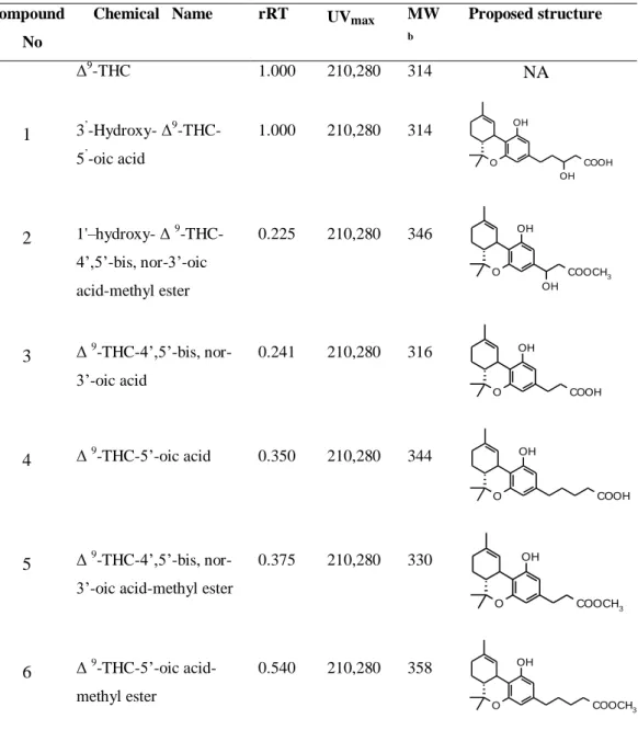
9α,10α-EHHC and 3'-hydroxy-∆9-THC (Table 3) are pharmacologically more
active than THC (Yamamoto et al. 2003).
Metabolism of THC in humans
The metabolism of ∆9
-THC and related cannabinoids in human has been studied
by utilizing parenteral, oral, and smoking as a route of administration. The
general pattern of metabolism is the same in all cases with as major metabolic
reactions the formation of an 11-hydroxy derivative and as minor product the
8-hydroxy derivative (Wall and Perez-Reyes 1981). The monocarboxylated
derivatives have been isolated from human urine (Table 3, compound 20 and
23), further side chain carboxylated and their hydroxylated derivatives occur
and were also found in urine (Table 4, compound 27, 28, 29, 30, 31, 32, 34, 35,
36, 37, 39) (Halldin et al. 1982a, Halldin et al. 1982b). Among the acid
derivatives, Δ9-THC-11-oic acid (Table 3, compound 20) (Halldin et al. 1982b,
Kanter and Hollister 1978), 4',5'-bisnor-Δ9-THC-11,3'-dioic acid (Table 4,
compound 35) (Halldin et al. 1982a) andΔ9-THC-11-oic acid glucoronide (Fig.
3a) (Williams and Moffat 1980) are identified as the major metabolites in
human urine. In vitro human liver metabolism yields 8α-hydroxy- (Table 1,
compound 2) (Bornheim et al. 1992, Widman et al. 1979), 11-hydroxy-,
8β-hydroxy- (Table 1, compound 1, 3) (Bornheim et al. 1992, Halldin et al. 1982c,
11-hydroxy-29 epoxyhexahydrocannabinol (11-OH-EHHC) (Fig. 3b) (Halldin et al. 1982c) and
8-keto-∆9-THC (Fig. 3c) (Bornheim et al. 1992).
OH
O R
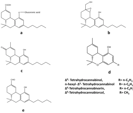
Δ9- Tetrahydrocannabinol, R= n-C 5H11
n-hexyl- Δ9- Tetrahydrocannabinol R= n-C
6H13
Δ9-Tetrahydrocannabivarin, R= n-C 3H7
Δ9-Tetrahydrocannabiorcol, R= CH 3
a b
OH CHO
OH O OH
OH O
COOH
O Glucoronic acid
c d
e
Figure 3:a; Δ9-THC-11-oic acid glucoronide b; 11-hydroxy-EHHC c; 8-keto-∆9-THC
d; Derivatives of THC e; 11-oxo- ∆9- THC.
Metabolism of THC in rabbits
After in vivo administration of the ∆9-THC to rabbits, a number of mono- and
dihydroxy derivatives (Table 1 ,2) (Nordqvist et al. 1979a), monocarboxylic
acid (Table 3, Compound 20) and side chain acids (Table 4, compound 26, 27,
29, 31, 32, 33, 34, 35, 38, 39) (Burstein et al. 1972, Nordqvist et al. 1979a) were
30
(Table 4, compound 35) as a major compound (Nordqvist et al. 1974).
Furthermore, three side-chain monocarboxylic acids hydroxylated in the allylic
position in the isoprene moiety were also identified as O-glucuronide in rabbit
urine (Nordqvist et al. 1979b). Rabbit in vitro liver microsomal metabolism of
∆9
-THC formed 11-hydroxy- (Table 1, compound 1) (Burstein and Kupfer
1971a, Nilsson et al. 1970; Wall et al. 1970) and 8α-hydroxy-∆9-THC (Table 1,
compound 2) (Benzvi and Burstein 1975). Microsomal oxygenase was found to
catalyze the oxidation of 11-hydroxy-Δ8-THC to 11-oxo-Δ8-THC (Watanabe et
al. 1979).
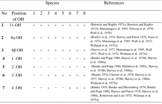
Table 1. Monohydroxylated metabolites of ∆9- THC. (1) man, (2) rhesus monkey, (3) mouse, (4) rat, (5) rabbit, (6) guinea pig, (7) dog, (8) microorganisms.
Species References
No Position
OH of OH
1 2 3 4 5 6 7 8
1 11-OH + + + + + + + + (Burstein and Kupfer 1971a, Burstein and Kupfer 1971b, Matsunaga et al. 1995, Nilsson et al. 1970, Wall et al. 1970)
2 8α-OH + + + + + + + + (BenZvi et al. 1974, Harvey and Paton 1976, Jones et al. 1974, Matsunaga et al. 1995, Wall et al. 1972, Widman et al. 1975a)
3 8β-OH + + + + + + + + (Harvey et al. 1977, Matsunaga et al. 1995, Wall 1971, Wall et al. 1972, Widman et al. 1975a)
4 1’-OH − − − − − + − + (Binder and Popp 1980, Harvey et al. 1978b, Harvey et al. 1980a)
5 2‘-OH + − − − − + − + (Binder and Popp 1980, Halldin et al. 1982c, Harvey et al. 1978b, Harvey et al. 1980a)
6 3‘-OH − − + + + + + + (Binder 1976, Christie et al. 1978, Harvey et al. 1977, Harvey et al. 1978b, Harvey et al. 1980a, Widman et al. 1975a)
7 4‘-OH − − + − − + + + (Binder 1976, Binder and Meisenberg 1978, Binder and Popp 1980, Harvey and Paton 1978, Harvey et al. 1980a, Robertson and Lyle 1975, Widman et al. 1975a)
Metabolism of THC in mouse
Harvey and Paton (1978) identified multiple substituted metabolites formed by
3'-31
Table 2. Di- and trihydroxy metabolites of ∆9-THC. (1) man, (2) rhesus monkey, (3) mouse, (4) rat, (5) rabbit, (6) guinea pig, (7) dog, (8) microorganisms.
Species References
No Position
of OH
Groups 1 2 3 4 5 6 7 8
9 8α , 11-di-OH + + + + + + − − (Benzvi Z. and Burstein 1975, Halldin et al. 1982c, Harvey and Paton 1976, Harvey et al. 1978b, Harvey et al. 1980a, Jones et al. 1974, Wall and Brine 1976, Wall et al. 1972)
10 8β, 11-di-OH + + + + − + + − (Halldin et al. 1982c, Harvey et al. 1977, Harvey et al. 1978b, Harvey et al. 1980a, Wall and Brine 1976)
11 1’, 11-di-OH − + − − − + − − (Harvey et al. 1980a, Wall and Brine 1976)
12 2’, 11-di-OH + + + − − + − − (Halldin et al. 1982c, Harvey et al. 1977, Harvey et al. 1978b, Harvey et al. 1980a, Wall and Brine 1976)
13 3’, 11-di-OH − + + + − + − + (Christie et al. 1978, Halldin et al. 1982c, Harvey et al. 1977, Harvey et al. 1978b, Wall and Brine 1976)
14 4’, 11-di-OH + + + − − + − + (Binder 1976, Halldin et al. 1982c, Harvey and Paton 1978, Harvey et al. 1980a, Wall and Brine 1976)
15 4’, 8α-di-OH − − − − − − − + (Binder 1976)
16 1’-4’, 8β-di-OH − − − − − + − − (Harvey et al. 1980a)
17 1’, 2’-4’-di-OH − − − − − + − − (Harvey et al. 1980a)
18 2’-4’ ,8α,11-tri- OH
− − + − − + − − (Harvey and Paton 1978, Harvey et al. 1977, Harvey et al. 1978b, Harvey et al. 1980a)
19 2’-4’, 8β,11-tri- OH
− − − − − + − − (Harvey et al. 1980a)
the monohydroxy derivatives (Table 1, compound 1, 5, 6), 8β, 11-; 2', 11-; and
3', 11-dihydroxy derivatives (Table 2, compound 10, 12, 13 ), 2', 8α, 11- and 3',
8α, 11- the most abundant trihydroxy derivatives (Table 2, compound 18).
Harvey and Paton (1978) also found the formation of 2',11-, and
3',11-dihydroxy-8-oxo-∆9-THC (Table 6, compound 48) in mouse liver and
4'-hydroxylation (Table 1, compound 7) was suggested as a major metabolic route
for the ∆9
-THC transformation (Harvey et al. 1977). Mono- and dihydroxy-
32
(Table 3, compound 20) was identified from the mouse liver together with its
8α- and 2'- monohydroxy derivative (Table 5, compound 41 and 43), 2',8α- and
3',8α-dihydroxy derivatives (Table 5, compound 45) (Harvey and Paton 1976;
Harvey et al. 1978b). In vitro mouse liver microsomes transformed ∆9-THC into
11-hydroxy- (Jones et al. 1974) and 8α-hydroxy-∆9-THC (BenZvi et al. 1974)
(Table 1, compound 1,2). Later on, transformation of 8α-; 8β- and
11-hydroxy-∆9
-THC was studied (Burstein and Shoupe 1981), metabolism of
8α-hydroxy-∆9
-THC was parallel to that of ∆9-THC but the presence of 8β-hydroxy group
was found to suppress the hydroxylation and oxidation at C-11 and to increase
the β-oxidation of the side-chain (Harvey et al. 1980b).
The in vivo metabolism of Δ8-THC produced three alcohols, five diols, six
triols, five monohydroxy acids, six dihydroxy acids, two substituted ketones, an
epoxide, three dihydroxy metabolites and a glucuronide conjugate (Harvey and
Paton 1980). Δ11-THC was transformed into 26 metabolites produced by
epoxidation among which 9α,11- and 9β,11-dihydroxyhexahydrocannabinols
were the key metabolites (Harvey et al. 1980c). 11-oxo-Δ8-tetrahydrocannabinol
(11-oxo- Δ8-THC) was identified from the mouse brain as a new in vivo
metabolite of Δ8-THC (Watanabe et al. 1980).
Metabolism of THC in monkey, guinea pig and dog
Purified Cytochrome P450 isozyme of monkey metabolized Δ9-THC to 11-;
8α-; 8β- and 3’- hydroxy-Δ9-THC (Table 1, compound 1, 2, 3, 6) (Matsunaga et al.
1995). Moreover, Δ9-THC was also metabolized to some dihydroxy derivatives,
such as 8α,11-; 8β,11-; 1’,11-; 2’,11-; 3’,11- and 4’,11-Δ9-THC (Table 2,
compound 9, 10, 11, 12, 13, 14) (Wall and Brine 1976).
Metabolism and autoradiography distribution showed the biotransformation of
∆8
33
Table 3. Mono-carboxylic acid metabolites of ∆9-THC. (1) man, (2) rhesus monkey, (3) mouse, (4) rat, (5) rabbit, (6) guinea pig, (7) dog, (8) microorganisms.
OH
R
2
O
R
1
metabolite in the monkey Callithrix jacchus (Just et al. 1975). The rhesus
monkey metabolized Δ8-THC to various monohydroxy and dihydroxy
metabolites. 11-hydroxy-Δ8-THC was the most abundant metabolite.
Furthermore, all the side-chain hydroxy metabolites except 5'-hydroxy- Δ8-THC
were identified. The total 4'-hydroxy- derivative was present in about one-third
and 3'-hydroxy- in about one-sixth of the amount of 11-hydroxy-Δ8-THC. Only
minor amounts of 1'- and 2'-hydroxy- Δ8-THC were isolated (Halldin et al.
1979, Widman et al. 1979).
No R1 R2 Species References 1 2 3 4 5 6 7 8
20 COOH C5H11 + + + + + + + - (BenZvi and Burstein 1974, Halldin et
al. 1982c, Harvey and Paton 1976, Harvey et al. 1978b, Harvey et al. 1980a, Kanter and Hollister 1978, Nordqvist et al. 1979a)
21 CH3 COOH - - - + - - (Harvey et al. 1980a)
22 CH3 CH2COOH - - - + - - (Harvey et al. 1978b, Harvey et al.
1980a)
23 CH3 C2H2COOH + + + - - + - + (Halldin et al. 1982b, Harvey et al.
1978b, Harvey et al. 1980a, Nordqvist et al. 1979a, Robertson et al. 1978a)
24 CH3 C3H6COOH - - - + - - (Harvey et al. 1978b, Harvey et al.
1980a)
34
Several mono-, di- and trihydroxy derivatives were formed by in vivo liver
metabolism of ∆9
-THC by guinea pigs. The 11-; 8α-; 8β-; 1’-; 2’- and 3’- were
the monohydroxy derivatives (Table 1, compound 1,2 ,3 4, 5, 6), 8α,11-; 8β,11-;
2,11-; and 3,11-dihydroxy derivatives (Table 2, compound 9, 10, 12, 13),
2,8α,11- and 3,8α,11- were major trihydroxy derivatives (Table 2, compound
18) (Harvey et al. 1978b). Guinea pigs also produced large amounts of the
monocarboyxlic acids (Table 3, compound 20, 21, 22, 23, 24), substituted
side-chain carboxylic acid (Table 4, compound 32), mono- and dihydroxy-
derivatives of ∆9
-THC-11-oic acid (Table 5, compound 41, 42, 43, 44). The
major metabolic pathways involved were allylic and aliphatic hydroxylations,
oxidation of alcohols to ketones and acids, β oxidative degradation of the pentyl
side chain and conjugation with glucuronic acid (Harvey et al. 1978a, Harvey et
al. 1980a).
Twenty-nine metabolites were reported in a study of ∆8-THC metabolism by
guinea pigs. The 1'-hydroxy- and 4'-oxo-Δ8-THC-11-oic acid were new
metabolites with other metabolites having β-oxidation at the side chain (Harvey
and Paton 1981). Hepatic microsomes of guinea pig oxidized ∆8-THC to
7α-OH- and 7β-7α-OH-∆8-THC which were further converted into 7-oxo-∆8-THC but
with different mechanisms in both cases (Narimatsu et al. 1988).
Isolated perfused dog lung metabolized ∆9
-THC into 3'-hydroxy- and
4'-hydroxy- derivatives (Table 1, compound 6, 7) as the major metabolites,
whereas small amounts of 8α- and 8β-hydroxy-∆9-THC (Table 1, compound 2,
3) were also produced which are predominant metabolites identified from in
35
Table 4. Substituted side-chain carboxylic acid metabolites of ∆9-THC (1) man, (2) rhesus monkey, (3) mouse, (4) rat, (5) rabbit, (6) guinea pig, (7) dog, (8)
microorganisms.
Species References
No Side- chain
Substituents
1 2 3 4 5 6 7 8
26 COOH 11-OH, 8α-OH, 8β-OH, 8β,
11-di-OH
− − − − + − − − (Nordqvist et al. 1979a)
27 COOH 9-COOH + − − − + − − − (Halldin et al. 1982a, Nordqvist et al. 1979a)
28 CH2COOH 11-OH + − − − − − − − (Halldin et al. 1982b) 29 CH2COOH 8β-OH + − − − + − − − (Halldin et al. 1982b,
Nordqvist et al. 1979a, Nordqvist et al. 1979b)
30 CH2COOH 9-COOH + − − − − − − − (Halldin et al. 1982a)
31 C2H4COOH 11-OH + − − − + − − − (Halldin et al. 1982b,
Nordqvist et al. 1979b)
32 C2H4COOH 8β-OH + − − − + + − − (Halldin et al. 1982b,
Harvey et al. 1980a, Nordqvist et al. 1979a, Nordqvist et al. 1979b)
33 C2H4COOH 8β, 11-di- OH − − − − + − − − (Nordqvist et al. 1979a)
34 C2H4COOH
35 C2H4COOH
9-COOH 11-COOH + + − − − − − − + + − − − − − −
(Halldin et al. 1982a, Nordqvist et al. 1979a, Nordqvist et al. 1974) (Halldin et al. 1982a, Nordqvist et al. 1974)
36 C3H6COOH 8β-OH + − − − − − − − (Halldin et al. 1982b)
37 C3H6COOH 9-COOH + − − − − − − − (Halldin et al. 1982a)
38C4H8COOH 9-COOH − − − − + − − − (Nordqvist et al. 1979a,
Nordqvist et al. 1979b)
39 CH=CH- COOH
9-COOH + − − − + − − − (Halldin et al. 1982a, Nordqvist M. et al. 1979a)
40 CH2- CH=CH-COOH
9-COOH + − − − − − − − (Halldin et al. 1982b)
Metabolism of THC in mice and rat
36
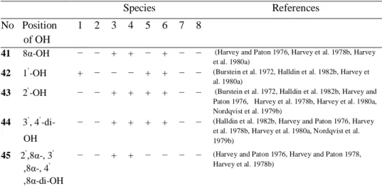
Table 5. Hydroxy and dihydroxy derivatives of ∆9-tetrahydrocannabinol-11-oic acid. (1) man, (2) rhesus monkey, (3) mouse, (4) rat, (5) rabbit, (6) guinea pig, (7) dog, (8) microorganisms
Species References
No Position of OH
1 2 3 4 5 6 7 8
41 8α-OH − − + + − + − − (Harvey and Paton 1976, Harvey et al. 1978b, Harvey et al. 1980a)
42 1‘-OH + − − − + + − − (Burstein et al. 1972, Halldin et al. 1982b, Harvey et al. 1980a)
43 2‘-OH − − + + + + − − (Burstein et al. 1972, Halldin et al. 1982b, Harvey and Paton 1976, Harvey et al. 1978b, Harvey et al. 1980a, Nordqvist et al. 1979b)
44 3‘, 4‘ -di-OH
− − + + + + − − (Halldin et al. 1982b, Harvey and Paton 1976, Harvey et al. 1978b, Harvey et al. 1980a, Nordqvist et al. 1979b)
45 2‘,8α-, 3‘ ,8α-, 4‘ ,8α-di-OH
− − + + − − − − (Harvey and Paton 1976, Harvey and Paton 1978, Harvey et al. 1978b)
Table 6. Mono- and di-hydroxy derivatives of 8-oxo-∆9-tetrahydrocannabinol. (1) man, (2)rhesus monkey, (3) mouse, (4) rat, (5) rabbit, (6) guinea pig, (7) dog, (8)
microorganisms.
Species References
No Position of OH 1 2 3 4 5 6 7 8
46 7-, 2‘-, 3‘-OH − + − − − + − − (Harvey et al. 1977, Harvey et al. 1978b, Harvey et al. 1980a)
47 4`-OH − − − − − + − + (Binder 1976, Harvey et al. 1980a)
48 2‘, 11-, 3‘, 11-, 4‘, 11-di-OH.
− − + − − − − − (Harvey and Paton 1978, Harvey et al. 1977, Harvey et al. 1978b)
male mice (Charles River CD-1) to predict metabolic pathways and to find new
metabolites. Brown and Harvey (1988a) reported the metabolism of ∆9
-tetrahydrocannabiorcol (Δ9-THCO) (Fig. 3d) and ∆8-Tetrahydrocannabiorcol
(Δ8-THCO) into ∆9-THC-11- and Δ8-THCO-11-oic acid respectively. Sixteen
metabolites were identified from n-hexyl-∆9-THC (Fig. 3d) and eleven from
n-hexyl-∆8-THC (Brown and Harvey 1988b). Seven metabolites were formed
-37 tetrahydrocannabivarin (∆8
-THCV). The major biotransformation pathway led
to the production of 11-hydroxy-tetrahydrocannabivarin and their further
oxidation to carboxylic acid metabolites, other metabolites were mainly the
hydroxy derivatives of these compounds (Brown and Harvey 1988c).
Metabolism of 2'-, 3'- and 4’-hydroxy-Δ9-THC were also studied in mice.
Hydroxylation at the allylic 11-position followed by oxidation to the carboxylic
acid was found to be an important metabolic pathway (Harvey 1990). Little
oxidative degradation of the pentyl side chain was found for 2'-hydroxy-∆9
-THC (Table 1, compound 5) but abundant metabolism occurred by oxidative
cleavage of the pentyl side chain from 3’ and 4’-hydroxy-∆9-THC (Table 1,
compound 6, 7) (Harvey 1989).
Harvey and Paton (1979) described the conversion of deuterium-labeled ∆8-,
∆11
- and ∆9-THC into a number of metabolites containing 1-3 additional groups
at positions 2'; 3'; 4'; 8α; 8β and 11 with the only difference that ∆11-THC did
not undergo allylic hydroxylation at position 11. In vivo liver metabolites of ∆9
-THC were extracted from rats. The 11-; 8α-; 8β- and 3’- (Table 1, compound 1,
2, 3 and 6) were the monohydroxy-, 8α,11-; 8β,11- and 3’,11- (Table 2,
compound 9, 10, 13 ) were the major dihydroxy- derivatives (Harvey et al.
1978b). Rat liver microsomes transformed ∆9-THC into 11-oxo- (Fig. 3e) and
further reduction of this derivative produced 11-hydroxy-∆9-THC (Table 1,
compound 1) (BenZvi and Burstein 1974, Burstein and Kupfer 1971a). Rat
lever homogenates led to the formation of 11-hydroxy- (Table 1, compound 1)
(Burstein and Kupfer 1971b) and 8α,11-dihydroxy-∆9-THC (Table 2, compound
9) (Burstein and Kupfer 1971a, Wall 1971, Wall et al. 1970).
Rat liver incubation of ∆8
-THC produced 11-hydroxy-; 7α,11- and
7β,11-dihydroxy-∆8-THC (Wall 1971). Purified cytochrome P450 isozymes from rat
11-oxo-Chapter 2
38 ∆8
-THC to ∆8-THC-11-oic acid (Watanabe et al. 1991). Sex related differences
were observed in the oxidative metabolism of ∆9
-THC between male and
female rats. Liver microsomes of male rats biotransformed ∆9
-THC into various
metabolites unlike female rats in which it was mainly oxidized selectively to
11-hydroxy-∆9-THC (Table 1, compound 1) (Burstein and Kupfer 1971a,
Narimatsu et al. 1991).
Biotransformation of THC in mammalian cells shows that cytochrome P450
enzymes play an important role in the hydroxylation of THC. THC undergoes
allylic hydroxylation at C-11 to form 11-hydroxy-THC which is the most
common metabolite in almost every species studied. Allylic hydroxylation is
also common at position 8 to give 8α- and 8β-hydroxy-THC. Other than allylic
hydroxylations there are a number of monohydroxy and dihydroxy metabolites
formed, being hydroxylated at all the positions of the alkane side chain of THC.
There is however considerable species variation in the position of substitution.
In addition to monohydroxy and dihydroxy, some polyhydroxylated metabolites
have also been found in mice with hydroxyl substitution at position 2’ and 3’. In
monkeys and dogs, hydroxyl substitution was more common on the positions 3’
and 4’ while the monkey also formed minor amounts of metabolites having
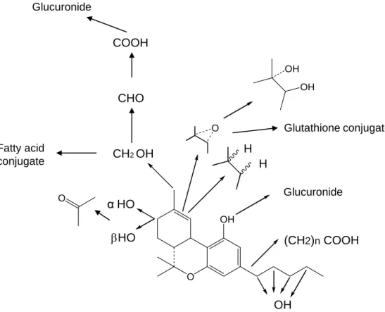
hydroxylation on position 1’ and 2’. The major metabolic pathway of THC (Fig.
4) after monohydroxy metabolism is further oxidation to either a carbonyl
compound or, in the case of the primary alcohols to a carboxylic acid, followed
by glucuronidation of the acid. Following this biotransformation pathway there
are a number of THC acids which are formed in man, rats, rabbits and guinea
pigs where the acids link with glucuronide and become the major urinary
39
Glucuronide
O
OH O
OH
OH
O
αHO
HO
Glucuronide
(CH2)nCOOH CH2 OH
CHO COOH
Fatty acid conjugate
OH
Glutathione conjugate
H H
Figure 4: Metabolic pathway of ∆9-THC
Biotransformation of Cannabidiol
Biotransformation of cannabidiol (CBD) has extensively been studied in mice
and rats. Fourteen metabolites were isolated from the liver of mice after the
administration of CBD. The most significant biotransformation step was
glucoronide conjugation and to a lesser extent CBD-11-oic-acid was formed
(Martin et al. 1977). Eight monohydroxy (Martin et al. 1976b) and ten
40
(Martin et al. 1976a), in both cases mainly 11-hydroxylation was formed with
further hydroxylation at C-4 of the side chain. Over fifty metabolites of CBD
were identified with considerable species variation in dog, rat and human urine.
Thirty three metabolites were identified from the urine of dystonic patients
treated with CBD. The major metabolic pathway was the hydroxylation and
oxidation at C-11 and further hydroxylation in the pentyl and propyl groups
produced 1'-; 2'-; 3'; 4'- and 10-hydroxy derivatives of CBD-11-oic acid. Acids
were formed by β-oxidation and further biotransformations from the pentyl
side-chain resulted in the formation of the oxidized metabolite CBD-11-oic acid
(Harvey and Mechoulam 1990). In dog unusual glucoside conjugates were
found of the metabolites 4’-; 5’-hydroxy- and 8-oxo-CBD. Other metabolites in
all three species were mainly acids. Side-chain hydroxylated derivatives of
CBD-11-oic acid were abundant in human urine (Harvey et al. 1991). CBD
followed almost the same pattern of biotransformation as observed in
Tetrahydrocannabinol, with several variations in pathways caused by different
species. The monohydroxylation of the side chain and also at position 11 and 8
found in CBD is quite similar to THC. Dihydroxy compounds are also reported
but unlike THC no trihydroxy metabolites are found in any species treated with
CBD. A large number of metabolites are formed by β-oxidation of the side
chain and Glucoronidation of CBD.
Biotransformation of Cannabinol
Cannabinol was metabolized by rat liver into side chain hydroxylated
compounds: the 2'-; 3'-; 4'- and 5'-hydroxy were isolated as minor metabolites
and 11-hydroxy-CBN was formed as the major metabolite (Widman et al.
1975b). Cannabinol-11-oic acid was found as a major compound from rat
feces (Yisak et al. 1977). The administration of 14C-labeled cannabinol to rats
4'-41 hydroxy- (Lindgren et al. 1977, Yisak et al. 1977) and the dihydroxy- 1',11-;
4',11-; 2’,11- and 3’,11-dihydroxycannabinol were the most abundant
compounds (Fonseka and Widman 1977, Yisak et al. 1977).
In rabbits, 4'-hydroxy-CBN was found as a major compound together with
smaller amounts of 5'- and 3'-hydroxy-CBN (Widman et al. 1975b). In mice,
the 11-position and the side chain of cannabinol was hydroxylated followed by
further oxidation to acidic metabolites (Burstein and Varanelli 1975).
Biotransformation of CBN also follows the same pattern like other
cannabinoids but unlike other cannabinoids allylic hydroxylation of the terpene
ring is absent in the metabolism of CBN. The 11-hydroxylation is the major
metabolic route and further oxidation of this monohydroxy compound produces
a common acidic metabolite, CBN-11-oic acid.
Biotransformation of cannabinoids by fungi and bacteria
Robertson et al. (1978a, 1978b) screened more than 100 species of fungi and
bacteria to investigate microbial transformations of the four common
cannabinoids, namely CBD, CBN, Δ8-THC and Δ9-THC. Syncephalastrum
racemosum and Mycobacterium rhodochrous partially degraded the n-pentyl
side chain of all these four compounds. Carboxylic acid and alcohol side chain
derivatives were found to be the major metabolites. Among side chain
derivatives, the 4’-hydroxy- metabolites were the most abundant compounds in
the transformation of CBD, CBN, Δ8-THC and Δ9-THC by Syncephalastrum
racemosum (Robertson and Lyle 1975).
Δ8
-Tetrahydrocannabinol was hydroxylated at the ring system and at the side
chain by fermentation with Pellicularia filamentosa, Streptomyces lavendulae
or P. filamentosa and yielded the compounds 7β,3'- and 7β,4'-dihydroxy-Δ8
42
7α,3'-; 7α,4'-dihydroxy- Δ8
-THC and 4'-hydroxy-7-oxo-Δ8-THC (Vidic et al.
1976).
Binder and Meisenberg (1978) indentified 51 fungal and bacterial strains which
actively transformed Δ9-THC into 11-; 8α-; 8β-; 3’- and 4’-hydroxy- (Table 1,
compound 1, 2, 3, 6) and 4’,11-dihydroxy-∆9-THC (Table 2, compound 14).
Cunninghamella blakesleeana also produced 4’-hydroxy-8-oxo-∆9-THC (Table
6, compound 47) (Binder 1976).
Binder and Popp (1980) further studied the metabolic transformations of 9
-THC by using cultures of Fusarium nivale, Gibberella fujikuroi (Ascomycetes)
and Thamnidium elegans (Phycomycetes). A number of metabolites were
isolated from these species after which they were partly purified and their
structures elucidated by combined gas chromatography/mass spectrometry.
Thamnidium elegans formed 11-; 8α-; 8β-; and 1’-hydroxy- (Table 1, compound
1, 2, 3, 4) and 2’,8β-dihydroxy- 9-THC (Table 2, compound 16). Fusarium
nivale and Gibberella fujikuroi both converted 9-THC to the metabolites 2’-,
3’- and 4’-hydroxy- 9-THC (Table 1, compound 5, 6, 7). These results show
that there are two different enzyme systems capable of hydroxylating the
substrate. System 1, which is common to the “Fusarium” type and
microorganisms are restricted in its hydroxylating capacity to the side chain
C-atoms 2’, 3’ and 4’ of cannabinoids. In addition to this ‘aliphatic hydroxylase’
Thamnidium elegans posseses an ‘allylic hydroxylase’ capable of hydroxylating
9
-THC in positions 1’, 8 and 11.
Microorganisms show similarities with mammalian hydroxylation of
cannabinoids. Cannabinoids undergo allylic hydroxylation at C-11 to form
11-hydroxy- derivatives. Allylic hydroxylation is also seen at position 8 to give 8α-
and 8β-hydroxy derivatives but unlike mammals, hydroxylation has also been
43 to form a carbonyl compound. In microorganisms, side chain degradation is
found as one of the major metabolic pathways of cannabinoids. Side chain
hydroxyl and carboxyl substitution is also common; hydroxylation is more
likely on C-atom 2’, 3’ and 4’ with their corresponding dihydroxy metabolites.
Trihydroxy derivatives of naturally occurring cannabinoids are not found in
bacteria or fungi. Side chain acid derivatives are present but carboxylation of
the monoterpene moiety and glucoronidation is not reported.
Plant biotransformation of cannabinoids
Cannabielsoin (CBE) and its diastereoisomers have been isolated from the
suspension cultures of C. sativa, Saccharum officinarum and tissue culture of C.
sativa, after the administration of cannabidiol (Braemer and Paris 1987, Tanaka
et al. 1997). ∆9-THC was converted into cannabicoumaronon by a cell
suspension culture of C. sativa (Braemer and Paris 1987). Tissue culture of
Pinelli ternatata transformed ∆8-THC and CBN into their glucopyranoside
derivatives ∆8-THC-2’-O-β-D-Glucopyranoside (Tanaka et al. 1997) and
CBN-2’-O-β-D-Glucopyranoside, respectively (Tanaka et al. 1993).
Discussion and conclusions
Despite having useful psychomimetic and pharmacological activities, research
into cannabis has been mainly focused on its use as a recreational drug and the
subsequent legal aspects controlling the possession of this material. Some of
this research has been conducted on the metabolism of cannabinoids in
mammalian systems, leading to a better understanding of their
pharmacodynamics and pharmacokinetics. A large library of THC isomers and
their derivatives have been developed by using mammalian cytochrome P450
systems, which is a successful approach to finding metabolic pathways but little
is known about the activity and interaction of these metabolites with the CB1
44
Perhaps the industrial scale production of potential compounds by means of
mammalian cells or tissue is an expensive tool and thus an obstacle to develop
cannabinoid based drugs. There are a number of other sources which can be
used to enlarge the library of active metabolites obtained from the
transformation of cannabinoids. Cannabinoids have been successfully
transformed by fungal and bacterial strains, while there are only a few reports
on the biotransformation with plant cell cultures. Microorganisms mimic some
of the mammalian biotransformation pathways and their enzyme systems have
great potential to produce improved or novel compounds in large quantities
keeping production at a low cost.
Bacterial cytochrome P450 (monooxygenases) enzymes have the ability to
hydroxylate exogenous and endogenous compounds at different positions. In
the case of cannabinoid transformations, hydroxylation is likely to produce
more polar cannabinoids which might be more suitable for medicinal use than
parent compounds, which generally tend to accumulate in fat tissues. The
significant role of the alkane side chain has been reported in the
pharmacological action of THC (Billy et al. 1995) and further oxidation may
change the chemistry of the side chain and lead to enhanced or modified activity
of the compound. By taking into account these findings, we used alkane
degrading bacterial strains for the transformation of ∆9
-THC and found eight
major metabolites produced in mg scale. All these transformations were limited
to the side chain and included two carboxylic acid derivatives formed by
oxidation and β-oxidation of the terminal hydroxyl group of 5’-hydroxy-Δ9
-THC (Rashidi et al. 2009). Numbers of new lead candidates are possible to
obtain by using the enzymatic systems of bacterial, yeast and plant cells in
cannabinoid transformation in combination with advanced chromatographic and