R E V I E W
Open Access
The role of leptin in the respiratory system:
an overview
Foteini Malli, Andriana I Papaioannou, Konstantinos I Gourgoulianis, Zoe Daniil
*Abstract
Since its cloning in 1994, leptin has emerged in the literature as a pleiotropic hormone whose actions extend from immune system homeostasis to reproduction and angiogenesis. Recent investigations have identified the lung as a leptin responsive and producing organ, while extensive research has been published concerning the role of leptin in the respiratory system. Animal studies have provided evidence indicating that leptin is a stimulant of ventilation, whereas researchers have proposed an important role for leptin in lung maturation and development. Studies further suggest a significant impact of leptin on specific respiratory diseases, including obstructive sleep apnoea-hypopnoea syndrome, asthma, COPD and lung cancer. However, as new investigations are under way, the picture is becoming more complex. The scope of this review is to decode the existing data concerning the actions of lep-tin in the lung and provide a detailed description of leplep-tin’s involvement in the most common disorders of the respiratory system.
Introduction
In the past years, a growing number of studies have examined the potential role of leptin in the respiratory system. Accumulative data have identified foetal and adult lung tissue as leptin responsive and producing organs, while leptin’s involvement in pulmonary home-ostasis has become increasingly evident (Table 1). On the basis of this conception, researchers have sought to determine the impact of leptin on specific respiratory disorders, including obstructive sleep apnoea-hypopnoea syndrome (OSAHS), asthma, chronic obstructive pul-monary disease (COPD) and lung cancer. We review herein the current understanding on the actions of lep-tin in the lung, and summarize the recent advances on its role in the pathophysiology of respiratory diseases.
Leptin and the Leptin Receptor at a glance
Leptin, a 16KDa protein of 167 amino acids, represents the product of theobgene which in humans is located on chromosome 7 [1]. The protein is synthesized and secreted mainly by white adipose tissue, apparently in proportion to fat stores, and thus is considered an adi-pokine [2]. However, leptin is produced in lower amounts by other tissues, such as the placenta [3],
gastric fundic mucosa [4], and pancreas [5]. Regarding the lung, theobgene is expressed in foetal lung tissue in baboons [6], and foetal rat lung fibroblasts [7] (Table 1). Interestingly, others have demonstrated the produc-tion of leptin in human peripheral lung tissue, namely bronchial epithelial cells, alveolar type II pneumocytes, and lung macrophages [8,9].
Accumulated evidence suggest that leptin production is mainly regulated by food intake; fasting reduces leptin levels while food consumption is associated with a tran-sient increase inobgene expression [10]. However, lep-tin levels can be influenced by other factors as well. Insulin and glucocorticoids can stimulate leptin secre-tion [11]. Leptin concentrasecre-tions are increased during infection and sepsis [12], in accordance with the obser-vation that leptin expression is up-regulated by various pro-inflammatory cytokines, including tumor necrosis factor-a (TNF-a) [13], interleukin-1 (IL-1) and leukae-mia inhibitory factor [14]. In contrast to acute stimula-tion of the inflammatory system, chronic inflammastimula-tion causes a reduction in leptin levels [15]. Moreover, the
obpromoter is induced by several transcription factors, such as hypoxia inducible factor-1 (HIF-1) [16], and suppressed by others, like peroxisome proliferators-acti-vated receptor-g agonists [17]. Leptin expression is inhibited by testosterone, whereas it is increased by ovarian sex steroids [14] in agreement with the strong
* Correspondence: [email protected]
Respiratory Medicine Department, University of Thessaly School of Medicine, University Hospital of Larissa, 41110, Greece
gender-related dimorphism of leptin levels (i.e. leptin is higher in females than age and body mass index (BMI) matched males) [12]. Finally, leptin concentrations are reduced by catecholamines [18].
The discovery of leptin was considered synonymous to the discovery of the antidote to obesity.ob/obmice have a single base pair mutation in the leptin gene that results in the absence of functional leptin, increased body weight, hyperphagia, impaired energy homeostasis, and low resting metabolic rate. Exogenous administra-tion of leptin reverses this phenotype [19]. Addiadministra-tional studies demonstrate that leptin crosses the blood brain barrier and serves as an afferent signal, originating from the adipose tissue, engaging distinct hypothalamic effec-tor pathways to suppress appetite and augment energy expenditure [20]. However, in humans, the action of lep-tin as an anorexigen is more complex. Human obesity is associated with increased circulating leptin levels and a relative leptin“insensitivity” [21]. Central resistance to leptin might be the result of diminished brain leptin transport [22] and/or down-regulation of the leptin receptor in the central nervous system (CNS) [23].
Beyond its metabolic functions, leptin is implicated in various other physiologic processes, including the immune response, with effects in both innate and adap-tive immunity. Indeed, leptin up-regulates the expres-sion of several pro-inflammatory cytokines, such as TNF-a, IL-6, and IL-12, while it increases chemotaxis and natural killer cells function [24,25]. Leptin enhances T helper (Th) 1 response and suppresses Th2 pathways, whereas it can exert direct effects on CD4+ T lympho-cyte proliferation and macrophage phagocytosis [12,25,26]. Moreover, leptin stimulates the proliferative activity of human monocytes in vitro and up-regulates the expression of several activation markers, like CD25 and CD38 [24].
The pleiotropy of leptin is reflected by the multiplicity of its biologic effects in other tissues. Leptin increases
sympathetic nervous system (SNS) activity [27,28], with possible implications in endothelial cell function and blood pressure homeostasis [29]. Furthermore, the adi-pokine up-regulates various pro-angiogenic factors, such as CC-chemokine ligand 2 (CCL2) [30], while synergisti-cally stimulates angiogenesis with vascular endothelial growth factor (VEGF) [31], indicating that it may contri-bute to the promotion of neo-vascularization processes [32]. Additionally, leptin has been proposed to mediate wound re-epithelization and healing [33], bone turn-over and skeletal development [34], as well as fertility [35]. Moreover, data suggest that leptin stimulates insu-lin secretion, regulates fatty acid oxidation [36] and reduces cortisol synthesis [37]. The implication of leptin in lung physiology and pathophysiology is discussed extensively below.
The leptin receptor (Ob-R) is a member of the class I cytokine receptor super-family, which includes the receptors of IL-1, IL-2, IL-6 and growth hormone [38]. Alternate splicing of the leptin receptor gene (dbgene) gives rise to six receptor isoforms that share a common extracellular and transmembrane domain, and a variable intracellular residue, characteristic for each type. The isoforms are classified according to the length of their cytoplasmic domain to four short (Ob-Ra, Ob-Rc, Ob-Rd and Ob-Rf) and one long form (Ob-Rb), while a soluble form (Ob-Re) also exists [26]. The long functional iso-form is expressed abundantly in the hypothalamus and is essential for signal transduction through Janus Kinase-signal transducer and activation of transcription factor (JAK-STAT) pathway [39]. The short isoforms are expressed in various tissues, such as the kidney, however their function has not been fully elucidated [38,40].
Importantly, dbgene is expressed in lung tissue; stu-dies in several animal models, including mice, rats, baboons and other animals, have identified Ob-R pre-sence in the lung (Table 2) [6,7,40-46]. Interestingly, other studies have localized the expression of Ob-R in
Table 1 Effects of leptin signaling in lung cells
Reference (year)
Effect Comments
Vernooy et al8 (2009)
Increased leptin and Ob-Rb expression in bronchial epithelial cells following smoke exposure
Leptin induces phosphorylation of STAT-3 in NCI-H292 and A549 cell lines
Cells obtained from lung cancer patients who underwent lung surgery (disease free areas)
A549 is a human alveolar epithelial cell line-NCI-H292 is a human bronchial epithelial cell line
Bruno et al9 (2009)
Leptin increases cell proliferation and decreases TGF-brelease in 16HBE cell line
TGF-bdecreases and fluticasone propionate increases leptin receptor expression in 16HBE cell line
16HBE is a human bronchial epithelial cell line
Nair et al47 (2008)
Leptin inhibits PDGF-airway smooth muscle migration and proliferation and IL-13-induced eotaxin production
Cells obtained from lung cancer patients who underwent lung surgery (disease free areas)
Tsuchiya et
al49(1999) Leptin induces cell proliferation in SQ-5 cells by increasing the MAPkinase activity SQ-5 is a clonal cell line derived from human lungsquamous cell cancer
human airway smooth muscle cells [47], epithelial cells and submucosa of lung tissue obtained by bronchial biopsies [48]. Of great importance is the expression of Ob-Rbin cells of the lung, like bronchial and alveolar epithelial cells, including type II pneumocytes [8,9,49]. Although the functional significance of the leptin recep-tors in the periphery is largely unknown, the existence of the functional receptor isoform indicates that the lung represents a target organ for leptin signaling.
The role of leptin in lung development
Evidence indicate that leptin can be synthesized by foe-tal adipose tissue, and the placenfoe-tal trophoblast, while leptin and Ob-R genes are expressed in foetal lung tis-sue, thus suggesting its novel role in foetal lung growth and development (Table 2 and Table 3) [6,7,43,45]. Importantly, researchers have reported enhanced leptin production by foetal rat lung fibroblasts during the per-iod of alveolar differentiation [7], while others have observed increased Ob-R abundance in foetal lung tissue in advanced gestation [6].
Studies of several models of pulmonary development suggest a modulatory role for leptin in foetal lung maturity. Antenatal administration of leptin results in a
significant increase of foetal rat lung weight, possibly due to an increase in the number and maturation of alveolar type II cells, accompanied by an induction in the expression of surfactant proteins B and C [50]. Interestingly, parathyroid hormone-related protein (PTHrP), an alveolar type II cell product that enhances type II cell differentiation, increases the production of leptin by lung lipofibroblasts [7,51]. Additionally, leptin stimulates surfactant protein synthesis when added to foetal rat lung explant culture [7,50], or foetal alveolar type II cell culture, thus suggesting the existence of a regulatory paracrine feedback loop in the foetal lung [45,51]. Further support is provided by studies demon-strating that cell stretch, known to stimulate the growth and differentiation of the alveolar septal wall, induces surfactant synthesis through enhancing the paracrine actions of leptin and PTHrP [51].
Accumulated evidence suggest a role for leptin in postnatal lung development. Interestingly, leptin concen-trations on the seventh day of life are positively corre-lated with lung weight in neonatal lambs receiving leptin intravenously, suggesting its potential role in lung growth [52]. The pulmonary phenotype of genetically obese mice provides supporting evidence to the hypothesized implication of leptin in lung development;
ob/ob mice exhibit significantly decreased lung volume
and lower alveolar surface area at 2 weeks of age, when compared to heterozygotes or control animals [53].
Despite the remarkable power of the aforementioned observations, which suggest that leptin enhances lung maturation, the fact that they derive from animal lung development models represents a major limitation in extrapolating the results to the human species.
Table 2 Lung cells as a source of leptin
Species Cell type (source) Reference
Baboon (foetal) NA [6]
Rat (foetal) Fibroblasts [7]
Human Type II pneumocytes [8]
Human Lung macrophages [8]
Human Bronchial epithelial cells [8,9]
Abbreviations: NA: Not applicable
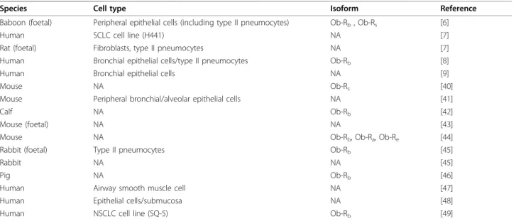
Table 3 Leptin Receptor expression in the lung
Species Cell type Isoform Reference
Baboon (foetal) Peripheral epithelial cells (including type II pneumocytes) Ob-Rb, Ob-Rs [6]
Human SCLC cell line (H441) NA [7]
Rat (foetal) Fibroblasts, type II pneumocytes NA [7]
Human Bronchial epithelial cells/type II pneumocytes Ob-Rb [8]
Human Bronchial epithelial cells NA [9]
Mouse NA Ob-Rs [40]
Mouse Peripheral bronchial/alveolar epithelial cells NA [41]
Calf NA Ob-Rb [42]
Mouse (foetal) NA NA [43]
Mouse NA Ob-Rb, Ob-Ra, Ob-Re [44]
Rabbit (foetal) Type II pneumocytes Ob-Rb [45]
Rabbit NA NA [45]
Pig NA Ob-Rb [46]
Human Airway smooth muscle cell NA [47]
Human Epithelial cells/submucosa NA [48]
Human NSCLC cell line (SQ-5) Ob-Rb [49]
Is leptin involved in Respiratory Control?
Studies in animal models have provided evidence indi-cating that leptin serves as a stimulant of ventilation.
ob/obmice exhibit increased breathing frequency,
min-ute ventilation and tidal volume, associated with signif-icantly elevated arterial PaCO2 and depressed hypercapnic ventilatory response (HCVR), present even before the onset of obesity, when compared to wild-type mice [54-57]. The aforementioned observations are evident during all sleep/wake states, although HCVR is more profoundly reduced during sleep [54]. Chronic leptin replacement restores the rapid breath-ing pattern and the diminished lung compliance asso-ciated with the obese phenotype [55]. To streamline these findings, leptin administration prevents weight gain in ob/ob mice, thus it is difficult to determine whether the attenuation of the respiratory complica-tions is caused by mechanical factors or by a direct effect of leptin on lung growth and respiration [55]. However, acute leptin replacement results in a signifi-cant increase in baseline ventilation and chemosensi-tivity during sleep, independent of weight gain [54]. Importantly, leptin microinjections into the tractus nucleus solitarius in the brain of rats is associated with increased pulmonary ventilation and respiratory volume and enhanced bioelectrical activity of the inspiratory muscles suggesting that leptin may be implicated in ventilatory control through direct effects on respiratory control centres [58].
At this point it should be mentioned that the ob/ob
model represents a model of obesity and systemic inflammation rather than a simple model of leptin defi-ciency with substantial diversities from human obesity that is associated with hyperleptinemia and central lep-tin resistance [59]. While clinical studies provide sup-porting evidence to the mouse-model observations indicating the critical role of leptin in ventilatory control (e.g. leptin is a predictor of lung function in various conditions, including asthma [60], heart failure [61] and is negatively correlated with lung volumes in COPD patients [62]) the pathophysiological significance of lep-tin regarding respiratory function in humans remains to be clarified.
The role of leptin in diseases of the lung
Over the past years, extensive research has been con-ducted concerning the impact of leptin on various respiratory disorders. Mounting evidence have been published, as the picture is becoming more complex. The scope of this review is to decode the existing data and provide a detailed description of the involvement of leptin in the most common disease entities associated with the respiratory system.
Obstructive sleep apnoea-hypopnoea syndrome (OSAHS) and obesity hypoventilation syndrome (OHS) (Table 4) OSAHS is a common disorder characterized by repeated episodes of partial or complete upper airway obstruction during sleep [63]. Approximately 90% of patients with OHS, a condition defined as a combination of obesity (i. e. BMI ≥ 30 Kg/m2) and sleep disordered breathing, have concurrent OSAHS (i.e. apnoea-hypopnoea index (AHI) > 5) [64], while 10-15% of patients with OSAHS develop hypoventilation and daytime hypercapnia [65].
Obesity is considered to be the most important risk factor of OSAHS [66]. The impact of obesity in sleep disordered breathing was originally reported to be mechanical but recent data suggest that adipose tissue can contribute to the genesis of the syndrome through its metabolic activity. The established role of leptin as a respiratory stimulant (discussed extensively above) raised the possibility that OSAHS may represent a lep-tin-deficient state. Inversely, several groups have demon-strated higher circulating leptin levels in OSAHS patients, when compared to age, sex, and weight-matched controls [67-72], while others have failed to document such a difference [73,74]. However, a collec-tive comparison of these findings is difficult, since many of the aforementioned studies have included patients with comorbid conditions (e.g. arterial hypertension) that could serve as confounding factors [68,74]. The preceding data, exhibit substantial weakness originating from the relatively small number of subjects included and, additionally, the male predominance in the majority of these reports raises difficulties in extrapolating the results to the female sex.
results should be interpreted with caution since the number of patients enrolled have been reported to be underpowered to detect a sufficient effect [81].
A subject of ongoing controversy is whether the pre-sence of hyperleptinemia in OSAHS derives from adip-osity or it reflects causality due to the effects of sleep-disordered breathing. Leptin levels are 50% higher in OSAHS patients than in controls, suggesting that other factors besides obesity contribute to the elevation of lep-tin [82]. In consistency with the previous results, leplep-tin levels are significantly correlated with several indices of OSAHS severity, i.e. AHI, percentage of sleep time with less than 90% hemoglobin saturation (%T90), oxygen desaturation index, as well as with a variety of anthropo-metric measurements, including BMI, waist-to-hip ratio (WHR), and skinfold thickness [68-70,72,75,83,84]. However, the data derived are rather contradictive; some researchers have documented a significant positive correlation of leptin levels with AHI, even when con-trolled for BMI [70], while others have reported no sig-nificant correlation between leptin values after adjustment for BMI, WHR and waist circumference, with measures of disease severity, although WHR and T %90 were found to be the most significant variables in a model predicting leptin [69]. In keeping with the afore-mentioned concepts, other researchers have documented that BMI is the only parameter significantly and inde-pendently associated with leptin concentrations [83]. Similarly, other groups have reported that adiposity measures are the only predictive factors of leptin levels, while AHI was not found to be significant [75].
To make matters more complicated, studies have documented significantly higher leptin levels in non-obese OSAHS patients versus controls [85,86]. Data sug-gest that repeated sleep hypoxemia may promote leptin production independently of the degree of obesity. How-ever, the authors provided evidence indicating that the
location of the body fat deposition (e.g. visceral fat accu-mulation) may account for the increased leptin concen-trations in non-obese OSAHS subjects [85]. Clearly, the aforementioned findings are inconclusive and due to their associative nature, cannot substantiate causality.
Additional studies examining the effects of nasal con-tinuous positive airway pressure (nCPAP) treatment were designed to elucidate the exact association of leptin with OSAHS. Leptin levels decrease significantly in OSAHS patients, treated with nCPAP for a period of 3 days to 6 months, without any significant change in BMI observed [68,83,87-89]. The significant reduction in circulating leptin following 1 to 4 days of nCPAP ther-apy [87,90] suggests that OSAHS itself may stimulate, at least in part, leptin production independently of obesity. However, the mechanisms responsible are yet unclear, and no definite conclusions can be made since several groups have reported no significant changes in leptin levels after the application of nCPAP [91,92]. Interest-ingly, Barcelo et al [86] documented a marginal, yet sig-nificant, decrease in leptin levels associated with nCPAP treatment in non-obese OSAHS patients, while leptin concentrations were reported unchanged in obese sub-jects. Similarly, others have illustrated a more pro-nounced reduction of leptin levels in non-obese patients versus obese OSAHS patients [89]. The physiological explanation has not been fully elucidated, but data in the literature suggest that the decrease in leptin might be explained by the effect of treatment on sympathetic nerve activation [90], or may be associated with changes in haemodynamics and visceral blood flow [83]. Other possible explanations include the reduction in visceral fat accumulation and stress levels [93], or a reverse in the Ob-R sensitivity [94], consistent with the hypothesis of leptin resistance discussed above.
Few studies in the literature have examined the possi-ble implication of leptin in OHS. As argued earlier,
Table 4 The role of leptin in OSAHS and OHS
Reference (year)
Main message Main limitations
Ip et al68 (2000)
Leptin significantly correlated with AHI Only males/Limited number of patients/Potential influence by comorbidities/No adjustment for FM
Campo et al78 (2007)
Higher leptin is associated with reduced respiratory drive and reduced hypercapnic response
Conditions of blood sampling unknown/Potential influence by comorbidities
Philips et al82 (2000)
Increased leptin in OSAHS Only males/Limited number of patients/Low statistical power
Barcelo et al86 (2005)
Decrease in leptin after nCPAP treatment in non-obese OSAHS Only males/Limited number of patients/No adjustment for FM
Shimizu et al90 (2002)
Significant decrease in leptin after 1 day of nCPAP The decrease of leptin correlated with cardiac sympathetic function
Only males/Limited number of patients/Potential influence by comorbidities
Low statistical power Phipps et al96
(2002)
Leptin is a predictor for the presence of hypercapnia Limited number of patients/Sex unknown
leptin deficient mice exhibit similar to OHS features, i.e. CO2 retention and depressed HCVR [95]. In obese patients, hyperleptinemia is associated with a reduction in respiratory drive and hypercapnic response, irrespec-tive of anthropometric measurements [78], while circu-lating leptin is a predictor for the presence of hypercapnia [76,96]. Leptin concentrations are statisti-cally significantly lower in OHS patients without OSAHS, when compared to BMI matched eucapnic obese subjects without OSAHS [97]. Additionally, the authors demonstrated a significant increase in leptin values following long-term non-invasive mechanical ven-tilation (NIVM), although the levels were still lower than those at the eucapnic group. Inversely, other researchers have reported a significant reduction in lep-tin levels in OHS patients receiving NIVM [98]. How-ever, a direct comparison of these results can be misleading, since Yee et al [98] enrolled subjects with OHS associated with OSAHS. In contrast, others have reported higher circulating levels of leptin in OHS when compared to eucapnic obese subjects despite similar degree of body fat [96]. Serum leptin served as a predic-tor for the presence of hypercapnia, suggesting that higher and not lower leptin levels predisposes to OHS. However, this study included patients with concurrent OSAHS that could serve as a confounding factor. In the light of these data, some have raised the possibility that OHS may be characterized by a more profound degree of leptin resistance than OSAHS, although this hypoth-esis requires further validation by more extensive studies [93].
Chronic Obstructive Pulmonary Disease (COPD) (Table 5) COPD is a disease state characterized by airflow limita-tion that is not fully reversible, usually progressive, and associated with an abnormal inflammatory response of the lung to noxious particles or gases [99]. Researchers have speculated that a potential link between obesity and COPD subsists since low BMI and weight loss is
associated with increased mortality in patients suffering from COPD [100]. However, the mechanisms underlying this association are not yet fully elucidated.
Studies in the literature have examined the hypothesis that underlying abnormalities in the leptin feedback mechanism might be involved in the impaired energy balance responsible for the cachexic status and muscle wasting commonly seen in COPD [101]. However, researchers have failed to demonstrate the presence of inappropriately increased leptin levels in cachexic stable COPD patients [102,103], while there is no statistically significant relationship detected between circulating lep-tin and the activated TNF-a system [102-105]. In con-trast, others have reported a significant partial correlation coefficient between leptin and soluble tumour necrosis factor receptor 55 (sTNF-R55), when adjusted for fat mass (FM) and oral corticosteroid use in the emphysematous subtype of COPD, but not in chronic bronchitis patients, while leptin levels were associated with FM in line with the reported feedback mechanism involved in the regulation of body weight [106]. Although leptin seems to be regulated physiologi-cally, low leptin levels may contribute to sexual distur-bances, impaired glucose tolerance, and higher frequency of pulmonary infection, observed in COPD patients [102], while leptin has been associated with the presence of osteoporosis in COPD subjects [62]. To gain a more comprehensive understanding, Takabatake et al [104] examined the circadian rhythm of circulating lep-tin in COPD and documented its absence in cachexic COPD patients, while it was preserved in normal weight COPD subjects. Interestingly, the very low frequency component of heart rate variability, which has been con-sidered to reflect neuroendocrine and thermoregulatory influences to the heart, showed similar diurnal rhythm with circulating leptin in all study groups [104]. These data suggest that the loss of the physiologic pattern of leptin release may have clinical importance in the patho-physiologic features in cachexic patients with COPD,
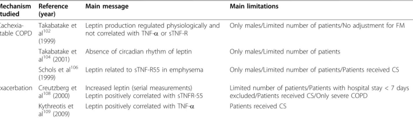
Table 5 The role of leptin in COPD
Mechanism studied
Reference (year)
Main message Main limitations
Cachexia-stable COPD
Takabatake et al102 (1999)
Leptin production regulated physiologically and not correlated with TNF-aor sTNF-R
Only males/Limited number of patients/No adjustment for FM
Takabatake et
al104(2001) Absence of circadian rhythm of leptin Only males/Limited number of patients Schols et al106
(1999)
Leptin related to sTNF-R55 in emphysema Only males/Limited number of patients/Patients received CS
Exacerbation Creutzberg et al108(2000)
Increased leptin (serial measurements) Leptin positively correlated with sTNFR-55
Limited number of patients/Patients with hospital stay < 7 days excluded/Patients received CS/Only severe COPD
Kythreotis et
al109(2009) Leptin positively correlated with TNF-a Patients received CS
such as abnormalities of the autonomous nervous sys-tem and the hypothalamic-pituitary axes, or may repre-sent a compensatory mechanism to maintain body fat content [104].
Researchers have investigated the possible involvement of leptin during the acute exacerbations of COPD. Mal-nourished patients experiencing exacerbation, exhibit sig-nificantly higher leptin levels, compared to normal-weight stable COPD patients, an observation not repli-cated when compared to malnourished stable COPD patients [107]. Similar results have been reported by other groups [103]. Importantly, leptin values, corrected for FM, are significantly elevated in COPD patients dur-ing acute exacerbation versus controls [108,109]. Leptin concentrations gradually decrease throughout the exacer-bation, but when corrected for FM, remain significantly elevated during hospitalization [108,109]. The normal feedback regulation of leptin by FM is preserved on Day 7 of the exacerbation, although dissociation has been reported on Day 1, possibly due to a temporary dysfunc-tion related to the event [108]. The natural logarithm (LN) of leptin is inversely correlated with the dietary intake/resting energy expenditure index (indicating the role of leptin in energy balance) and positively correlated with sTNF-R55 (after correction for FM) [108]. Other researchers have reported a positive correlation between TNF-a and leptin on Day 1 of admission [109]. sTNF-R55 significantly explains 66% of the variation in energy balance in Day 7 of the exacerbation, while leptin is excluded, suggesting that the influence of leptin is under the control of the systemic inflammatory response [108].
The airflow limitation in COPD is linked to structural changes, including the presence of an abnormal inflam-matory pattern detected in each lung compartment [110]. AKR/J mice (i.e. a strain that presents similar to COPD anatomic abnormalities following cigarette smoke exposure for 4 months) exhibit reduced Ob-R expression in the airway wall, upon smoke exposure [111]. Inversely, stimulation of bronchial epithelial cells and alveolar type II pneumocytes, isolated from human lung tissue, with increasing doses of cigarette smoke condensate results in a significant induction of leptin and Ob-Rb m-RNA, suggesting that smoking itself may increase the expression of the leptin/leptin receptor sys-tem in lung tissue [8]. However, others have demon-strated down-regulation of leptin/leptin receptor system in bronchial epithelial cells of proximal airways of mild-to-severe COPD patients, when compared to tissues obtained from non-smoking subjects [48], while immu-nohistochemical studies show that leptin expression is increased in bronchial epithelial cells and alveolar macrophages in the peripheral lung of COPD patients (GOLD stage 4) [8]. Additionally, leptin is over-expressed in the submucosa of proximal airways of
COPD patients [48]. The diversities observed in pul-monary leptin/leptin-receptor system expression among COPD patients, symptomatic smokers and never-smo-kers despite similar anthropometric measurements, lend further support to the concept of local production of leptin in the lung [8].
Accumulated evidence suggest that leptin may be involved in the local inflammatory response seen in the airways of COPD patients, hypothetically regulating the infiltration and the survival of inflammatory cells in the submucosa of COPD patients [48]. Interestingly, leptin’s up-regulation in the proximal airways correlates to the expression of activated T lymphocytes (mainly CD8+) and to the absence of apoptotic T cells [48]. In addition, leptin is detected in induced sputum of patients with COPD, whereas it is significantly positively correlated with inflammatory markers measured in induced spu-tum, such as CRP and TNF-a [112]. Importantly, plasma and sputum leptin levels are inversely correlated. In harmony with the previous results, the presence of Ob-Rbin lung epithelium and inflammatory cells com-bined with the fact that the lung is a source of leptin, suggests the existence of a paracrine cross-talk between resident pulmonary epithelial cells and immune cells in response to noxious particles [8]. This hypothesis needs further validation by subsequent studies, enrolling a lar-ger number of patients and including experiments that will shed further light to the pathophysiological role of leptin in the pathogenesis of COPD.
Recently, researchers have reported that COPD patients carrying minor alleles of polymorphisms in the Ob-R gene are less susceptible to loss of lung function, as indicated by %FEV1decline [111]. Although the func-tional significance is not known, these data have led to the hypothesis that the Ob-R gene may serve as a novel candidate gene for COPD.
Asthma (Table 6)
Asthma represents a chronic inflammatory disorder of the airways associated with airway hyper-responsiveness that leads to recurrent episodes of widespread, and often reversible, airflow obstruction within the lung [113]. Obesity is a risk factor for asthma, while studies indicate that adiposity may increase disease severity in asthmatic subjects and possibly alter the efficacy of stan-dard asthma medications [114-116]. The mechanisms underlying the relationship between obesity and asthma have not been fully established yet, however, experimen-tal evidence suggests that changes in adipose-tissue derived hormones, including leptin, as well as other fac-tors, are possibly implicated.
ob/ob mice exhibit significantly elevated pulmonary
in greater increase in these two parameters, associated with an enhanced expression of bronchoalveolar alveo-lar lavage fluid (BALF) protein, eotaxin, and IL-6 when compared to lean controls [117]. Acute leptin replace-ment in chronically leptin-deficient mice cannot reverse the enhanced inflammatory response. However, mice fasted overnight exhibit reduced leptin levels, associated with a significant increase in RL and airway responsiveness following O3 exposure, as compared to fed mice [118]. The restoration of leptin to fed levels prevented the fasting induced changes in response to O3. Exogenous leptin administration in wild-type mice results in increased O3-induced cytokine and protein release into BALF [117]. Similarly to the ob murine model, db/dbmice (i.e. mice that lack functional Ob-Rb isoform due to a mutation in the cytoplasmic domain of the receptor) and carboxypeptidase E-defi-cient (CPEfat) mice (i.e. a strain characterized by obe-sity, resulting from a functional mutation in the gene encoding carboxypeptidase, and increased leptin levels) present increased baseline airway responsiveness, as well as augmented responses to O3 exposure, when compared to their lean controls [119,120]. In harmony with the latter results, mice with diet-induced obesity exhibit innate AHR and enhanced O3-induced pulmon-ary inflammation, similar to that observed in geneti-cally obese mice [121]. Collectively, the aforementioned findings suggest that leptin may have the potential to augment the pulmonary response to acute O3 exposure, but other effects of obesity may also play an important role [122]. Since innate AHR is a common feature of leptin and leptin receptor defi-cient mice, as well as CPEfat mice and mice with diet induced obesity (i.e. mice with reduced and mice with increased leptin concentrations) it seems unlikely that the adipokine can act as an intermediary in the causal pathway [122].
Clinical studies provide confounding evidence to the mouse-model observation regarding the role of leptin in asthma. Overweight asthmatic children present twice as high leptin levels as those without asthma, despite no differences in BMI [123]. Similar results are documented by other researchers; asthmatic children, especially asth-matic boys, exhibit higher leptin levels compared to controls [124]. Leptin concentrations are significantly associated with bronchodilator response in overweight/ obese men, but not in overweight/obese women [125]. Furthermore, leptin levels, even when adjusted for BMI, are predictive of asthma in male subjects [124]. Addi-tionally, increased BMI and leptin concentrations are associated with asthma in adults, but when adjusted for leptin, no effect is observed in the association among BMI and asthma, indicating that the association is not mediated by the leptin pathway alone [126]. In contrast, others have failed to document any direct association between leptin and the presence of asthma [60].
Increasing evidence suggest that the pro-inflammatory effects of leptin may contribute to the higher incidence of asthma in the obese population. As discussed previously, administration of leptin to wild-type mice enhances O3-induced airway inflammation [117], while ovalbumin sensitization and challenge increases serum leptin levels in mice [127]. Additionally, in animal models, exogenous leptin enhances the phagocytosis by macrophages and the production of TNF-a, IL-6 and IL-12 [124]. Adminis-tration of pro-inflammatory cytokines, such as TNF-a and IL-1, in mice results in a dose-dependent increase in leptin concentrations [126]. However, since these cyto-kines have been implicated in the pathophysiology of asthma [124] it is conceivable that the disease-related inflammation induces the release of leptin from the adi-pose tissue or the lung itself, which may in turn increase airway inflammation and hyper-responsiveness through a continuous interaction [122,126,128].
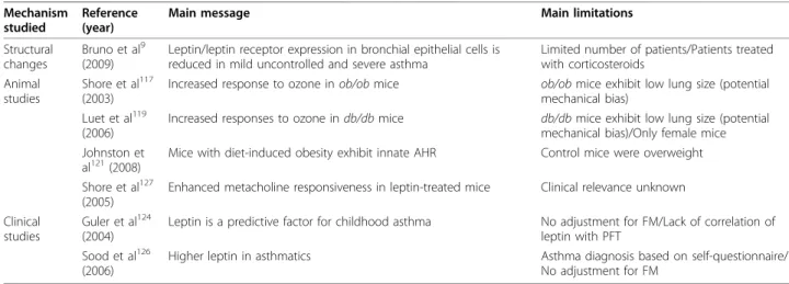
Table 6 The role of leptin in asthma
Mechanism studied
Reference (year)
Main message Main limitations
Structural changes
Bruno et al9 (2009)
Leptin/leptin receptor expression in bronchial epithelial cells is reduced in mild uncontrolled and severe asthma
Limited number of patients/Patients treated with corticosteroids
Animal studies
Shore et al117 (2003)
Increased response to ozone inob/obmice ob/obmice exhibit low lung size (potential mechanical bias)
Luet et al119 (2006)
Increased responses to ozone indb/dbmice db/dbmice exhibit low lung size (potential mechanical bias)/Only female mice Johnston et
al121(2008) Mice with diet-induced obesity exhibit innate AHR Control mice were overweight Shore et al127
(2005)
Enhanced metacholine responsiveness in leptin-treated mice Clinical relevance unknown
Clinical studies
Guler et al124 (2004)
Leptin is a predictive factor for childhood asthma No adjustment for FM/Lack of correlation of leptin with PFT
Sood et al126 (2006)
Higher leptin in asthmatics Asthma diagnosis based on self-questionnaire/
No adjustment for FM
Over the past few years, researchers have hypothesized that decreased immunological tolerance, as a conse-quence of immunological changes induced by adipo-kines, may be implicated in the pathogenesis of allergic asthma [129]. As argued above, leptin-treated animals exhibit augmented responses to metacholine and increased levels of IgE, following ovalbumin challenge, when compared to saline-infused mice [127]. No differ-ence on the inflammatory response in the airways was observed between the two study groups. In keeping with the aforementioned results, leptin and IgE levels are sig-nificantly correlated in asthmatic children [124]. Inter-estingly, atopic asthmatic boys have significantly higher leptin levels than non-atopic asthmatic subjects. Addi-tionally, in vitro studies have documented that leptin can significantly up-regulate the cell surface expression of intracellular adhesion molecule (ICAM)-1 and CD18 and suppress those of ICAM-3 and L-selectin in eosino-phils [130], while it augments alveolar macrophage leu-kotriene synthesis [131]. The latter results suggest that leptin may induce accumulation of eosinophils and may enhance inflammatory processes at sites such as the lung or the airways, and thereby augment allergic airway responses, at least in part [130,131].
Additionally, studies have raised the issue whether lep-tin may play an important role on asthma pathophysiol-ogy through its ability to activate SNS. Leptin increases the activity of the adrenal medulla and sympathetic nerves in various organs, although its impact on the sympathetic nerves of the lung is unknown [132,133]. On the basis of this conception, researchers have exam-ined the effects of leptin on human airway smooth mus-cle cells and airway remodeling associated with asthma; leptin itself cannot promote muscle proliferation, migra-tion or cytokine synthesis, suggesting that the effects of obesity on asthma may not be attributed to a direct effect of leptin on airway smooth muscle [47]. Leptin has no proliferative effect when administered in a human airway smooth muscle cell line culture, although it stimulates the release of VEGF by these cells [134]. However, the expression of leptin/leptin receptor in bronchial epithelial cells is significantly reduced in patients with mild uncontrolled asthma and severe trea-ted asthma versus patients with mild controlled treatrea-ted asthma and healthy volunteers, while leptin and leptin receptor expression are inversely correlated with reticu-lar basement membrane thickness suggesting that lep-tin/leptin receptor expression may be associated with the airway remodeling observed in asthma, implicating the adipokine in the homeostasis of lung tissue [9].
Lung Cancer (Table 7)
Increased BMI is significantly associated with higher death rates due to cancer [135], and it is well established
that obesity increases the risk of cancer developing in numerous sites [136,137]. Can leptin be the mediator linking obesity with cancer?
A functional polymorphism in the promoter region of leptin gene is associated with a threefold increased risk of developing non-small cell lung cancer (NSCLC) [138]. The over-expressing variant is associated with earlier onset of lung cancer, but not with advanced metastatic disease, suggesting that continuous exposure to higher leptin concentrations due to the polymorphism in the leptin gene may accelerate cancer initiation [138]. This hypothesis is further strengthened by other groups who observed increased leptin levels in NSCLC patients and recognized leptin as a risk factor for cancer, even after controlling for BMI and recent weight loss [139].
In accordance with the previous studies, primary cul-tures of tracheal epithelial cells of db/db mice demon-strate significantly lower cell proliferation versus those of their lean litternates, while administration of leptin significantly increased cell proliferative ability in lean mice, but not indb/dbmice [49]. Leptin has a stimula-tory action on a clonal cell line derived from human lung squamous cell cancer (SQ5 cells), an effect mediated through mitogen activated protein (MAP) kinase activity, indicating that leptin may act as a growth factor. On the contrary, in an experimental pul-monary metastasis model,ob/ob anddb/dbmice present a remarkably increased number of metastatic colonies when compared to wild-type mice [140]. Administration of leptin inob/obmice abolished the increase in metas-tasis, indicating a rather prophylactic role of leptin. However, when cancer cells were inoculated orthotopi-cally, through a chest incision, tumor growth at the implanted site was comparable among the groups.
Studies have led to the hypothesis that leptin contri-butes in cancer development, at least in part, through its up-regulatory role in the inflammatory system [141]. Leptin affects both innate and adaptive immunity by sti-mulating and activating neutrophils, macrophages, blood mononuclear cells, dendritic cells and T cells, and con-secutively their products, which may induce chronic inflammation and lung carcinogenesis [141]. However, until today, this complex interplay between leptin, immune system, and cancer has received only some experimental support and further investigations are required.
progression, and there were no differences presented between patients with and without weight loss. There-fore, leptin cannot serve as a diagnostic or prognostic factor in advanced NSCLC. Moreover, these results sug-gest that cancer anorexia and cachexia are not due to a dysregulation of leptin production. The aforementioned observations are in contrast with those reported by other researchers, who observed higher concentrations of leptin in NSCLC patientsvs. controls [147]. Patients recruited in the latter study had mainly non-advanced disease and there was no adjustment of leptin levels for FM, factors that can attribute to the discrepancies among studies.
Infectious diseases of the lung (Table 8) Pneumonia
Recently, several reports have identified a role for leptin in regulating immune function [24,25] while leptin levels acutely increase during inflammation, infection and sep-sis [12]. Furthermore, leptin deficiency has been asso-ciated with an increased frequency of infection [148,149]. Interestingly, leptin levels in serum, BALF and whole lung homogenates are elevated in wild-type mice, following intra-tracheal challenge with Klebsiella
pneumoniae [150]. Additionally, ob/ob mice exhibit
increased susceptibility and enhanced lethality following
K. pneumoniaeadministration, as compared to wild-type
mice, associated with impaired macrophage and neutro-phil phagocytosis of the microorganism, and reduced macrophage leukotriene synthesis in vitro [150,151].
Concerning the impact of chronic leptin deficiency on gram-positive pneumonia, ob/obmice display reduced survival following intra-tracheal challenge with
Strepto-coccus pneumoniae[152]. This impairment is associated
with increased pulmonary cytokine and lipid mediator levels, and defective alveolar macrophage phagocytosis and neutrophil polymononuclear (PMN) leukocyte kill-ing in vitro. However, leptin administration to ob/ob
mice in vivo improved pulmonary bacterial clearance and survival [152]. Furthermore, a physiologic reduction in leptin, induced by acute starvation, in a murine model of pneumococcal pneumonia, was associated with reduced PMN accumulation, IL-6 and macrophage inflammatory protein (MIP)-2 levels in BALF, impair-ment of leukotriene B4 (LTB4) synthesis and phagocyto-sis, and killing ofS. pneumoniae in vitro [153]. Leptin administration to fasted mice corrects these defects. In contrast, others have failed to detect differences
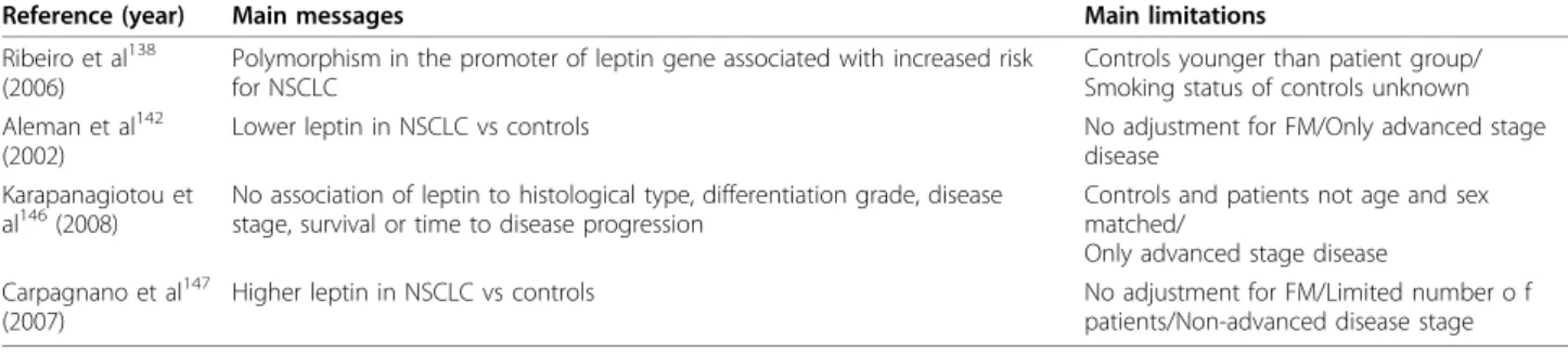
Table 7 The role of leptin in lung cancer
Reference (year) Main messages Main limitations
Ribeiro et al138 (2006)
Polymorphism in the promoter of leptin gene associated with increased risk for NSCLC
Controls younger than patient group/ Smoking status of controls unknown Aleman et al142
(2002)
Lower leptin in NSCLC vs controls No adjustment for FM/Only advanced stage
disease Karapanagiotou et
al146(2008)
No association of leptin to histological type, differentiation grade, disease stage, survival or time to disease progression
Controls and patients not age and sex matched/
Only advanced stage disease Carpagnano et al147
(2007)
Higher leptin in NSCLC vs controls No adjustment for FM/Limited number o f
patients/Non-advanced disease stage
Abbreviations: NSCLC: Non small cell lung cancer, FM: Fat mass
Table 8 The role of leptin in infectious diseases of the lung
Infectious disease
Reference (year)
Main message Main limitations
Pneumonia Mancuso et
al150(2002) Klebsiella pneumonia(WT mice) and increased mortality (ob/obadministration results in increased leptinmice) Experimental condition not well correspondingwith clinical pneumonia/Only female mice Hsu et al152
(2007)
Increased mortality following pneumonococcal pneumonia (ob/ obmice)
Leptin administration improves survival
Experimental conditions not well corresponding with clinical pneumonia/Only female mice
Diez et al155 (2008)
No differences in leptin in pneumonia vs controls Leptin lacks prognostic value for pneumonia lethality
Possible influence by comorbidities/Only hospitalized patients included
Tuberculosis Buyukoglan et
al159(2007) Lower leptin in tuberculosis No adjustment for FM/Higher BMI in controls/Limited number of patients van Crevel et
al161(2002) Leptin increases during antituberculous treatment No adjustment for FM Cakir et al163
(1999)
Higher leptin in tuberculosis
No significant difference in leptin before and after antituberculous treatment
No adjustment for FM/Limited number of patients
concerning the extent and severity of lung inflammation, and the bacterial outgrowth in the lung, during either gram-positive or gram-negative pneumonia, in ob/obor wild-type mice [154].
To gain a more comprehensive understanding concern-ing the role of leptin in human lung infections, Diez et al [155], compared leptin levels in patients hospitalized for community acquired pneumonia and healthy controls and reported no significant differences in the two study groups, after adjusting for BMI, whereas leptin was inver-sely correlated with inflammatory markers. Interestingly, patients who died exhibited significantly lower leptin levelsvs. controls. One of the most remarkable ascertain-ments of this study was the observation that leptin lacked independent prognostic value, since it was displaced by nutritional status on multiple logistic regression analysis, suggesting that leptin cannot act as an inflammatory reactant, but as a nutritional marker. Collectively, the few data that are present in the literature need further valida-tion before any definite conclusions can be made. Tuberculosis (TB)
Obesity is associated with lower risk of pulmonary TB [156]. Thin subjects are more likely to develop active TB and this may be a result of a relative deficiency of leptin [157,158]. A number of studies report that leptin levels are suppressed in tuberculous patients versus con-trols [159-161]. TB associated reductions of leptin are mediated independently by weight loss and prolonged inflammation [161], while leptin cannot account for the weight loss and anorexia associated with the disease [162]. Interestingly, leptin levels show no significant dif-ference, corrected for energy balance and FM, at base-line and after TB treatment, in all but one study [159,161,162]. One may hypothesize that the prolonged inflammatory response in TB down-regulates or exhausts leptin production [161]. Since leptin is impor-tant for cell mediated immunity, low leptin concentra-tions during active TB may contribute to increased infection susceptibility, disease severity, and recovery with sequelae lesions [159,161]. However, the reduction of leptin levels may represent a protective component of the immune response in pulmonary TB [159]. The pre-vious findings are not replicated in two studies, which report higher leptin concentrations in patients with active pulmonary TB versus controls [163,164].
Evidence in the literature demonstrates the presence of lower pleural fluid leptin levels in tuberculous pleural effusions when compared to other exudates [160,165]. Pleural fluid leptin levels may be used for the diagnosis of tuberculous pleural effusions (sensitivity 82,1%, speci-ficity 82,4% for cut-off value of 9,85 ng/ml), however, the diagnostic value of low pleural fluid leptin was not as good as that of conventional methods, like adenosine deaminase [160].
Data from animal models suggest that leptin plays a role in the early immune response to pulmonary infec-tion with Mycobacterium tuberculosis, most likely by mediating an effective interferon-gdriven Th1 response, adequate lymphocyte trafficking and granuloma forma-tion [166]. ob/ob mice intra-nasally infected with live virulent M. tuberculosis display a transiently reduced host defense that is partially restored after leptin repla-cement [166]. Additionally, leptin deficient mice exhibit delayed mycobacterial elimination when challenged with high-dose aerosol of Mycobacterium asbcessus, when compared to wild-type controls [167]. Clearly, the latter hypothesis needs further confirmation from clinical studies.
Acute Lung Injury (ALI)
Most of our knowledge regarding the role of leptin in ALI results from experimental animal models studies. The few data available are rather controversial and inconclusive. Researchers have demonstrated thatdb/db
mice develop less edema and injury, whereas exhibit lower mortality in response to hyperoxia, when com-pared to control animals [41]. In addition, intratracheal instillation of leptin produces lung edema in wild-type mice, but not in db/db mice, suggesting that leptin induces ALI-related changes [41]. In contrast, exogenous leptin administration in rats significantly abrogates ALI and reduces mortality in cerulein-induced acute pan-creatitis [168]. To make matters more complicated, ob/ ob mice exhibit increased resistance to hyperoxia-induced ALI, when compared to control animals, while leptin or anti-leptin antibody administration to wild-type mice has no effect on the course of the hyperoxic injury [169]. The latter findings suggest that the adipo-kine itself does not play an essential role in oxygen-induced alveolar injury [169]. More studies are definitely required to assess the implication of leptin in ALI.
Diffuse Parenchymal Lung Diseases (DPLDs)
To our knowledge, there are no studies in the literature examining the association of leptin with DPLDs. Investi-gations should be designed in order to examine the pos-sible role of leptin on DPLDs pathogenesis.
Conclusions
system remains controversial, possibly due to the fact that much of the existing knowledge derives from ani-mal models of obesity (e.g. ob/ob model) that cannot identically represent the complex biological state of human obesity.
The presence of the functional leptin receptor in the lung recognizes the potential involvement of leptin in the pathogenesis of respiratory disorders, however, whether it represents a friend or a foe is not yet eluci-dated. Although animal studies provide direct indica-tions that leptin enhances lung maturation and stimulates ventilation, further clinical studies are war-ranted in order to evaluate its significance in humans. The increased leptin levels observed in OSAHS cannot exclude the possible involvement of leptin in the depressed respiratory response during sleep since studies have not yet examined whether the disease is a leptin-resistant state, like obesity per se. Research data suggest that leptin/leptin receptor system expression and signal-ing is altered in the airways of patients with asthma and COPD. However, whether it represents an epiphenome-non or a pathogenetic mechanism remains poorly defined, revealing the need for further basic research studies. As for lung cancer, the role of leptin as a growth factor, derived by data examining the effects of leptin in cell lines, requires validation by experimental studies examining its pathophysiological impact on can-cer development.
The boundary from research to clinical application is far from being crossed, as the current data have not revealed an exact role for leptin in the diagnosis, man-agement or follow-up of patients with diseases of the respiratory system. As new investigations are under way, additional consequences of the action of leptin will emerge, adding more information to the already large body of knowledge and thus provide, possibly unex-pected, answers to the questions that remain to be answered to date.
Acknowledgements
The authors thank Violet Stathopoulou (University of Cambridge ESOL Oral Examiner) and Dr Dimosthenis Makris for improvements in the quality of written English and their assistance in editing the manuscript.
Authors’contributions
FM, ZD and AP were involved in the study conception and design. FM performed the acquisition and interpretation of data and prepared the manuscript. ZD and KG were involved in revising the manuscript for important intellectual content. All authors read and approved the final manuscript.
Competing interests
The authors declare that they have no competing interests.
Received: 19 January 2010 Accepted: 31 October 2010 Published: 31 October 2010
References
1. Zhang Y, Proenca R, Maffei M, Barone M, Leopold L, Friedman JM:
Positional cloning of the mouse obese gene and its human homologue.
Nature1994,372(6505):425-432.
2. Friedman JM, Halaas JL:Leptin and the regulation of body weight in mammals.Nature1998,395(6704):763-770.
3. Masuzaki H, Ogawa Y, Sagawa N, Hosoda K, Matsumoto T, Mise H, Nishimura H, Yoshimasa Y, Tanaka I, Mori T,et al:Non-adipose tissue production of leptin: leptin as a novel placenta-derived hormone in humans.Nat Med1997,3(9):1029-1033.
4. Bado A, Levasseur S, Attoub S, Kermorgant S, Laigneau JP, Bortoluzzi MN, Moizo L, Lehy T, Guerre-Millo M, Le Marchand-Brustel Y,et al:The stomach is a source of leptin.Nature1998,394(6695):790-793.
5. Larsson H, Ahren B:Short-term dexamethasone treatment increases plasma leptin independently of changes in insulin sensitivity in healthy women.J Clin Endocrinol Metab1996,81(12):4428-4432.
6. Henson MC, Swan KF, Edwards DE, Hoyle GW, Purcell J, Castracane VD:
Leptin receptor expression in fetal lung increases in late gestation in the baboon: a model for human pregnancy.Reproduction2004,127(1):87-94. 7. Torday JS, Sun H, Wang L, Torres E, Sunday ME, Rubin LP:Leptin mediates the parathyroid hormone-related protein paracrine stimulation of fetal lung maturation.Am J Physiol Lung Cell Mol Physiol2002,282(3):L405-410. 8. Vernooy JH, Drummen NE, van Suylen RJ, Cloots RH, Moller GM, Bracke KR, Zuyderduyn S, Dentener MA, Brusselle GG, Hiemstra PS,et al:Enhanced pulmonary leptin expression in patients with severe COPD and asymptomatic smokers.Thorax2009,64(1):26-32.
9. Bruno A, Pace E, Chanez P, Gras D, Vachier I, Chiappara G, La Guardia M, Gerbino S, Profita M, Gjomarkaj M:Leptin and leptin receptor expression in asthma.J Allergy Clin Immunol2009,124(2):230-237, 237 e231-234. 10. Saladin R, De Vos P, Guerre-Millo M, Leturque A, Girard J, Staels B, Auwerx J:
Transient increase in obese gene expression after food intake or insulin administration.Nature1995,377(6549):527-529.
11. Widjaja A, Schurmeyer TH, Von zur Muhlen A, Brabant G:Determinants of serum leptin levels in Cushing’s syndrome.J Clin Endocrinol Metab1998,
83(2):600-603.
12. La Cava A, Alviggi C, Matarese G:Unravelling the multiple roles of leptin in inflammation and autoimmunity.J Mol Med2004,82(1):4-11. 13. Zhang HH, Kumar S, Barnett AH, Eggo MC:Tumour necrosis factor-alpha
exerts dual effects on human adipose leptin synthesis and release.Mol Cell Endocrinol2000,159(1-2):79-88.
14. Otero M, Lago R, Lago F, Casanueva FF, Dieguez C, Gomez-Reino JJ, Gualillo O:Leptin, from fat to inflammation: old questions and new insights.FEBS Lett2005,579(2):295-301.
15. Popa C, Netea MG, Radstake TR, van Riel PL, Barrera P, van der Meer JW:
Markers of inflammation are negatively correlated with serum leptin in rheumatoid arthritis.Ann Rheum Dis2005,64(8):1195-1198.
16. Raguso CA, Guinot SL, Janssens JP, Kayser B, Pichard C:Chronic hypoxia: common traits between chronic obstructive pulmonary disease and altitude.Curr Opin Clin Nutr Metab Care2004,7(4):411-417.
17. Kallen CB, Lazar MA:Antidiabetic thiazolidinediones inhibit leptin (ob) gene expression in 3T3-L1 adipocytes.Proc Natl Acad Sci USA1996,
93(12):5793-5796.
18. Baratta M:Leptin–from a signal of adiposity to a hormonal mediator in peripheral tissues.Med Sci Monit2002,8(12):RA282-292.
19. Pelleymounter MA, Cullen MJ, Baker MB, Hecht R, Winters D, Boone T, Collins F:Effects of the obese gene product on body weight regulation in ob/ob mice.Science1995,269(5223):540-543.
20. Rajala MW, Scherer PE:Minireview: The adipocyte - at the crossroads of energy homeostasis, inflammation, and atherosclerosis.Endocrinology 2003,144(9):3765-3773.
21. Ahima RS:Central actions of adipocyte hormones.Trends Endocrinol Metab2005,16(7):307-313.
22. Van Heek M, Compton DS, France CF, Tedesco RP, Fawzi AB, Graziano MP, Sybertz EJ, Strader CD, Davis HR Jr:Diet-induced obese mice develop peripheral, but not central, resistance to leptin.J Clin Invest1997,
99(3):385-390.
23. Bjorbaek C, Elmquist JK, Frantz JD, Shoelson SE, Flier JS:Identification of SOCS-3 as a potential mediator of central leptin resistance.Mol Cell1998,
1(4):619-625.
25. Tilg H, Moschen AR:Adipocytokines: mediators linking adipose tissue, inflammation and immunity.Nat Rev Immunol2006,6(10):772-783. 26. Otero M, Lago R, Gomez R, Dieguez C, Lago F, Gomez-Reino J, Gualillo O:
Towards a pro-inflammatory and immunomodulatory emerging role of leptin.Rheumatology (Oxford)2006,45(8):944-950.
27. Cao GY, Considine RV, Lynn RB:Leptin receptors in the adrenal medulla of the rat.Am J Physiol1997,273(2 Pt 1):E448-452.
28. Dunbar JC, Hu Y, Lu H:Intracerebroventricular leptin increases lumbar and renal sympathetic nerve activity and blood pressure in normal rats.
Diabetes1997,46(12):2040-2043.
29. Fruhbeck G:Peripheral actions of leptin and its involvement in disease.
Nutr Rev2002,60(10 Pt 2):S47-55, discussion S68-84, 85-47.
30. Kiguchi N, Maeda T, Kobayashi Y, Fukazawa Y, Kishioka S:Leptin enhances CC-chemokine ligand expression in cultured murine macrophage.
Biochem Biophys Res Commun2009,384(3):311-315.
31. Cao R, Brakenhielm E, Wahlestedt C, Thyberg J, Cao Y:Leptin induces vascular permeability and synergistically stimulates angiogenesis with FGF-2 and VEGF.Proc Natl Acad Sci USA2001,98(11):6390-6395. 32. Bouloumie A, Drexler HC, Lafontan M, Busse R:Leptin, the product of Ob
gene, promotes angiogenesis.Circ Res1998,83(10):1059-1066. 33. Frank S, Stallmeyer B, Kampfer H, Kolb N, Pfeilschifter J:Leptin enhances
wound re-epithelialisation and constitutes a direct function of leptin in skin repair.J Clin Invest2000,106(4):501-509.
34. Gordeladze JO, Reseland JE:A unified model for the action of leptin on bone turnover.J Cell Biochem2003,88(4):706-712.
35. Keisler DH, Daniel JA, Morrison CD:The role of leptin in nutritional status and reproductive function.J Reprod Fertil Suppl1999,54:425-435. 36. Muoio DM, Lynis Dohm G:Peripheral metabolic actions of leptin.Best
Pract Res Clin Endocrinol Metab2002,16(4):653-666.
37. Kruse M, Bornstein SR, Uhlmann K, Paeth G, Scherbaum WA:Leptin down-regulates the steroid producing system in the adrenal.Endocr Res1998,
24(3-4):587-590.
38. Fruhbeck G:Intracellular signalling pathways activated by leptin.Biochem J2006,393(Pt 1):7-20.
39. Badman MK, Flier JS:The adipocyte as an active participant in energy balance and metabolism.Gastroenterology2007,132(6):2103-2115. 40. Hoggard N, Mercer JG, Rayner DV, Moar K, Trayhurn P, Williams LM:
Localization of leptin receptor mRNA splice variants in murine peripheral tissues by RT-PCR and in situ hybridization.Biochem Biophys Res Commun1997,232(2):383-387.
41. Bellmeyer A, Martino JM, Chandel NS, Scott Budinger GR, Dean DA, Mutlu GM:Leptin resistance protects mice from hyperoxia-induced acute lung injury.Am J Respir Crit Care Med2007,175(6):587-594.
42. Chelikani PK, Glimm DR, Kennelly JJ:Short communication: Tissue distribution of leptin and leptin receptor mRNA in the bovine.J Dairy Sci 2003,86(7):2369-2372.
43. Hoggard N, Hunter L, Duncan JS, Williams LM, Trayhurn P, Mercer JG:
Leptin and leptin receptor mRNA and protein expression in the murine foetus and placenta.Proc Natl Acad Sci USA1997,94(20):11073-11078. 44. Lollmann B, Gruninger S, Stricker-Krongrad A, Chiesi M:Detection and
quantification of the leptin receptor splice variants Ob-Ra, b, and, e in different mouse tissues.Biochem Biophys Res Commun1997,
238(2):648-652.
45. Bergen HT, Cherlet TC, Manuel P, Scott JE:Identification of leptin receptors in lung and isolated foetal type II cells.Am J Respir Cell Mol Biol 2002,27(1):71-77.
46. Lin J, Barb CR, Matteri RL, Kraeling RR, Chen X, Meinersmann RJ, Rampacek GB:Long form leptin receptor mRNA expression in the brain, pituitary, and other tissues in the pig.Domest Anim Endocrinol2000,
19(1):53-61.
47. Nair P, Radford K, Fanat A, Janssen LJ, Peters-Golden M, Cox PG:The effects of leptin on airway smooth muscle responses.Am J Respir Cell Mol Biol 2008,39(4):475-481.
48. Bruno A, Chanez P, Chiappara G, Siena L, Giammanco S, Gjomarkaj M, Bonsignore G, Bousquet J, Vignola AM:Does leptin play a cytokine-like role within the airways of COPD patients?Eur Respir J2005,26(3):398-405. 49. Tsuchiya T, Shimizu H, Horie T, Mori M:Expression of leptin receptor in
lung: leptin as a growth factor.Eur J Pharmacol1999,365(2-3):273-279. 50. Kirwin SM, Bhandari V, Dimatteo D, Barone C, Johnson L, Paul S, Spitzer AR,
Chander A, Hassink SG, Funanage VL:Leptin enhances lung maturity in the foetal rat.Pediatr Res2006,60(2):200-204.
51. Torday JS, Rehan VK:Stretch-stimulated surfactant synthesis is coordinated by the paracrine actions of PTHrP and leptin.Am J Physiol Lung Cell Mol Physiol2002,283(1):L130-135.
52. Gnanalingham MG, Mostyn A, Gardner DS, Stephenson T, Symonds ME:
Developmental regulation of the lung in preparation for life after birth: hormonal and nutritional manipulation of local glucocorticoid action and uncoupling protein-2.J Endocrinol2006,188(3):375-386. 53. Huang K, Rabold R, Abston E, Schofield B, Misra V, Galdzicka E, Lee H,
Biswal S, Mitzner W, Tankersley CG:Effects of leptin deficiency on postnatal lung development in mice.J Appl Physiol2008,105(1):249-259. 54. O’Donnell CP, Schaub CD, Haines AS, Berkowitz DE, Tankersley CG,
Schwartz AR, Smith PL:Leptin prevents respiratory depression in obesity.
Am J Respir Crit Care Med1999,159(5 Pt 1):1477-1484.
55. Tankersley CG, O’Donnell C, Daood MJ, Watchko JF, Mitzner W, Schwartz A, Smith P:Leptin attenuates respiratory complications associated with the obese phenotype.J Appl Physiol1998,85(6):2261-2269.
56. Groeben H, Meier S, Brown RH, O’Donnell CP, Mitzner W, Tankersley CG:
The effect of leptin on the ventilatory response to hyperoxia.Exp Lung Res2004,30(7):559-570.
57. Tankersley C, Kleeberger S, Russ B, Schwartz A, Smith P:Modified control of breathing in genetically obese (ob/ob) mice.J Appl Physiol1996,
81(2):716-723.
58. Inyushkin AN, Inyushkina EM, Merkulova NA:Respiratory responses to microinjections of leptin into the solitary tract nucleus.Neurosci Behav Physiol2009,39(3):231-240.
59. Caro JF, Kolaczynski JW, Nyce MR, Ohannesian JP, Opentanova I, Goldman WH, Lynn RB, Zhang PL, Sinha MK, Considine RV:Decreased cerebrospinal-fluid/serum leptin ratio in obesity: a possible mechanism for leptin resistance.Lancet1996,348(9021):159-161.
60. Kim KW, Shin YH, Lee KE, Kim ES, Sohn MH, Kim KE:Relationship between adipokines and manifestations of childhood asthma.Pediatr Allergy Immunol2008,19(6):535-540.
61. Sierra-Johnson J, Romero-Corral A, Somers VK, Olson LJ, Johnson BD:
Leptin, a novel predictor of lung function in heart failure.Chest2008,
134(2):346-350.
62. Vondracek SF, Voelkel NF, McDermott MT, Valdez C:The relationship between adipokines, body composition, and bone density in men with chronic obstructive pulmonary disease.Int J Chron Obstruct Pulmon Dis 2009,4(2):267-277.
63. Sleep-related breathing disorders in adults: recommendations for syndrome definition and measurement techniques in clinical research. The Report of an American Academy of Sleep Medicine Task Force.
Sleep1999,22(5):667-689.
64. Mokhlesi B, Tulaimat A:Recent advances in obesity hypoventilation syndrome.Chest2007,132(4):1322-1336.
65. Olson AL, Zwillich C:The obesity hypoventilation syndrome.Am J Med 2005,118(9):948-956.
66. Malhotra A, White DP:Obstructive sleep apnoea.Lancet2002,
360(9328):237-245.
67. Antonopoulou S, Loukides S, Papatheodorou G, Roussos C, Alchanatis M:
Airway inflammation in obstructive sleep apnoea: is leptin the missing link?Respir Med2008,102(10):1399-1405.
68. Ip MS, Lam KS, Ho C, Tsang KW, Lam W:Serum leptin and vascular risk factors in obstructive sleep apnoea.Chest2000,118(3):580-586. 69. Kapsimalis F, Varouchakis G, Manousaki A, Daskas S, Nikita D, Kryger M,
Gourgoulianis K:Association of sleep apnoea severity and obesity with insulin resistance, C-reactive protein, and leptin levels in male patients with obstructive sleep apnoea.Lung2008,186(4):209-217.
70. Ozturk L, Unal M, Tamer L, Celikoglu F:The association of the severity of obstructive sleep apnoea with plasma leptin levels.Arch Otolaryngol Head Neck Surg2003,129(5):538-540.
71. Vgontzas AN, Papanicolaou DA, Bixler EO, Hopper K, Lotsikas A, Lin HM, Kales A, Chrousos GP:Sleep apnoea and daytime sleepiness and fatigue: relation to visceral obesity, insulin resistance, and hypercytokinemia.J Clin Endocrinol Metab2000,85(3):1151-1158.
72. Ulukavak Ciftci T, Kokturk O, Bukan N, Bilgihan A:Leptin and ghrelin levels in patients with obstructive sleep apnoea syndrome.Respiration2005,
72(4):395-401.