The World Health Organization recognizes Chagas disease, which is caused by the kinetoplastid protozoan Trypanosoma cruzi, as “one of the world’s 10 most neglected tropical diseases.”18 Pri-marily seen in Central and South America, T. cruzi is now recog-nized as endemic in various areas of the southern United States.5 Transmission usually occurs through the bite of hematophagous triatomine bugs but can also occur through the oral route,5 due to blood transfusion4 or organ transplantation,3,8 and through verti-cal transmission.9
Clinical signs of Chagas vary between the acute and chronic phases of disease. During the acute phase, symptoms can include swelling at the inoculation site, lymphoadenopathy, fever, my-algia, and rash. More rare severe acute disease can include me-ningoencephalitis,12 acute myocarditis, and pericardial effusion.15 In humans, the chronic phase usually occurs 10 to 30 years after the initial infection in untreated patients.13 Symptoms include anemia; cardiomyopathy involving cardiac insufficiency, myocar-ditis, or cardiac arrhythmia; and digestive disease such as mega-colon and megaesophagus.18
Case Report
History. A 5-y-old, male, research-naïve cynomolgus macaque (Macaca fascicularis)was enrolled in a cardiac allograft
transplan-tation study under a protocol approved by the IACUC at the Uni-versity of Maryland School of Medicine (Baltimore, MD). This macaque originated from a facility in the southern United States, where he was born, raised, and housed in indoor–outdoor group housing. The dam’s origin was Chinese; sire’s identity and origin were unknown. The heart donor was a 6-y-old, male cynomolgus macaque that was born and raised in China prior to importation into the United States; his parents were born and raised in China.
Upon arrival, both macaques underwent standard facility quarantine for 13 wk. During quarantine, each animal had a total of 6 consecutive negative tuberculosis tests at 2-wk intervals as well as 2 consecutive negative examinations for intestinal para-sites and pathogenic intestinal bacteria (Salmonella spp., Shigella spp, Campyolbacter spp.,and Yersinia spp.). After week 13, the macaques were released from quarantine for use by the inves-tigator. For the duration of their stay at the School of Medicine, the animals were housed indoors and maintained in accordance with the Guide for the Care and Use of Laboratory Animals10 in our AAALAC-accredited facility. All procedures were approved by the IACUC of the University of Maryland School of Medicine (protocol #1013002).
The recipient macaque underwent surgical transplantation of a heterotopic cardiac allograft. Both the donor and the recipient ma-caques received heparin (200 IU/kg) intravenously prior to graft removal and implantation. Briefly, the donor heart was harvested by transection of the great vessels and by cutting the left atrial wall along the entrance of the pulmonary veins. The left atrium was closed after creation of an atrial septal defect by fossa ovalis resection. The donor heart was implanted into the recipient’s ab-domen by connecting the donor aorta to the recipient’s infrarenal
Case Report
Clinical
Trypanosoma cruzi
Disease after Cardiac
Transplantation in a Cynomolgus Macaque
(
Macaca fascicularis
)
Elana R Rybak,1,4,5,† Steve Shipley,2,3,† Ivan Tatarov,1,2,* Tianshu Zhang,1 Wenji Sun,1 Gheorghe Braileanu,1 Lars Burdorf,1
Evelyn Sievert,1 Agnes M Azimzadeh,1 Louis J DeTolla,2 and Richard N Pierson III1
A cynomolgus macaque received a heterotopic cardiac allograft as part of a transplant study, with monoclonal antibodies targeted to specific immune costimulation molecules (CD154, CD28) but no traditional immunosuppressive therapy after surgery. Clinical anemia was detected on postoperative day (POD) 35 and had worsened (Hgb, 2.3 g/dL; Hct = 7.3%) by POD 47, despite type-matched whole-blood transfusions. After a total of 4 blood transfusions, hematologic parameters were improved (Hgb, 5.9 g/dL; Hct, 18.7%). On POD 50, a peripheral blood smear revealed trypomastigotes, and qualitative RT-PCR of whole blood identified the organism as
Trypanosoma cruzi. Although clinically stable initially, the macaque soon developed sufficient weight loss to necessitate euthanasia on POD 64. The final diagnosis was clinical anemia due to T. cruzi infection. This study represents the first reported case of Chagas disease after heart transplant in a NHP.
Abbreviation: POD, postoperative day
Received: 11 Jan 2016. Revision requested: 15 Feb 2016. Accepted: 26 May 2016.
1Department of Surgery and 2Comparative Medicine Program and Veterinary Resources,
University of Maryland School of Medicine, Baltimore, Maryland; 3Division of Laboratory
Animal Medicine, University of North Carolina, Chapel Hill, North Carolina; 4First
Equine, Dover, Delaware; and 5Banfield Pet Hospital, Laurel, Maryland
495
organ donor, including archived serum, plasma, and snap-frozen tissue (colon, kidney, spleen, liver) were sent for testing. All sam-ples from blood donors and from the organ donor were negative for T. cruzi by RT-PCR.Pathologic findings. Spleen, lymph node and esophageal sam-ples from the recipient animal were free of noteworthy gross and histologic lesions. On necropsy, multiple firm, nodular lung le-sions were noted that were approximately 0.5 to 1 cm in diameter with circumferential erythema. These lesions corresponded to focal areas of moderate to severe chronic pneumonia on histo-pathology. All other tissues were grossly normal on necropsy. Mild myeloid hyperplasia of the bone marrow, consistent with a mildly regenerative anemia, was present. The liver demonstrated evidence of mild chronic hepatitis, cholestasis, and hemosidero-sis. The colon and skeletal muscle showed histologic inflamma-tion, presenting with chronic serositis and with chronic myositis and cellulitis, respectively. Samples of the native heart had mod-erate myocarditis with lymphocytic and monocytic infiltrates. The most striking finding involved the transplanted heart, which demonstrated severe myocarditis and epicarditis with moderate fibrosis, and numerous protozoa consistent with T. cruzi in mul-tiple tissue sections (Figure 2).
A complete profile of blood cell populations is shown in Figure 3. WBC, neutrophil, and lymphocyte counts remained quite stable throughout the posttransplantation and infection periods. The CD4/CD8 ratio decreased, probably as a result of the alloimmune response. However, blood eosinophils increased (2- to 3-fold) during the weeks after transplantation, suddenly decreased on POD 35, and then remained very low until euthanasia.
Discussion
The literature contains several reports of asymptomatic (but se-rologic or PCR-positive) T. cruzi in NHP. Reports of symptomatic clinical disease include sudden death in a chimpanzee (P. trog-lodytes),6 encephalitis in a Celebes macaque (M. nigra),15 natural Chagas infection in baboons,20 and reactivation of T. cruzi after experimental infection with SIV.11 What makes the presented case unique is that this animal succumbed to T. cruzi after treatment with an alternative immunosuppressive regimen in which no glucocorticoids or calcineurin inhibitors were used. Specifically, costimulation blockade consists of the selective inhibition of re-ceptor–ligand interactions involved in the activation of T and B cells. The CD28–B7 and CD40–CD154 receptor–ligand pairs play major roles in the initiation of immune responses, and inhibition of one or both of these pathways (by using antibodies or fusion proteins) is associated with significantly decreased responses to pathogens, vaccines, and transplant antigens.2,16
Although we were unable to determine the source of the dis-ease, 2 scenarios might explain the occurrence. The most like-ly case is that the donor animal was the source of the T. cruzi. Given that histopathologic changes were present only in the do-nor heart, the immunomodulation provided by costimulation blockade likely reactivated latent disease in the transplanted heart, which then shed organisms that were seen on peripheral blood smears and ultimately led to widespread Chagas disease. The other possibility is that the recipient animal was actually previously infected with T. cruzi but was asymptomatic, such that immunotherapy led to reactivation of the disease and clinical Chagas disease.
aorta and the donor pulmonary artery to the recipient’s infrare-nal vena cava. The recipient macaque recovered normally from surgery and received an immunosuppressive regimen compris-ing costimulation blockade with monoclonal antibodies specific for human CD154 (hu5c8 mouse–human chimeric antibody [re-combinant], NHP Reagent Resource, Worcester, MA) and human CD28 (FR104, monovalent PEGylated Fab antibody antagonist, Effimune, Nantes, France).
The recipient macaque was doing well until postoperative day (POD) 35, when CBC analysis revealed mild anemia (Table 1). Subsequent CBC panels showed progression of the anemia, and type-matched whole blood transfusions were started on POD 47. The macaque was bright and alert at this time. A total of 4 blood transfusions on POD 47, 49, 50, and 56 were provided for clinical support according to the approved IACUC protocol. Af-ter these 4 blood transfusions, the animal’s anemia showed mild regeneration as evidenced by anisocytosis and reticulocytosis on blood smears. Throughout this time, the animal was bright initially but became progressively lethargic and weak. Because the macaque’s weight subsequently dropped below the eutha-nasia criteria of 20% weight loss, he was euthanized on POD 64. The transplanted graft had normal function at the time of euthanasia. A necropsy was performed, and tissues were sent for histopathology.
Hematology and PCR testing. Immune monitoring of the recipi-ent included standard hematologic and flow cytometric analysis of peripheral blood samples at regular intervals. Absolute cell counts with differentials were obtained (Antech Diagnostics, Ir-vine, CA). In addition, blood samples (50 μL) were mixed with the fluorescently labeled antibodies CD45–PerCP (clone D058-1283), CD2–FITC (clone RPA-2.10), CD3–BV450 (clone SP34-2), and CD20–PE (clone 2H7; all labeled antibodies were obtained from BD Biosciences, San Jose, CA) in specialized tubes (BD Tru-count, BD Biosciences). The absolute count of each cell population was calculated as the number of positive cell events divided by the number of bead events and then multiplied by the bead con-centration. For phenotypic analysis, 50 μL of blood was stained with CD3–BV450 (clone SP34-2, BD Biosciences, San Jose, CA), CD4–BV500 (clone L200, BD Biosciences, San Jose, CA), or CD8– PerCP (clone SK1, BD Biosciences Pharmingen, San Diego, CA), and the CD4:CD8 ratio of the CD3+ population was calculated. All samples were evaluated by flow cytometry (Verse, BD Biosci-ences), and results were analyzed by using Flowjo (version 0.6; Tree Star, Ashland OR).
Because of the macaque’s persistent anemia and our previous experience with simian parvovirus as a cause of posttransplanta-tion anemia,19 POD 50 blood was submitted for RT-PCR testing for simian parvovirus (Zoologix, Chatsworth, CA); the assay re-sults were negative for this virus. In these studies, mild anemia is common and routinely addressed with iron dextran injections, but this animal’s anemia was persistent and not responsive to transfusion. In addition, after the second blood transfusion, the reference laboratory measuring CBC values noted few to moder-ate numbers of trypomastigotes (most closely resembling T. cruzi) on blood smears (Figure 1). At this time, whole blood sent for RT-PCR testing was positive for T, cruzi.
Table 1. Results of CBC analyses
POD Hgb (g/dL) Hct (%) (× 103WBC cells/μL) (x 106 cells/mL)RBC Neutrophils (cells/μL) Lymphocytes (cells/μL) Eosinophils (cells/μL) (× 10Platelets 3 cells/μL)
35 9.0 24.4 5.3 3.82 2968 2067 53 849
47 2.3 7.3 4.6 0.96 1380 2898 0 859
50 5.6 17.8 9.0 2.27 3780 4860 180 385
54 5.9 18.7 8.3 2.26 3071 4814 0 451
56 4.5 14.1 11 1.90 — — 0 —
64 5.1 14.4 9.9 1.77 2871 6237 0 903
POD, postoperative day
All CBC results are from ANTECH Diagnostics (Irvine, CA), except for those on POD 56, which were obtained inhouse.
Normal reference ranges: Hgb, 9.6–13.3 g/dL; Hct, 24%–41%; WBC, 4.5–18.3 ×10³ cells/μL; RBC, 3.5–6.9 × 10⁶ cells/mL; neutrophils, 4800–12,000
cells/μL; lymphocytes, 999–10,551 cells/μL; eosinophils, (80–800 cells/μL)
Figure 1. This peripheral blood smear from POD 50 contains trypomas-tigotes (red arrows), with anisocytosis and polychromasia due to the poorly regenerative anemia
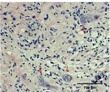
Figure 2. Transplanted heart, right ventricle. Clusters of protozoa, con-sistent with T. cruzi (red arrows), severe myocarditis, and fibrosis are present.
Although pharmacologic therapies for T. cruzi infections in humans exist, few resources for treatment of NHP are available. Medications used to treat humans include benznidazole, which
has the highest efficacy, and nifurtinox.18 Both of these drugs can have serious side effects, are not fully effective, and are only avail-able through the Centers for Disease Control and Prevention in the United States. In humans, these drugs are used only during acute infection (which is usually not recognized) or reactivation; other than organ transplant in the face of organ failure, there is no specific treatment for the end-organ damage caused by infec-tion. Drug unavailability and humane experimental endpoints precluded treatment of this animal after diagnosis. Because treat-ment is usually prolonged (60 to 90 d) and is most effective dur-ing acute or reactivated phases, therapy is not a viable option in NHP used for research. Finally, the efficacy of treatment against T. cruzi has not been validated in the context of immunosuppres-sion7. Therefore, careful screening is the key to prevent use of NHP that may have been infected with this organism.
Routine screening testing should be performed on all NHP en-tering research facilities when the animals have originated from a geographic location where Chagas is known to be endemic. This is particularly true of NHP that will be used in cardiac, gastroin-testinal, or immunosuppressive studies, such as those involving transplantation or myelosuppressive irradiation. Testing methods for T. cruzi detection include RT-PCR, dipstick assay (ELISA, rap-id immunochromotographic strip assay), and peripheral blood smears. Although PCR testing is typically considered the ‘gold standard,’14 even this method can miss animals that have been infected with T. cruzi, especially chronically ill animals. In a re-cent study, 23% of macaques that were IgG ELISA-positive tested negative by RT-PCR, and of these animals, 70% had previously been positive for T. cruzi by PCR assay.17 Given the inconsistency of these tests, multimodal testing by PCR analysis and ELISA and multiple testing of quarantined animals may be the most accurate method for detecting prior or active infection with this organ-ism. Because of the associated cost, we recommend this approach only for animals from endemic areas and their first-generation offspring and when T. cruzi would likely have a significant effect on the study to be performed.
497
of Th1 responses by costimulation blockade-based immuno-modulation, allowing ongoing alloimmune responses to favor an immune shift toward Th2 responses. Therefore, the initial in-crease in eosinophils in our macaque may be related to the im-munosuppression and transplantation events rather than to the parasitic infection. However, uninfected control animals did not demonstrate a decrease in eosinophil counts (Figure 3, open sym-bols). Therefore we infer that the dramatic decrease in peripheral blood eosinophils might be an early marker of infection with T. cruzi (or other parasites), and we therefore recommend initiating specific diagnostic tests in response, especially when this obser-vation is combined with decreased RBC numbers and anemia in NHP.
Acknowledgments
This work was supported by the NIH grant Immunomodulation for Heart Allograft Tolerance (U01 AI066719 to RNP).
We thank Dr Charles McLeod, Jr for histopathology evaluation and the Cardiac Surgery Laboratory and Veterinary Resources at UMSOM for their professional work and support.
References
1. AcquatellaH. 2007. Echocardiography in Chagas heart disease. Circulation 115:1124–1131.
2. Adams AB, Ford ML, Larsen CP. 2016. Costimulation blockade in autoimmunity andtransplantation: the CD28 pathway. J Immunol
197:2045–2050.
3. BarcánL,LunaC,ClaraL,SinagraA,ValledorA,De RissioAM, GadanoA,GarciaMM,de SantibanesE,RiarteA. 2005. Transmis-sion of T. cruzi infection via liver transplantation to a nonreactive recipient for Chagas’ disease. Liver Transpl 11:1112–1116.
4. BenjaminRJ,StramerSL,LeibyDA,DoddRY,FearonM,Castro E. 2012. Trypanosoma cruzi infection in North America and Spain: evidence in support of transfusion transmission. Transfusion
52:1913–1921.
5. BernC,KjosS,YabsleyMJ,MontgomerySP. 2011. Trypanosoma cruzi and Chagas disease in the United States. Clin Microbiol Rev
24:655–681.
6. BommineniYR,DickEJJr,EstepJS,Van de BergJL,HubbardGB.
2009. Fatal acute Chagas disease in a chimpanzee. J Med Primatol
38:247–251.
7. Cicora F, Paz M, Mos FA, Petroni J, Roberti JE. 2014. Belatacept-based immunosuppression in a chagasic adult recipient of en bloc pediatric kidneys. Transplantation 98:e34–e35.
8. FigueiredoJF,MartinezR,da CostaJC,Moyses NetoM,Suaid HJ,FerrazAS. 1990. Transmission of Chagas disease through renal transplantation: report of a case. Trans R Soc Trop Med Hyg 84:
61–62.
9. HowardEJ,XiongX,CarlierY,Sosa-EstaniS,BuekensP. 2013. Frequency of the congenital transmission of Trypanosoma cruzi: a systematic review and meta-analysis. BJOG 121:22–33.
10. Institute for Laboratory Animal Research. 2011. Guide for the care and use of laboratory animals, 8th ed. Washington (DC): National Academies Press.
11. KunzE,Matz-RensingK,StolteN,HamiltonPB,KaupFJ. 2002. Reactivation of a Trypanosoma cruzi infection in a rhesus monkey (Macaca mulatta) experimentally infected with SIV. Vet Pathol 39:
721–725.
12. MarchioriPE,AlexandrePL,BrittoN,PatzinaRA,FiorelliAA, LucatoLT,RosembergS,PereiraSL,StolfNG,ScaffM. 2007. Late reactivation of Chagas disease presenting in a recipient as an expan-sive mass lesion in the brain after heart transplantation of chagasic myocardiopathy. J Heart Lung Transplant 26:1091–1096.
13. NakhleMC,de Menezes MdaC,IruleguiI. 1989. Eosinophil levels in the acute phase of experimental Chagas disease. Rev Inst Med Trop Sao Paulo 31:384–391.
14. NdaoM,KellyN,NormandinD,MacleanJD,WhitemanA, Ko-koskinE,ArevaloI,WardBJ. 2000. Trypanosoma cruzi infection of squirrel monkeys: comparison of blood-smear examination, com-mercial enzyme-linked immunosorbent assay, and polymerase chain reaction analysis as screening tests for evaluation of monkey-related injuries. Comp Med 50:658–665.
15. OlsonLC,SkinnerSF,PalotayJL,McGheeGE. 1986. Encephalitis associated with Trypanosoma cruzi in a Celebes black macaque. Lab Anim Sci 36:667–670.
16. Pierson RN 3rd, Crowe JE Jr, Pfeiffer S, Atkinson J, Azimzadeh A, Miller GG. 2001. CD40 ligand in primate cardiac allograft and viral immunity. Immunol Res 23:253–262.
17. PisharathH,ZaoCL,KreegerJ,PortugalS,KawabeT,Burton T,TomaeckL,ShoiebA,CampbellBM,FrancoJ. 2013. Immuno-pathologic characterization of naturally acquired Trypanosoma cruzi
infection and cardiac sequalae in cynomolgus macaques (Macaca fascicularis). J Am Assoc Lab Anim Sci 52:545–552.
18. RassiAJr,RassiA,Marin-NetoJA. 2010. Chagas disease. Lancet
375:1388–1402.
19. SchröderC,PfeifferS,WuG,AzimzadehAM,AberA,Pierson RN3rd,O’SullivanMG. 2006. Simian parvovirus infection in cy-nomolgus monkey heart transplant recipients causes death related to severe anemia. Transplantation 81:1165–1170.