COMPARING ANKLE RANGE OF MOTION, ARTHROKINEMATIC POSTERIOR GLIDE MOTION, AND MUSCULAR STIFFNESS OF THE TRICEPS SURAE IN DIVISION 1
FEMALE GYMNASTS TO DIVISION 1 FEMALE NON-JUMPING ATHLETES
Morgan Langton
A thesis submitted to the faculty at the University of North Carolina at Chapel Hill in partial fulfillment of the requirements for the degree of Masters of the Arts in the Exercise and Sport
Science.
Chapel Hill 2016
Approved by:
Meredith Petschauer
Troy Blackburn
iii
ABSTRACT
Morgan Langton: Comparing ankle range of motion, arthrokinematic posterior glide motion, and muscular stiffness of the triceps surae in Division 1 female gymnasts to Division 1 female
non-jumping athletes
(Under the direction of Meredith Petschauer)
iv
TABLE OF CONTENTS
ABSTRACT ... iii
LIST OF FIGURES ... vi
LIST OF TABLES ... vii
LIST OF ABBREVIATIONS ... viii
CHAPTER 1 INTRODUCTION ... 1
Research Questions: ... 4
Research Variables: ... 5
LITERATURE REVIEW ... 7
Epidemiology ... 7
Anatomy and Biomechanics ... 9
Stiffness... 13
Injury Risk Factors ... 16
Instrumentation ... 18
METHODS ... 22
Participants ... 22
General Description ... 22
Procedures ... 23
Demographic Survey ... 23
v
Non Weight Bearing ROM Measurements ... 24
Triceps Surae Muscle Stiffness Measurements ... 25
Talocural Joint Motion Measurements ... 28
Statistical Analysis ... 31
MANUSCRIPT ... 33
Introduction ... 33
Methods... 35
Participants ... 35
Procedures ... 36
Data reduction ... 41
Statistical analysis ... 42
Results ... 42
vi
LIST OF FIGURES
Figure 3.1 WBL ROM measurement set up ... 24
Figure 3.2 MVIC set up. ... 26
Figure 3.3 Muscle stiffness set up... 28
Figure 3.4 Talar glide set up ... 29
Figure 3.5 Hollis ankle arthrometer ... 30
Figure 3.6 Medial view of ankle arthometer set up ... 31
Figure 3.7 Lateral view of ankle arthrometer set up ... 31
Figure 4.1 WBL ROM measurement set up ... 37
Figure 4.2 .MVIC set up ... 38
Figure 4.3 Muscle stiffness set up... 39
Figure 4.4 Talar glide set up ... 40
vii
LIST OF TABLES
Table 4.1 ICC and SEM ... 43
Table 4.2 Ankle ROM, muscle stiffness, and arthrokinematic mean comparisons ... 43
Table 4.3 Ankle ROM, muscle stiffness, and arthrokinematic correlations ... 44
viii
LIST OF ABBREVIATIONS
ACL Anterior cruciate ligament
AP Anterior to posterior
DFROM Dorsiflexion range of motion
GRF Ground reaction force
ICC Intraclass correlation coefficient
MVIC Maximal voluntary isometric contraction
NCAA National Collegiate Athletic Association
ROM Range of motion
SEM Standard error of measurement
SPSS Statistical package for the social sciences
1
CHAPTER 1 INTRODUCTION
Ankle and foot injuries are among the most common in physically active individuals. Collegiate female gymnasts sustain ankle and foot injuries at a rate of 4.41 injuries per 1000 competition athlete exposures and 1.18 injuries per 1000 practice athlete exposures.1 These rates are higher than in other female collegiate sports such as lacrosse, volleyball, softball, and field hockey which occur at 1.8, 1.51, 0.44, and 0.92 injuries per 1000 athlete exposures respectively during competition.2-5 Why these numbers are higher for gymnasts than in individuals who participate in sports that do not routinely involve jumping and landing (i.e. non-jumping athletes) is not understood.
2
This study will measure dorsiflexion range of motion (ROM) in weight bearing and non-weight bearing positions as well as the factors affecting ROM such as triceps surae muscle stiffness and talocrural joint glide. There is a lack of research in the gymnastics population to assess these factors which have been shown in both the general and athletic population to increase ankle injury risk.9-11,17 Gymnasts are a unique athletic population as they require
extreme flexibility along with strong and powerful muscles to compete in their sport. Comparing these factors together in gymnasts and non-jumping athletes who do not have those same muscle requirements may elucidate contributions to the greater risk of ankle injury in gymnasts. This may then inform preseason screenings and lead to the development of preventative intervention as range of motion can be altered depending on the cause of the motion limitation. A lack of dorsiflexion ROM has been seen empirically in female college gymnasts by athletic trainers who work with them, but the cause of this decrease in motion is yet to be determined. Restrictions in ankle mobility coming from arthrokinematic factors could be due to the nature of the sport and the repetitive pounding of the ankle during landing causing scar tissue build up in the joint capsule.13,15 This could cause bony changes in the ankle similar to that which occurs with humeral torsion in the shoulder as the repetitive pounding occurs during the formative years. This will then decrease the amount of motion at the joint due to capsular restrictions.14,18 Greater muscular stiffness can decrease ROM at the ankle due to a higher resistance to change in
motion.19 This is expected in gymnasts because they are constantly landing and using their triceps surae eccentrically to control the landing. Greater injury risk has been reported in
3
restricted dorsiflexion ROM may be present, thus contributing to the heightened risk of ankle injury.
While external injury risk factors cannot easily be modified, internal risk factors are potentially targets for rehabilitation interventions designed to reduce injury risk. If gymnasts have a decreased ROM due to decreased arthrokinematic motion, joint mobilizations could be utilized to normalize the motion.21 Muscle stiffness can be increased by performing isometric or eccentric exercises.22 In a sport plagued with ankle injury there is a need to establish preventative measures and manage the high injury risk which can only be done once targets for intervention are identified.
This study will compare dorsiflexion ROM in a weight bearing lunge, non-weight bearing dorsiflexion ROM, non-weight bearing plantarflexion ROM, and a ratio of dorsiflexion ROM compared to total ankle ROM in non-weight bearing between Division 1 female gymnasts and non-jumping athletes. These measurements will assess possible differences in ROM and the underlying contributors to these differences. Restricted dorsiflexion ROM has been shown to increase injury risk in other populations and if seen in gymnasts as compared to a control group of division 1 athletes this information could be used to provide opportunities for future research in this area. Identifying difference between these groups could lead to development of
4
compared to an ankle arthrometer. Not many athletic trainers have access to an ankle arthrometer due to the expense and training needed to use it, therefore if the talar glide method is comparable to the ankle arthrometer, it will be a useful clinical measurement. These assessments will
hopefully lead to future research in both the gymnastics and physically active population to help decrease the risk of injury.
Research Questions:
1. Do ankle non weight bearing dorsiflexion ROM, weight bearing lunge dorsiflexion ROM, arthrokinematic posterior glide, and triceps surae muscle stiffness differ between collegiate gymnasts and female collegiate field non-jumping athletes?
a. Gymnasts will have less weight bearing lunge dorsiflexion ROM compared to non-jumping athletes.
b. Gymnasts will have a less non-weight bearing dorsiflexion ROM as compared to non-jumping athletes.
c. Gymnasts will have a smaller ratio of non-weight bearing dorsiflexion ROM to total ankle ROM as compared to non-jumping athletes.
d. Gymnasts will have greater triceps surae muscle stiffness compared to non-jumping athletes.
e. Gymnasts will have less talocrural joint posterior glide compared to non-jumping athletes.
5
a. There will be a negative correlation between talar glide motion and normalized triceps surae muscle stiffness.
b. There will be a positive correlation between talar glide motion and ratio of dorsiflexion ROM to total ankle non-weight bearing ROM.
c. There will be a negative correlation between triceps surae muscle stiffness and ratio of dorsiflexion ROM to total ankle non-weight bearing ROM.
3. Is the talar glide method of measuring posterior glide restriction a valid clinical test in comparison to the ankle arthrometer measurement of posterior glide?
a. The talar glide method will have moderate positive correlation with the ankle arthrometer posterior glide measurements.
Research Variables:
1. Independent variables a. Group:
i. Gymnasts
ii. Softball, Tennis, and Field Hockey Athletes 2. Dependent Variables
a. Ankle ROM
b. Triceps Surae Stiffness
c. Arthrokinematic posterior glide
Assumptions:
6
2. The participants will perform a maximum contraction for the maximal voluntary isometric contraction (MVIC) measures.
3. The participants accurately relay previous injury history and history of stress fractures to their lower extremity.
4. The participants will go through full ROM in a weight bearing lunge (WBL).
5. The participants will relax lower extremity musculature on the tested leg during the talar glide assessment to allow adequate assessment of motion.
7
CHAPTER 2 LITERATURE REVIEW
Epidemiology
According to NCAA surveillance survey data, which measures injury rate per 1000 athlete exposures, collegiate gymnasts are injured at an overall rate of 15.19 injuries per 1000 athlete exposures in competition and 6.07 injuries per 1000 athlete exposures in practice.1 This was twice the rate of injury that occurred in collegiate women’s lacrosse, 7.15 and 3.30 injuries per 1000 athlete exposures in competition and practice respectively.5 Collegiate field hockey had similar injury rates to lacrosse with 7.87 and 3.70 injuries per 1000 athlete exposures in
competition and practice respectively.2 Collegiate women’s softball athletes had an injury rate of 2.64 and 1.63 injuries per 1000 athlete exposures in competition and practice respectively.4 Another study showed that elite gymnasts were injured at a rate of 6.29 injuries per 18 month period per gymnast.23 A third study showed that 109 injuries occurred in 87 gymnasts with 62% of athletes injured at least once24. Based on this evidence, ankle injuries in gymnasts occur at a higher rate than other women’s collegiate sports.
8
in competition and 0.70 during practice.5 In collegiate women’s field hockey there was a rate of 0.92 in competition and 0.50 during practice.2 Collegiate women’s softball teams are injured at a rate of 0.58 in competition and 0.33 during practice.4 In comparison to the injury rates of non-jumping sports such as field hockey or softball, even a jumping sport like women’s volleyball had an injury rate of 1.51 in competition and 0.94 during practice which was still lower than women’s gymnastics.3
Although injury rates differ among the types of female collegiate athletes described above, for this study gymnasts will be compared to female
collegiate softball, tennis, and field hockey athletes who all participate in a sport where jumping does not regularly occur. Comparisons will be made between women gymnasts, field hockey, softball, and tennis athletes as they participate in non-jumping sports. Differences in height and weight will be present between these athletes, but controls for height and weight will be taken into account in stiffness measurements where differences have been shown to create different measurements.
9
showed that 84% of injuries occurred during practices even though they occurred at a rate of 1.0 injury per 1000 exposure hours in practice compared to 196.1 injuries per 1000 exposure hours during competition.27 This is important because the landing surface used in practice often consists of a 4 or 8 inch landing mat which provides additional cushioning.13 Even with the use of additional mats and padding, injuries still occur at rates anywhere from 1 to 26 injuries per 1000 hours of gymnastics training.24,27,31 These mats are utilized when learning new techniques to decrease injury risk and increase safety measures.13 While gymnasts have higher injury rates than non-jumping athletes despite altered training techniques to lower the external risks, this study aims to determine any possible internal injury risk factors that could be altered to decrease injury risk.
Anatomy and Biomechanics
Gymnasts are typically injured during the landing aspect of the sport. Therefore, it is important to understand what is happening to the body when this occurs.20,24,26,27,37 The foot and ankle complex serves a dual purpose to absorb the forces and to propel the body to the next motion during walking or jumping.14 The foot and ankle complex combines for a total of 28 bones, 25 joints, 23 muscle tendon units, and ligaments that provide the stability and mobility needed to walk.14 The foot is broken up into sections (rearfoot, midfoot, and forefoot) depending on the bony anatomy of each structure. The tarsals are split between the rearfoot (calcaneus and talus) and midfoot (cuneiforms, navicular, and cuboid) but each section is connected during closed chain movement. Therefore, when pathology occurs in one part of the chain, it affects the entire complex as dissipation of forces cannot occur as seamlessly.14
10
metatarsals.14 These bones, therefore, absorb a large amount of the force as stress is consistently placed on them, even when doing simple tasks.14 For example during gait, the height of the medial longitudinal arch decreases by 15% of its tallest height as the foot moves from heel strike to the stance phase.14 The arch decreases in height due to calcaneal eversion with adduction and plantar flexion of the talus joint during what is known as pronation of the foot in a closed kinetic chain.14 During this motion, much of the force of the body is distributed through the deformation of the arch.14 As this occurs, the tibialis anterior, extensor digitorum longus, and extensor
hallucis longus move eccentrically to lower the foot to the ground and move from neutral foot position to 15 degrees of plantar flexion to also aid in the distribution of forces through the leg and foot.18 Force distribution continues as the foot moves into the stance phase and pronation and eccentric loading of the gastroc soleus complex occurs.18 After midstance, the foot and ankle complex move into supination during the push off phase of gait.14,18 During this phase the soleus and flexor hallucis longus and brevis all initially continue to move eccentrically and then switch to a concentric contraction.18 This concentric contraction causes the foot to become a rigid lever which is used to then propel the body forward.18 Then the foot continues to move from heel off to toe off as the plantar flexors including the gastrocnemius, soleus, peroneals, and toe flexors, all concentrically contract to force the foot into plantar flexion.18 Then as the foot moves through the swing phase the tibialis anterior, extensor digitorum longus, and extensor hallicus longus all work concentrically to dorsiflex the foot and then work isometrically to hold the foot in that position through heel contact.18 The multitude of parts which have to work together seamlessly create many opportunities for imbalances if one part is not working properly.
11
helps to transfer the loads more effectively and with the best distribution patterns of stress on the structures of the foot.14 The intrinsic muscles and ligaments on the medial side of the foot, including the deltoid ligament, tibialis anterior, tibialis posterior, flexor digitorum longus, flexor hallucis longus, and spring ligament, also aid in the distribution of forces.14 The availability of movement in each structure of the foot is important, because an overly rigid structure does not allow for force distribution. However, a foot that is too mobile also puts additional stress on the bony structures of the foot and other structures higher in the chain.14 This occurs when excessive pronation occurs which causes the force to be distributed through the second metatarsal.14
Pronation can also be seen with knee valgus which then puts stress on the ligamentous structures of the knee and the muscular structures responsible for stability.14 There is no concrete definition of how much motion is ideal and how this leads to decreased injury risk. Normal dorsiflexion ROM is defined at 20 degrees according to multiple sources.14,18 Motion at the talocrural joint is typically restricted by the posterior musculature of the leg, mainly the triceps surae, which acts as the check reign for end ROM.18 If the triceps surae complex is flexible enough, the posterior joint capsule itself may be the check reign.18 When that occurs, the joint capsule is more prone to injury which can then cause joint or ligamentous damage such as sprains. When the muscle is too tight and restricts motion, the injury typically occurs to the muscle itself in the form of strains.
In gymnastics, the landing and push off phases are important motions used to perform their maneuvers during routines. Therefore, the ankle experiences the greatest loads during these phases.14 When going into a tumble pass or when going to vault, the gymnast pushes off or ‘punches’ to propel themselves into the air.13,15
12
feet to propel the gymnast into the air to then perform a tumbling pass, a beam dismount, or a vault skill.15 A quick eccentric motion first occurs to increase the power in the take off by eliciting the stretch reflex.15 Then the feet must become supinated to form a rigid lever to propel the gymnast into the air, the gastrocnemius soleus complex must contract concentrically to generate the force needed to propel the body into the air.15,18 During vault this is done by increasing forward velocity by sprinting. Forward velocity is partially converted to vertical velocity with a tumbling skill like a handspring onto the springboard where the landing and concurrent punching motion transitions into the take-off.15
During landings, gymnasts must work to stop all momentum and force of their moving bodies.15 Young gymnasts are taught the importance of ‘sticking’ a landing.15 This is crucial to the sport, because there are point deductions for any ensuing steps or hops taken after the landing.15 There are also point deductions for going into full flexion at any lower extremity joint.15 In order to stick a landing in the safest way the gymnast must land with the ankle dorsiflexed, the knee slightly flexed, the hip slightly flexed, and the shoulders flexed and
13
due to bone spurs forming on the talus.33 This happens due to the calcification of areas where the talus is jammed into the syndesmosis between the tibia and fibula.
Proper biomechanics of punching, gait, and landing are critical to gymnasts because they perform barefoot.13 Barefoot participation can lead to multiple problems. The main issue from a prevention standpoint is the inability to correct any naturally occurring biomechanical pathology. Gymnasts with forefoot or rearfoot varus cannot simply wear an orthotic in a shoe to correct the problem and create ideal alignment. In runners those with uncorrected forefoot or rearfoot varus are more prone to stress fractures of the 2nd metatarsal and the same is true for gymnasts due to the hours of compounded stress that occurs to the area. These biomechanics and the inability for gymnasts to use protective equipment compound to raise the question of is there anything that can then be done for gymnasts to decrease injury risk? This question can be answered by looking at measures of triceps surae stiffness, dorsiflexion ROM, and posterior talar motion. If there are differences in these measurements between female gymnasts and non-jumping athletes, then further research can be done to assess why these differences occur.38-40
Stiffness
Another factor affecting ROM and injury rates is muscular stiffness. According to Foure, stiffness is the “degree of resistance offered by tissues in response to lengthening.”41
14
between having muscles that are stiff enough to assist with stability and function, but not so stiff that they become injured.
Hamstring stiffness, has been researched in regards to anterior cruciate ligament (ACL) injury and anterior tibial translation. The posterior attachment of the hamstring tendons on to the tibia produce posterior tibial translation and provide a protective mechanism from ACL injury. It has been seen that increased hamstring stiffness was correlated with decreased anterior tibial translation.42 However, hamstring strength was not correlated to either measure. Clinically this is important to prevent excessive joint motion leading to injury. Another study by Blackburn showed a trend towards significance utilizing isometric interventions to increase stiffness. However, the result was likely not significant due to small sample size.22
As much of the research on muscle stiffness has been done in relationship to ACL injury, another common ideology is that the phase of the menstrual cycle has an effect on the likelihood of ACL injury. This has been researched in comparison to muscle stiffness at different phases of the menstrual cycle to see how the hormones of the female body affect hamstring muscle
stiffness. There are inconsistent results for the correlation between hamstring stiffness and the menstrual cycle phases. Multiple studies showed that both the phase of the menstrual cycle and the differences between taking oral contraceptives and having normal hormonal changes during regular menstrual cycles did not significantly change muscle stiffness in the hamstring.8,43 In contrast women with decreased estrogen levels from the lack of a menstrual cycle had
15
research indicating that there should be any concern about the part of the menstrual cycle the participants are measured during. However, for extreme caution, the participants could be measured during the same period of the menstrual cycle.
Gender differences and anatomical differences are other factors that affect stiffness measures of hamstring muscles. Foure et al. demonstrated that men had stiffer hamstrings compared to women, but after correcting for height and anatomical differences, the stiffness measures were the same.41 Other studies showed that height and weight are factors contributing to stiffness.44-46 This factor is very important when gymnasts are compared to athletes such as women’s lacrosse and field hockey players as their heights and weights can be more variable in those sports as compared to gymnastics. Gymnasts tend to be shorter and weigh less due to the nature of the sport, whereas lacrosse and field hockey players are more varied in size.
16 Injury Risk Factors
When muscular imbalance or pathology occurs, biomechanics change which can lead to injury.10,14 More research on common risk factors for ankle and foot injuries is needed, as current research has varied findings. Risk factors evaluated include ROM measurements, strength
measurements of the ankle, proprioception measures, hormone levels and part of the menstrual cycle, landing times, landing surfaces, footwear, and concentration levels. Mahieu’s prospective study of risk factors for Achilles injuries showed that decreased plantar flexor strength and excessive dorsiflexion ROM were significant indicators of injury.10 Biomechanically, decreases in dorsiflexion ROM area are associated with greater biomechanical errors in completing a double leg squat.48 The double leg squat is also a part of jump landing mechanics for gymnasts. Carcia’s literature review on Achilles pain indicated the factors that lead to injury include abnormal dorsiflexion or subtalar joint ROM, decreased plantar flexion strength, increased foot pronation, and training errors.9 However, Baumhaue found no significant changes in risk of injury with dorsiflexion ROM differences, but increased risk with greater eversion ROM and strength.11
17
optimal ROM so that the muscle can also absorb a portion of the force and it is not solely being absorbed by the bones and joints.14 Fong et al. showed that greater passive dorsiflexion ROM with drop landings lead to decreased ground reaction forces. However, it was not a significant finding due to small sample size.17 However, another study looking at ROM compared to motor task performance indicated that while there was less dorsiflexion in walking down stairs, there was no significant difference in total net moment, joint angle, or timing of peak muscle
activation.50 As can be seen there is limited and conflicting research in risk factors for ankle injuries in gymnastics as they have a unique mechanism of injury due to landings. There is a need for research in ankle ROM and strength to allow health care professionals, strength and conditioning coaches, and coaches to create preventative systems to decrease injury rates.
Other injury risk factors to gymnasts that have been researched include landing surface, time of training, and type of landing. Little consistency has been found with these factors with some studies finding that neither a softer nor a harder landing surface alters ground reaction forces.51 However, Zhang showed that when comparing three different heights and three different landing surfaces, the stiffer landing surfaces required more eccentric contraction from the triceps surae and caused increased loading to the ankle joint.12 This was correlated to gymnasts who train doing landings from higher heights and practice on softer landing surfaces but then compete on stiffer surfaces which may be a cause of the increased injury rate during competition.12 Another study compared the GRF of recreational athletes and gymnasts dropping from a height of 30, 60, and 90 cm and showed that gymnasts have higher GRFs at 60 and 90 cm than recreational athletes do with landing.7
18
more pounding on their body when they land.52 The same can be said with an increase in training time as a gymnast progresses to higher levels of gymnastics.52 The more a gymnast practices, the more opportunities there are for her to become injured.52 However, there are interventions that can be taken to decrease the risk of injury. External interventions such as using softer landing surfaces or foam pits will increase the safety of training for gymnasts. Other interventions can include muscle stiffness interventions or doing joint mobilizations and soft tissue massage to increase ROM measures.
Instrumentation
Measurements will be taken to evaluate the dorsiflexion ROM in weight bearing, talar glide tibial angle, ankle arthrometer joint motion measurement, and the stiffness of the triceps surae muscle. All ROM measures will be taken with a digital inclinometer to decrease the amount of human error which can occur with manual goniometric measurements.53 In weight bearing, the proper placement of the digital inclinometer is along the tibial tuberosity and lined up distally with the shaft of the tibia.53 The intraclass correlation coefficient (ICC), a
measurement of reliability, of the digital inclinometer in measuring ankle dorsiflexion ROM in a weight bearing lunge is 0.96.53 The closer to 1 the ICC measure, the better the correlation and reliability of the technique or tool. The inclinometer also had the lowest mean detectable change meaning it was more sensitive and able to measure differences in ROM more easily than other measures including a manual goniometer.53
19
posterior aspect of the leg.54 This tool had high success at measuring dorsiflexion ROM compared to camera angle measures and had an ICC of 0.97. 54 However, this tool lacks the ability to be clinically applicable because it is out dated and not readily available.54 The Iowa ankle measure device also has an ICC of 0.95 or higher in all measurements. Like the Lidcombe template apparatus, the Iowa ankle measure is a homemade device for the lab that is not feasible for use in the clinic.55 Another limitation of both of these measuring devices is the lack of measuring while Participants are in a weight bearing maneuver. As has been previously stated it is important to measure ROM in a weight bearing position for gymnasts because most of the injuries occur in the landing phase with weight bearing.26 Measuring dorsiflexion ROM in a weight bearing lunge with a digital inclinometer has a high ICC rating of 0.97 and thus has high reliability and validity.56
The talar glide measurement and the ankle arthrometer posterior assessment will both be used to assess arthrokinematic gliding motion at the talocrural joint. The talar glide measurement has been used previously in multiple studies to determine where dorsiflexion stops due to lack of talar motion.57 This method has been shown to have a high ICC3,1 = 0.88 which assesses the
reliability when each subject is assessed by each examiner of interest and an ICC3,3 = 0.99 which
20
throughout ankle research to demonstrate the amount of motion in an anterior and posterior direction at the talocrural joint. Reliability of this tool has been much more variable with ICC measures of 0.91 for interrater measurement with an SEM = 1.02.59 However, these
measurements were taken in a cadaveric study which brings to question the reliability in a living participant.59 The sensitivity and specificity of measurements were measured in another
cadaveric study lead by Nauck.60 The sensitivity of the ankle arthrometer was 96.3 while the specificity was measured to be 44.4.60 This indicates that the arthrometer helps to rule out hypermobility at the talocrural joint in the anterior and posterior directions.60 Schwarz
determined normal measurements for the ankle arthrometer in women aged 19-25, similar to the age of participants in the current study.61 Schwarz showed that normal total anteroposterior displacement of the talocrural joint was 18.79 mm and that posterior displacement averages were 8.82 mm in females in the specific age range.61 However, distributions showed that ‘normal’ mobility in a posterior direction is anywhere from 6.34 – 11.3 mm and for total displacement can vary from 14.67 – 22.91 mm.61
21
22
CHAPTER 3 METHODS
Participants
Gymnasts from the University of North Carolina at Chapel Hill and North Carolina State University will participate in this study. Division I collegiate female non jumping athletes will also be selected from the women’s teams at the University of North Carolina at Chapel Hill and will serve as control participants. Each group will be comprised of 20 participants as determined by power analysis (P=0.80) of pilot testing data. Participants from all sports will be selected by voluntary participation. Participants will be between 18 and 23 years old. Exclusion criteria for all participants include restriction from activity within two months prior to data collection due to a lower extremity injury or bilateral ankle surgery. Control group participants will be excluded if they have a history of competitive participation (at the high school or college level) in a jumping sport including gymnastics, volleyball, basketball, field events, and hurdles in the past 3 years.
General Description
This study will utilize a cross-sectional experimental design. Subjects will report to the Neuromuscular Research Lab for a single testing session, lasting approximately 45 minutes. The participant’s will first complete a health history questionnaire (Appendix A). They will then complete ROM measurements of the ankle, including both weight-bearing lunge ankle ROM measurements and non-weight-bearing seated plantarflexion and dorsiflexion ROM
23 Procedures
Demographic Survey
When participants arrive for their testing session they will read and sign the consent form approved by the Institutional Review Board at the University of North Carolina at Chapel Hill. Participants will complete a questionnaire about their medical history (Appendix A). The questionnaire includes questions to gain insight to previous lower extremity injury, surgical history, menstruation history (if they regularly have a period and when the last one began) and which foot will be measured. The foot measured in gymnasts will be the opposite of their lead leg. The lead leg is the leg which they hurdle with and the opposite foot is the one which is important for balance and push off on single limb events. In non-jumping athletes, the measured foot will be the stance leg when they kick a ball to mimic the stance leg in a gymnast.
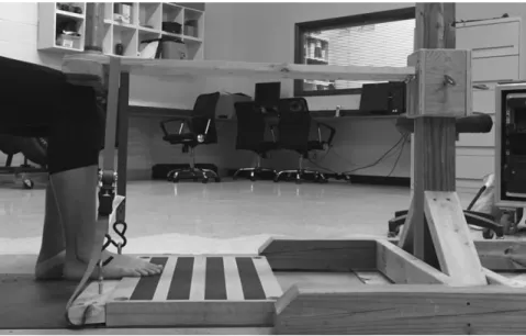
Weight Bearing Lunge ROM Measurements
The weight bearing lunge measurement will assess the functional flexibility of the ankle joint in dorsiflexion using a digital inclinometer (Saunders Group, Inc., Chaska, MN). There is excellent intrarater reliability (ICC = 0.96-0.97) using the digital inclinometer to measure active and passive ROM while participants are in a weight bearing lunge.53 Intrarater reliability for the weight bearing lunge ROM measurement were calculated with ICC2,k=0.976 and SEM=0.297
24
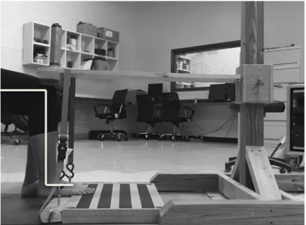
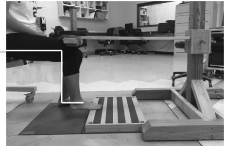
front foot as she can without lifting the heel of the front foot off of the ground (Figure 3.1). A digital inclinometer, zeroed to the vertical prior to use, will be placed on the shank segment just distal to the tibial plateau to measure the angle between the true vertical and the tibial shaft.
Figure 3.1: Weight bearing lunge ROM measurement using digital inclinometer
This will be completed in a quick, fluid motion to minimize the chance of the Golgi tendon organs eliciting a relaxation reflex, which would allow a further stretch in the muscle. This process will be completed three times and the average of the three consecutive measurements will be used as a measure of the weight bearing dorsiflexion ROM.
Non Weight Bearing ROM Measurements
Non weight-bearing ROM measurements will be obtained with the participant supine on a plinth with a foam roller under the knee and the ankle 2-3 inches off the end of the table. A digital inclinometer aligned parallel to the participant’s 5th
metatarsal will be used to measure ankle ROM as the participant actively plantarflexes and then dorsiflexes her foot. The
25
plantarflexion and dorsiflexion. Intrarater reliability for plantarflexion and dorsiflexion ROM were calculated with intraclass correlations at ICC2,k=0.953-0.954 and a SEM=0.325 degrees.
This measurement will be completed five times and the average will be used. A ratio of
dorsiflexion ROM compared to total ROM will also be used to assess how much of the available motion in the ankle is due to dorsiflexion.
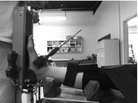
Triceps Surae Muscle Stiffness Measurements
To measure triceps surae muscle stiffness, the participant will be placed into the perturbation device with her hip, knee, and ankle all at 90 degrees and the metatarsal heads resting on two blocks that are secured on top of a force plate (Bertec 4060, Columbus, OH). The computer samples ground reaction force data at 1000Hz during MVIC and muscle stiffness measurements. Triceps surae stiffness will be obtained via the damped oscillatory technique. A perturbation will be applied to the knee which will create an oscillation of the ankle. The force plate below will record the vertical ground reaction force from the oscillation. The frequency of the oscillation will be calculated from the inverse of the interval between the first two oscillatory peaks in the vertical ground reaction force. Stiffness will be calculated using the equation
k=4π2
mf2 where k is stiffness, m is the mass of the system (applied load + mass of the shank and foot segment (Dempster et al)), and f is the frequency of oscillation.
MVIC
26
which is securely anchored to the ground (Figure 3.2). The force plates will be zeroed after this set up. The participant will then maximally contract her plantar flexors for five seconds. Three trials will be performed with a 90 second rest interval in between trials. Similar to previous research, twenty percent of the highest force found from the three measurements will be used as the load during triceps surae stiffness measures.66
Figure 3.2 Experimental setup for collecting MVIC data. The participant’s hip, knee, and ankle will be flexed to 90 degrees. A strap will hold the perturbation lever in place while the participant maximally
contracts their plantarflexor muscles forcing their knee into the lever.
Triceps Surae Muscle Stiffness Measure
27
will be zeroed prior to obtaining the stiffness measure. The participant will close her eyes to reduce anticipation of perturbation. The examiner will apply a manual perturbation to the device at a random time within 5 seconds of the participant closing her eyes. The perturbation will create an oscillation of the shank about the ankle in plantarflexion and dorsiflexion which will be reflected in the vertical ground reaction force on the force plate. The first two peaks in vertical ground reaction force will be used to measure the time in between them. This time (t2-t1) will be
used to calculate the frequency by using the equation f=1/(t2-t1). This frequency will then be used
to calculate muscle stiffness by entering it into the following equation: k=4π2
28
Figure 3.3 Triceps surae muscle stiffness perturbation device and set up with hip, knee, and ankle at 90 degrees and perturbation lever parallel with ground.
Talocural Joint Motion Measurements
Talar Glide Measurement
29
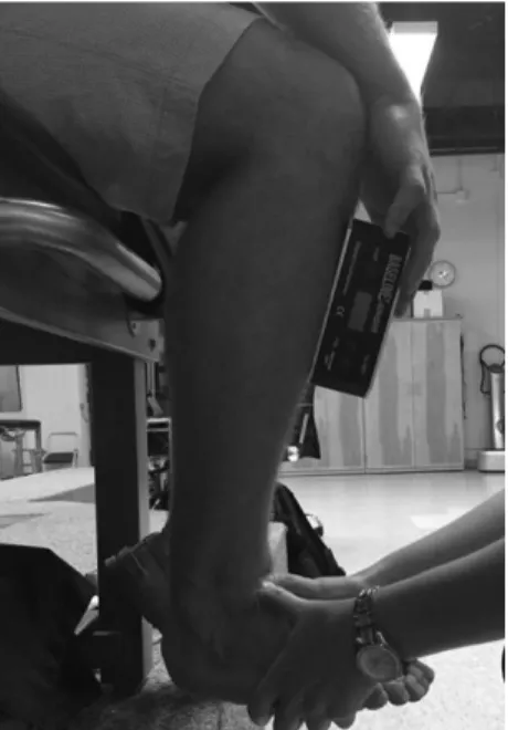
Intra-class correlation coefficients of the talar glide after pilot testing was calculated at ICC2,k= 0.981.
Figure 3.4: Talar glide measurement with foot in neutral eversion/inversion and plantarflexion/dorsiflexion and digital inclinometer below tibial tuberosity.
Ankle Arthrometer Posterior Glide Measurement
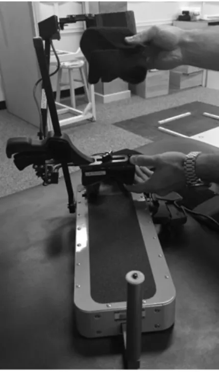
The Hollis ankle arthrometer will be utilized to measure talocrural anteroposterior (AP) glide. This equipment has been used in previous studies and similar methods will be used to collect arthrokinematic motion.59-61 The arthrometer is a unique tool that has been created
30
Figure 3.5: Hollis Ankle Arthrometer
31
Figure 3.6: Medial view of Hollis ankle arthrometer set up with straps securing knee and ankle
Figure 3.7: Lateral view of Hollis ankle arthrometer set up.
Statistical Analysis
32
33
CHAPTER 4 MANUSCRIPT
Introduction
Ankle injuries plague the athletic population each year, particularly in female
gymnasts.1,20,29,37 Gymnasts sustain ankle and foot injuries at a rate of 4.41 injuries per 1000 athletic exposures in competition and 1.18 injuries per 1000 practice exposures, representing a 2.5 to 3 times greater risk of ankle injury compared to other sports such as volleyball or
lacrosse.1 Volleyball and lacrosse have injury incidence rates of 1.513 and 1.85 injuries per 1000 competition exposures. However, there is limited research evaluating this heightened risk of injury.
A number of factors could contribute to greater ankle injury risk in gymnasts. External factors that play a role in greater ankle injury risk include greater height of jump or fall, greater velocity of landing, type of landing surface, and greater ground reaction force.9,11,17,23,67 Over the years, gymnastics has adapted to these findings in research by adding padding to landing
34
through full plantarflexion ROM. Landing from those take offs requires a large amount of dorsiflexion range of motion (DFROM) and eccentric plantar flexor strength to dissipate forces. As previous literature demonstrates that greater dorsiflexion ROM is associated with smaller ground reaction forces, restricted dorsiflexion ROM may contribute to the heightened risk of ankle injury in gymnasts.17
The need to determine internal ankle injury risk factors for gymnasts differs from other sports because of the increased need for both flexibility and strength.34 To perform well in sport, gymnasts need an increased plantarflexion ROM to maintain pointed toes throughout routines. They also need increased strength in the plantar flexors as well as the ankle dorsiflexors to both push off and propel themselves high into the air, and for eccentric control of the body while landing. Maintenance of both strength and flexibility requires a demand on the body rarely achieved in other sports. Another potential contributor to the heightened risk of ankle injury in gymnasts is arthrokinematic motion at the talocrural joint, particularly in a posterior glide. This is currently best measured with an arthrometer, an expensive piece of equipment used in
35
was done by using the measurements for each participant and running a correlation between the two measurements.
The primamry purpose of this study was to compare ankle ROM, talar glide
arthrokinematic motion, and triceps surae muscle stiffness between Division 1 female gymnasts and Division 1 female non-jumping athletes. A secondary purpose of this study was to evaluate relationships between the ratio of dorsiflexion ROM to total ROM, arthrokinematic motion, and triceps surae muscle stiffness. Finally, we also sought to evaluate the validity of the clinical talar glide assessment relative to the ankle arthrometer.
Methods
Participants
A total of 40 female participants volunteered for this study. The experimental group consisted of a total of 20 gymnasts; 12 from The University of North Carolina at Chapel Hill (UNC-CH) and eight from North Carolina State University (NCSU). The non-jumping control group consisted of 20 females split between softball (10), field hockey (8), and tennis (2) athletes at UNC-CH. Participants from both groups were excluded if they were limited during practice due to a lower extremity injury in the two months prior to participation or had a history of bilateral ankle surgery. Participants were excluded from the control group if they participated competitively in any jumping sport (e.g. basketball, volleyball, gymnastics, hurdling, or jumping field events for track) over the past 3 years.
36
exclusion criteria and leg dominance. The dominant leg was defined as the stance leg when kicking a soccer ball for maximum distance for control participants and the stance leg in gymnasts during single leg skill.
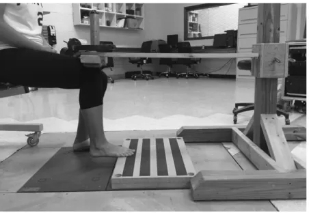
Procedures
Range of motion
Ankle ROM was measured first with a digital inclinometer (Saunders Group, Inc., Chaska, MN). Good intra-rater reliability was established prior to data collection (Table 4.1). Participants performed a weight bearing lunge (WBL) to assess dorsiflexion ROM. The participant stood with her dominant foot placed in front and moved in a lunge position as far as forward as possible, moving the knee over the toes without lifting the heel off the floor (Figure 4.1). Trials were repeated if the heel came off the floor. A digital inclinometer placed on the tibial tuberosity was used to assess the angle between the tibia and the vertical.53 An observer ensured there was no noticeable knee valgus or varus during the lunge. Non-weight bearing plantarflexion and dorsiflexion ROM were assessed with the participant seated on a plinth with the knee flexed over a foam roller. The digital inclinometer was placed parallel to the 5th
37
Figure 4.1: Weight bearing lunge ROM measurement using digital inclinometer
Muscle stiffness
38
Figure 4.2 Experimental setup for collecting MVIC data. The participant’s hip, knee, and ankle will be flexed to 90 degrees. A strap will hold the perturbation lever in place while the participant maximally
contracts their plantarflexor muscles forcing their knee into the lever
Set up of the muscle stiffness trials was similar to the MVIC setup, but the plank under the foot and the strap were removed. A load equal to 20% MVIC was placed on the perturbation device directly in line with the tibia. The force plate was zeroed with the participant’s heel on the ground and metatarsal heads rested on the force plate. The participant was instructed to lift her heel off the ground so her foot was parallel to the ground and her ankle was at 90 degrees (Figure 4.3). The participant then closed her eyes and the examiner applied a manual force to the
39
Figure 4.3: Triceps surae muscle stiffness perturbation device and set up with hip, knee, and ankle at 90 degrees and perturbation lever parallel with ground.
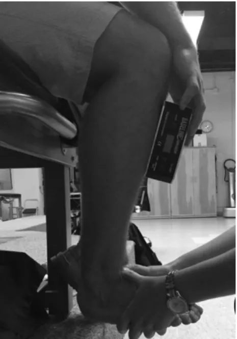
Arthrokinematic measurements
40
Figure 4.4: Talar glide measurement with foot in neutral eversion/inversion and
plantarflexion/dorsiflexion. A digital inclinometer placed along the tibia inferior to the tibial tuberosity to measure the angle at the restriction point.
41
Figure 4.5: Medial view of Hollis ankle arthrometer set up with straps securing knee and ankle
Data reduction
Ground reaction force data were lowpass filtered at 10Hz (4th order Butterworth). The time between the first two oscillatory peaks of vertical ground reaction force was used to
calculate oscillation frequency (f=1/(t2-t1). The frequency was then used to calculate triceps surae
muscle stiffness by entering it into the following equation: k=4π2
mf2 where k was stiffness, m was mass of the system (applied load + mass of the shank and foot segment (6.1% bodyweight)), and f is the frequency at which the oscillation occurs. Stiffness was normalized to the mass of each participant.
42
Statistical analysis
Minimum sample size was estimated at 19 participants in each group using G*Power 3 based on an alpha level of 0.05 and a power of 0.80.68 Independent t-tests were used to compare height, weight, ankle ROM, triceps surae stiffness, and arthrokinematic motion between
gymnasts and non-jumping athletes. Bivariate correlations were used to assess the relationship between the ratio of dorsiflexion to total ROM, muscle stiffness, and talar glide. A bivariate correlation assessed the relationship between the arthrometer and talar glide measurements to assess clinical utility of the talar glide method. All statistical analyses were performed using SPSS 23.0 statistical software (SPSS, Inc., Chicago, IL) with statistical significance set at α<0.05.
Results
Gymnasts had significantly lower weight (59.48 + 5.91 vs 67.52 + 7.90 kg, P =.001) and height (159.18 + 4.72 vs 167.28 + 6.07 cm, P <0.001) than non-jumping athletes. ICC values were calculated for each of the following assessments and ranged from 0.82 to 0.98 which are all good to excellent values (Table 4.1). Gymnasts had significantly less non-weight bearing
dorsiflexion ROM (15.19 + 4.59 vs 19.47 + 5.93 degrees, P =0.015) and a significantly smaller ratio of non-weight bearing dorsiflexion ROM to total ROM (0.18 + .05 vs 0.26 + 0.08, P
<0.001) than non-jumping athletes. Gymnasts had significantly more non-weight bearing plantar flexion ROM than non-jumping athletes (68.43 + 6.36 vs 55.1 + 9.53 degrees, P <0.001).
43
data, that was not lost, was compared between groups for the 10 gymnasts and 6 non-jumping athletes. Even with the small sample size, gymnasts had a significantly greater amount of arthrometric motion (9.31+3.6 vs 5.22+3.61 mm, P=0.045).
Neither triceps surae stiffness nor athrometric motion assessed with the talar glide method significantly correlated with the ratio of dorsiflexion ROM to total ROM (Table 4.3). Additionally, the talar glide measurement did not significantly correlate with the available arthrometer data we had (Table 4.4).
Table 4.1 Intraclass Correlation Coefficient (ICC) and Standard Error of Measurements (SEM)
Measure ICC (2,5) SEM
Weight bearing lunge(o) 0.976 + 0.297 Non-weight bearing ROM(o) 0.953 + 0.325
Talar glide(o) 0.981 + 0.263
Normalized stiffness (N m-1*kg-1) 0.846 + 30.538
Arthrometer(mm) 0.822 + 0.746
Table 4.2 Mean (Standard Deviation) of ROM Measurements, Triceps Surae Muscle Stiffness, and Arthrokinematic Motion in Gymnasts Compared to Non-jumping Athletes
Test Gymnasts Mean (SD) Non-Jumping Athletes Mean (SD) 95% Confidence
Interval P
Weight bearing lunge (o) 37.49 (6.66) 37.39 (5.64) (-3.85, 4.04) 0.961 Non-weight bearing
dorsiflexion ROM (o) 15.19 (4.59) 19.47 (5.93) (-7.67, -0.88) 0.015 Non-weight bearing
plantarflexion ROM (o) 68.43 (6.36) 55.1 (9.53) (8.14, 18.51) <0.001 Ratio of dorsiflexion to
total ankle ROM 0.18 (.05) 0.26 (0.08) (-.12, -.04) <0.001 Talar glide (o) 26.15 (5.49) 25.47 (6.79) (-3.27, 4.63) 0.73 Normalized stiffness (N
m-1*kg-1)
91.13 (30.96)
103.55
44
Table 4.3 Correlation Between Ratio of NWB DF ROM to Total ROM, Talar Glide, and Normalized Triceps Surae Stiffness
Ratio of NWB DFROM to Total ROM
Talar Glide Normalized Triceps Surae Stiffness Ratio of NWB DFROM to Total ROM
r 1
Significance -
Talar Glide r 0.072 1
Significance 0.651 -
Normalized Triceps Surae Stiffness
r -0.114 -0.054 1
Significance 0.483 0.739 -
Table 4.4 Correlation of Ankle Arthrometer Measurements and Talar Glide Measurement Talar
Glide
Arthrometer
Talar Glide r 1
Significance -
Arthrometer r -0.19 1
Significance 0.481 -
Discussion
The primary purpose of this study was to compare ankle ROM, muscle stiffness, and arthrokinematics between gymnasts and non-jumping athletes. The secondary purpose was to assess how ankle measurements are related to each other. We hypothesized that gymnasts would display less dorsiflexion ROM, dorsiflexion ROM to total ankle ROM ratio, weight bearing lunge dorsiflexion ROM, and talar glide motion than non-jumping athletes. We also
45
talar glide test relative to the instrumented ankle arthrometer. We hypothesized that the talar glide methods would have a positive correlation with the arthrometer measurements.
Gymnasts had a significantly smaller height and weight when compared to the non-jumping athletes. This was expected to be the case as gymnasts tend to be smaller than other female athletes. This could potentially play a role in some of the results shown in this study.
The primary findings were that gymnasts displayed lesser dorsiflexion ROM, a smaller ratio of dorsiflexion ROM to total ankle ROM, and greater plantar flexion ROM compared to non-jumping athletes. However, gymnasts did not significantly differ on triceps surae muscle stiffness, arthrokinematic motion assessed with the talar glide, or a weight bearing lunge dorsiflexion ROM. Furthermore, triceps surae muscle stiffness and arthrokinematic motion assessments with the talar glide were not significantly correlated with the ratio to dorsiflexion ROM.
46
arc of motion is measured at the shoulder. In the shoulder the arc is measured to compare bilaterally as a right handed baseball pitcher through repetitions increases the available external rotation motion, while then decreasing the internal rotation available. However, the whole arc of motion is measured bilaterally to make sure the amount of motion is still the same. This could be similar at the ankle for gymnasts as they increase the plantar flexion ROM and thus decrease the available motion for dorsiflexion.
This difference in ROM is important because it has been shown that those with decreased dorsiflexion ROM are at a greater risk of ankle injury. In previous literature, it has been stated that decreased dorsiflexion ROM causes an alteration of kinematics at both the ankle and joints higher up the chain.16,17,48,69 This then leads to increased injury risk not only at the knee but also at the ankle because of a compensatory knee valgus or pronation maneuver to make up for the lacking dorsiflexion ROM.16,17,48,69
There was no significant difference between groups in weight bearing lunge ROM, even though there were non-weight bearing differences. Both groups, however, had more ankle ROM while in the weight bearing lunge compared to the non-weight bearing ROM. The increase in dorsiflexion ROM from non-weight bearing to a weight bearing lunge has been seen in previous studies.16,48 In those studies, an even greater amount of dorsiflexion ROM was seen with jump landing.16,48 Ankle ROM differences from weight bearing to non-weight bearing can be attributed to a few things. First, a greater knee flexion angle at the time of measurement can decrease motion limitations caused by gastrocnemius tightness. As the gastrocnemius crosses the knee as well as the ankle, the angle of the knee affects available motion at the ankle. The
47
not control the maximum knee flexion angle between participants or trials, while the non-weight bearing measures remained the same. The momentum of the body leaning forward into the lunge could have added a passive component forcing the participants into greater ROM. This is
because body weight is greater than the force production of the ankle dorsiflexion musculature. The lack of difference seen in the weight bearing lunge could also be explained by
compensations at other joints further up the chain which were not controlled for. The knee could have collapsed into valgus or the hip could have internally rotated allowing for more apparent motion at the ankle. The foot could have also pronated to allow for more dorsiflexion ROM motion in weight bearing that would not have been seen in non-weight bearing.
Gymnasts had significantly more arthrokinematic motion at the ankle when measured with the arthrometer than non-jumping athletes. This rejected our hypothesis that gymnasts would have less arthrokinematic motion than non-jumping athletes. A possible explanation for this could be differences in treatment protocols and use of joint mobilizations for their ankle which would increase the posterior arthrokinematic motion available at the ankle. This is
considered a better tool for measurement of posterior ankle arthrokinematic motion as compared to the talar glide method which has little evidence to support its use. The talar glide method addressed the posterior talar glide at the ankle, but does so by moving the foot into dorsiflexion with a bent knee. As the ankle arthrometer requires the knee to be extended and locked into place, this could have inherently created a difference between the two measurements as structures other than the capsule, such as the gastroc-soleus complex, could have caused
48
The lack of a significant difference between groups in muscle stiffness and talar glide however is not as straight forward. There is not one distinct reason as to why this might be the case. We hypothesized that there would be greater muscle stiffness and less talar glide motion in gymnasts, and that this would contribute to less dorsiflexion ROM. This was also expected because of previous literature showing an increase in muscle stiffness in an injured population and most gymnasts have a history of ankle injury.66 The values for muscle stiffness however are overall lower in this study than they were in Pamukoff’s study of runners with and without stress history of stress fracture.66 However, the participants in that study were all male which has been shown previously to have different stiffness measures even when controlled for weight.63,66 Gymnasts lack dorsiflexion ROM, but what causes those restrictions could vary by the
individual. Some have increased triceps surae stiffness, while others that are lacking dorsiflexion ROM are due to increased talocrural capsule tightness. Another component that was not assessed in the study was the bony alignment in the ankle joint. As gymnasts are constantly pointing their toes for both aesthetics and to propel themselves off the ground, they do not as consistently go into full dorsiflexion ROM. This repetitive motion, along with the constant pounding from landings on the ankle joint, could cause bony blocks that restrict dorsiflexion ROM. Future studies could take x-rays or bone scans to assess abnormalities in bony alignment in combination with joint capsular and musculotendinous facets which could explain alterations in ROM.
49
that can affect dorsiflexion ROM. Those previously injured individuals are likely to have less ROM. This has been seen in runners with a history of 2nd metatarsal stress fractures.66
There were no significant relationships between the ratio of dorsiflexion ROM to total ankle ROM, talar glide, and triceps surae muscle stiffness. This further supports our rationale of the lack of significance between groups for muscle stiffness and talar glide. No relationship between ROM and stiffness or joint motion could mean the differences in ROM between groups cannot be attributed to one specific restriction. They instead could come from a combination of the many restrictions and one individual may have restricted motion due to either muscle stiffness or capsular restrictions, however, overall the trend is that there are differences due to variations in both measurements.
The secondary finding was that there is no significant relationship between the talar glide and posterior glide ankle arthrometer measurements. The relationship was being assessed to see if there was a clinical tool to similarly assess posterior talocrural glide motion with an ankle arthrometer. The ankle arthrometer is an expensive piece of laboratory equipment that is somewhat fragile. The talar glide is a measurement that can be completed easily with a
goniometer and a plinth in the clinic. However, as the talar glide had a very weak correlation to the arthrometer measurements, this assessment cannot be recommended to measure the amount of posterior joint glide.
50
magnitude of posterior glide clinically. Not controlling for knee valgus, knee flexion angle, or foot collapse during the weight bearing lunge could have also played a role in the lack of significance between the two groups, while a significant difference was seen for dorsiflexion ROM in a non-weight bearing stance. As gymnasts are subject to constant pounding of the ankle from landings, there is the possibility they have bone spurs on the talus or navicular which could restrict motion. No x-rays were taken in this study to assess the presence of these abnormalities, but future research could assess those differences.
In conclusion, gymnasts have significantly altered non-weight bearing ROM as compared to non-jumping athletes, but triceps surae stiffness and talar glide measurements do not show any significance. This could be due to previous injury history or bony abnormalities that were not taken into account in the current study. Further prospective studies need to be conducted to assess how these ROM differences affect injury risk in a population that is known to have an increased risk of ankle injury. The relationship between ROM, muscle stiffness, and
51
APPENDIX A: HEALTH HISTORY QUESTIONNAIRE
Health History Questionnaire
1. Have you competed in any jumping sport (basketball, gymnastics, hurdling, volleyball, or jumping field events in track) during high school or currently? YES NO 2. Have you ever had surgery on your ankle or foot? YES NO
a. If yes explain what procedure you had done, which leg, and when
_________________________________________________________________ _________________________________________________________________ __
3. Have you had an ankle or foot injury in the past year? YES NO
a. If yes how long did it restrict you from participation and when were you cleared? _________________________________________________________________ _________________________________________________________________ __
4. Do you have a history of stress fracture in your shin or foot? YES NO a. If yes where and when did this occur?
_________________________________________________________________ _
5. Do you regularly experience a menstrual cycle? YES NO
52 REFERENCES
1. Marshall SW, Covassin T, Dick R, Nassar LG, Agel J. Descriptive epidemiology of
collegiate women's gymnastics injuries: National Collegiate Athletic Association Injury Surveillance System, 1988-1989 through 2003-2004. Journal of athletic training.
2007;42(2):234-240.
2. Dick R, Hootman JM, Agel J, Vela L, Marshall SW, Messina R. Descriptive epidemiology of collegiate women's field hockey injuries: National Collegiate Athletic Association Injury Surveillance System, 1988-1989 through 2002-2003. Journal of athletic training.
2007;42(2):211-220.
3. Agel J, Palmieri-Smith RM, Dick R, Wojtys EM, Marshall SW. Descriptive epidemiology of collegiate women's volleyball injuries: National Collegiate Athletic Association Injury Surveillance System, 1988-1989 through 2003-2004. Journal of athletic training.
2007;42(2):295-302.
4. Marshall SW, Hamstra-Wright KL, Dick R, Grove KA, Agel J. Descriptive epidemiology of collegiate women's softball injuries: National Collegiate Athletic Association Injury Surveillance System, 1988-1989 through 2003-2004. Journal of athletic training.
2007;42(2):286-294.
5. Dick R, Lincoln AE, Agel J, Carter EA, Marshall SW, Hinton RY. Descriptive epidemiology of collegiate women's lacrosse injuries: National Collegiate Athletic Association Injury Surveillance System, 1988-1989 through 2003-2004. Journal of athletic training.
2007;42(2):262-269.
6. Yeow CH, Lee PV, Goh JC. Effect of landing height on frontal plane kinematics, kinetics and energy dissipation at lower extremity joints. Journal of biomechanics.
2009;42(12):1967-1973.
7. Seegmiller JG, McCaw ST. Ground Reaction Forces Among Gymnasts and Recreational
Athletes in Drop Landings. Journal of athletic training. 2003;38(4):311-314.
8. Bell DR, Blackburn JT, Ondrak KS, et al. The effects of oral contraceptive use on muscle stiffness across the menstrual cycle. Clinical journal of sport medicine : official journal of the Canadian Academy of Sport Medicine. 2011;21(6):467-473.
9. Carcia CR, Martin RL, Houck J, Wukich DK. Achilles pain, stiffness, and muscle power deficits: achilles tendinitis. The Journal of orthopaedic and sports physical therapy.
53
10. Mahieu NN, Witvrouw E, Stevens V, Van Tiggelen D, Roget P. Intrinsic risk factors for the development of achilles tendon overuse injury: a prospective study. The American journal of sports medicine. 2006;34(2):226-235.
11. Baumhauer JF, Alosa DM, Renstrom AF, Trevino S, Beynnon B. A prospective study of
ankle injury risk factors. The American journal of sports medicine. 1995;23(5):564-570.
12. Zhang SN, Bates BT, Dufek JS. Contributions of lower extremity joints to energy
dissipation during landings. Medicine and science in sports and exercise. 2000;32(4):812-819.
13. Sands WA. Injury prevention in women's gymnastics. Sports medicine (Auckland, N.Z.).
2000;30(5):359-373.
14. Neumann D. Kinesiology of the musculoskeletal system: foundations for physical rehabilitation. 1 ed. St Louis, Missouri: Mosby, Inc; 2002.
15. Hay JG. Gymnastics. The biomechanics of sports techniques. 4 ed. New Jersey Prentice Hall; 1993:296-337.
16. Whitting JW, Steele JR, McGhee DE, Munro BJ. Dorsiflexion capacity affects achilles tendon loading during drop landings. Medicine and science in sports and exercise.
2011;43(4):706-713.
17. Fong CM, Blackburn JT, Norcross MF, McGrath M, Padua DA. Ankle-dorsiflexion range of
motion and landing biomechanics. Journal of athletic training. 2011;46(1):5-10.
18. Levangie PK, Norkin CC. Joint structure and function: a comprehensive analysis. 3rd ed. Philadelphia, PA F. A. Davis 2001.
19. Kay AD, Blazevich AJ. Isometric contractions reduce plantar flexor moment, Achilles tendon stiffness, and neuromuscular activity but remove the subsequent effects of stretch. Journal of applied physiology (Bethesda, Md. : 1985). 2009;107(4):1181-1189.
20. Harringe ML, Renstrom P, Werner S. Injury incidence, mechanism and diagnosis in
top-level teamgym: a prospective study conducted over one season. Scandinavian journal of medicine & science in sports. 2007;17(2):115-119.
21. Collins N, Teys P, Vicenzino B. The initial effects of a Mulligan's mobilization with movement technique on dorsiflexion and pain in subacute ankle sprains. Manual therapy. 2004;9(2):77-82.
54
electromyography and kinesiology : official journal of the International Society of Electrophysiological Kinesiology. 2014;24(1):98-103.
23. Kolt GS, Kirkby RJ. Epidemiology of injury in elite and subelite female gymnasts: a comparison of retrospective and prospective findings. British journal of sports medicine.
1999;33(5):312-318.
24. Kirialanis P, Malliou P, Beneka A, Gourgoulis V, Giofstidou A, Godolias G. Injuries in artistic gymnastic elite adolescent male and female athletes. Journal of back and musculoskeletal rehabilitation. 2002;16(4):145-151.
25. Grapton X, Lion A, Gauchard GC, Barrault D, Perrin PP. Specific injuries induced by the practice of trampoline, tumbling and acrobatic gymnastics. Knee surgery, sports traumatology, arthroscopy : official journal of the ESSKA. 2013;21(2):494-499.
26. Kirialanis P, Malliou P, Beneka A, Giannakopoulos K. Occurrence of acute lower limb injuries in artistic gymnasts in relation to event and exercise phase. British journal of sports medicine. 2003;37(2):137-139.
27. O'Kane JW, Levy MR, Pietila KE, Caine DJ, Schiff MA. Survey of injuries in Seattle area levels 4 to 10 female club gymnasts. Clinical journal of sport medicine : official journal of the Canadian Academy of Sport Medicine. 2011;21(6):486-492.
28. Caine D, Cochrane B, Caine C, Zemper E. An epidemiologic investigation of injuries affecting young competitive female gymnasts. The American journal of sports medicine.
1989;17(6):811-820.
29. Caine DJ, Nassar L. Gymnastics injuries. Medicine and sport science. 2005;48:18-58.
30. Dixon M, Fricker P. Injuries to elite gymnasts over 10 yr. Medicine and science in sports and exercise. 1993;25(12):1322-1329.
31. Lund SS, Myklebust G. High injury incidence in TeamGym competition: a prospective
cohort study. Scandinavian journal of medicine & science in sports. 2011;21(6):e439-444.
32. Purnell M, Shirley D, Nicholson L, Adams R. Acrobatic gymnastics injury: occurrence, site and training risk factors. Physical therapy in sport : official journal of the Association of Chartered Physiotherapists in Sports Medicine. 2010;11(2):40-46.
33. Wadley GH, Albright JP. Women's intercollegiate gymnastics. Injury patterns and "permanent" medical disability. The American journal of sports medicine.
55
34. Sands WA, Shultz BB, Newman AP. Women's gymnastics injuries. A 5-year study. The
American journal of sports medicine. 1993;21(2):271-276.
35. McAuley E, Hudash G, Shields K, et al. Injuries in women's gymnastics. The state of the art. The American journal of sports medicine. 1988;16 Suppl 1:S124-131.
36. Garrick JG, Requa RK. Epidemiology of women's gymnastics injuries. The American journal of sports medicine. 1980;8(4):261-264.
37. Vormittag K, Calonje R, Briner WW. Foot and ankle injuries in the barefoot sports.
Current sports medicine reports. 2009;8(5):262-266.
38. Harvey L, Herbert R, Crosbie J. Does stretching induce lasting increases in joint ROM? A systematic review. Physiotherapy research international : the journal for researchers and clinicians in physical therapy. 2002;7(1):1-13.
39. Radford JA, Burns J, Buchbinder R, Landorf KB, Cook C. Does stretching increase ankle dorsiflexion range of motion? A systematic review. British journal of sports medicine.
2006;40(10):870-875; discussion 875.
40. Etnyre BR, Abraham LD. Gains in range of ankle dorsiflexion using three popular stretching techniques. American journal of physical medicine. 1986;65(4):189-196.
41. Foure A, Cornu C, McNair PJ, Nordez A. Gender differences in both active and passive parts of the plantar flexors series elastic component stiffness and geometrical
parameters of the muscle-tendon complex. Journal of orthopaedic research : official publication of the Orthopaedic Research Society. 2012;30(5):707-712.
42. Blackburn JT, Norcross MF, Padua DA. Influences of hamstring stiffness and strength on anterior knee joint stability. Clinical biomechanics (Bristol, Avon). 2011;26(3):278-283.
43. Bell DR, Myrick MP, Blackburn JT, Shultz SJ, Guskiewicz KM, Padua DA. The effect of menstrual-cycle phase on hamstring extensibility and muscle stiffness. Journal of sport rehabilitation. 2009;18(4):553-563.
44. Faria A, Gabriel R, Abrantes J, Bras R, Moreira H. Biomechanical properties of the triceps surae muscle-tendon unit in young and postmenopausal women. Clinical biomechanics (Bristol, Avon). 2011;26(5):523-528.