The Role of Phosphodiesterase 12 (PDE12) as a Negative
Regulator of the Innate Immune Response and the Discovery
of Antiviral Inhibitors
*
Received for publication, March 29, 2015, and in revised form, May 20, 2015 Published, JBC Papers in Press, June 8, 2015, DOI 10.1074/jbc.M115.653113
Edgar R. Wood‡1, Randy Bledsoe‡, Jing Chai§, Philias Daka¶, Hongfeng Deng§, Yun Ding§, Sarah Harris-Gurley‡,
Luz Helena Kryn‡, Eldridge Nartey‡, James Nichols‡, Robert T. Nolte储, Ninad Prabhu§, Cecil Rise§, Timothy Sheahan¶,
J. Brad Shotwell¶, Danielle Smith‡, Vince Tai¶, J. David Taylor‡, Ginger Tomberlin‡, Liping Wang储, Bruce Wisely‡,
Shihyun You¶, Bing Xia§, and Hamilton Dickson¶
From the Departments of‡Biological Sciences and储Chemical Sciences,¶Antiviral Discovery Performance Unit, GlaxoSmithKline, Research Triangle Park, North Carolina 27709 and§ELT Boston, GlaxoSmithKline, Waltham, Massachusetts 02451
Background:PDE12 degrades 2⬘,5⬘-oligoadenylate, a second messenger involved in the antiviral action of interferon. Results:Inactivation of thePDE12gene and novel inhibitors of the enzyme render cells resistant to more than one virus. Conclusion:PDE12 negatively regulates the innate immune response, and inhibitors of PDE12 have antiviral activity. Significance:PDE12 inhibitors have the potential to be broadly acting antiviral medicines.
2ⴕ,5ⴕ-Oligoadenylate synthetase (OAS) enzymes and RNase-L constitute a major effector arm of interferon (IFN)-mediated antiviral defense. OAS produces a unique oligonucleotide sec-ond messenger, 2ⴕ,5ⴕ-oligoadenylate (2–5A), that binds and activates RNase-L. This pathway is down-regulated by virus-and host-encoded enzymes that degrade 2–5A. Phosphodiester-ase 12 (PDE12) was the first cellular 2–5A- degrading enzyme to be purified and described at a molecular level. Inhibition of PDE12 may up-regulate the OAS/RNase-L pathway in re-sponse to viral infection resulting in increased resistance to a variety of viral pathogens. We generated a PDE12-null cell line, HeLa⌬PDE12, using transcription activator-like effec-tor nuclease-mediated gene inactivation. This cell line has increased 2–5A levels in response to IFN and poly(I-C), a double-stranded RNA mimic compared with the parental cell line. Moreover, HeLa⌬PDE12 cells were resistant to viral pathogens, including encephalomyocarditis virus, human rhinovirus, and respiratory syncytial virus. Based on these results, we used DNA-encoded chemical library screening to identify starting points for inhibitor lead optimization. Com-pounds derived from this effort raise 2–5A levels and exhibit antiviral activity comparable with the effects observed with
PDE12 gene inactivation. The crystal structure of PDE12 complexed with an inhibitor was solved providing insights into the structure-activity relationships of inhibitor potency and selectivity.
Conventional antiviral drugs target essential viral proteins such as the RNA or DNA polymerase or protease. Such agents have provided highly effective medicines for pathogens such as
human immunodeficiency virus and hepatitis C. Drawbacks to these types of direct acting antiviral agents can be the emer-gence of resistance or efficacy against a narrow range of strains within a divergent family. Alternative approaches that augment host innate defense or target essential host pathways required by the virus may provide benefit by increasing the barrier to resistance or by acting against a broad array of viral pathogens. The discovery of agents with activity against multiple viral pathogens is particularly appealing to help combat the threat of emerging diseases like pandemic influenza, severe acute respi-ratory syndrome, Middle East respirespi-ratory syndrome, and Ebola (1).
The first line of defense against viral infections occurs within the epithelial cell layer invaded by the pathogen. This function is regulated in mammalian cells through the biological activity of type 1 interferon (IFN). The enzymes oligoadenylate
synthe-tase (OAS)2and RNase-L constitute one of the major effector
pathways of IFN action (2– 4). IFN induces the expression of certain OAS enzymes. The activity of OAS is tightly regulated at the post-translational level by the binding of pathogen-associ-ated double-stranded RNA intermediates. Once activpathogen-associ-ated, OAS polymerizes ATP in a template-independent fashion to a
length of 4 –10 nucleotides. Interestingly, OAS produces 2⬘,5⬘
-linked adenylate polymers (2–5A) as opposed to the 3⬘,5⬘
-link-age found in typical nucleic acids like mRNA and DNA. RNase-L normally exists in an inactive monomeric state. 2–5A binds to an ankyrin repeat regulatory domain that triggers the dimerization and activation of the RNase. RNase-L promotes an antiviral state within the cell through direct cleavage of viral genomes and intermediates as well as through the regulation of
*All authors were employees of GlaxoSmithKline at the time the work was performed.
The atomic coordinates and structure factors (code4Z0V) have been deposited in the Protein Data Bank (http://wwpdb.org/).
1To whom correspondence may be addressed: Biological Sciences, Glaxo-SmithKline, Five Moore Dr., Research Triangle Park, NC 27709. Tel.: 919-539-7086; E-mail: [email protected].
acting antiviral agents that augment host innate immune defense.
Experimental Procedures
Inactivation of the PDE12 Gene—A PDE12-deficient cell line was generated in a HeLa cell background using TALEN nuclease-directed genomic breaks (Transposagen Biopharma-ceuticals, Inc., Lexington, KY). Nonhomologous end-joining of these breaks generates small deletions leading to frameshift mutations (20). A cell clone with frameshift mutations in all
alleles ofPDE12 was desired, although the exact number of
alleles was unknown. Two TALEN-encoding cDNA constructs
were designed to cleave the first exon of the humanPDE12gene
downstream of the last alternate ATG start codon at base pair
646 (Homo sapienschromosome 3, GRCh38 Primary
Assem-bly, NCBI Reference Sequence: NC_000003.12). The target sites were as follows with the TALEN-binding sites
underlined: (a)
TCTTCTTCTTGGACTGAGACTGAT-GTGGAGGAGCGTGTCTACACCCCGTCCA; (b)
TAG- AGGCTGGGCCTGGCACCTGCACTTTTGACCACCGG-CATCTCTACACGA. The TALEN constructs were co-transfected into HeLa cells (American Type Culture Collection, CCL-2). After 1 week of culture, the transfection was repeated, and the population was sorted to single cells and expanded for screening. Genomic DNA from candidate clones was isolated and subjected to quantitative PCR using a probe containing the targeted region of the TALENs. Clones with no quantitative PCR signal were further characterized. For these clones, a stan-dard PCR using primers that span the region targeted by the TALENs was conducted to amplify the genomic DNA. Several bacterial clones from each cell clone were obtained and sequenced to determine the nature of the genomic alteration at the cut site. One cell clone was identified that showed four
possible frameshift mutations in thePDE12gene. This clone
was selected for further genomic sequencing and Western blot analysis. PDE12 protein levels were determined using anti-PDE12 (Abcam, catalog no. ab87738, Cambridge, UK) at a
1:1000 dilution loading 40g of total cell protein per lane. This
antibody was generated using a synthetic peptide representing PDE12 residues 560 – 609.
Determination of Cellular 2–5A and RNase-L Activation— Functional 2–5A was measured in cell lysates using purified RNase-L as a biosensor as described (21). To induce 2–5A
pro-duction, cells were treated with IFN␣and the double-stranded
RNA mimetic polyinosinic-polycytidylic acid (poly(I-C)). IFN␣
(Sigma) at 15 units/ml was added to HeLa cells in Minimum Essential Media, 10% fetal bovine serum, nonessential amino acids, glutamine, sodium pyruvate, penicillin, and streptomy-cin. The following day, the cells were detached from the flask with Cell Dissociation Buffer (Gibco, Life Technologies, Inc.), counted, and diluted into antibiotic-free media to 500,000 cells/
ml. The transfection mixture containing 0.8g/ml poly(I-C)
(Sigma) and 5l/ml Lipofectamine 2000 in Opti-MEM (Gibco,
Life Technologies, Inc.) media was mixed one to one with the
cells, and 20l of this mixture was distributed to each well of a
384-well cell culture plate (Greiner Bio-One, Monroe, NC). The plate was placed in a 37 °C incubator for 3 h. The
transfec-tion mixture was aspirated, and 25l of ice-cold cell lysis buffer,
specifichostmRNAs.HighlevelsofprolongedRNase-Lactivity
leadtocell death(5), so itisimportant that theenzymebe
tightly regulated. In addition to regulating the synthesis of
2–5A through OAS, host cells down-regulate the ligand
through cleavage by 2⬘,5⬘-specific phosphodiesterase (2⬘5⬘
-PDE)(6,7).
Viralpathogenshaveevolvedseveralmechanismsto
over-cometheOAS/RNA-Lpathway(4,8).Forexample,aconserved
regionintheRNAofpoliovirusbindsdirectlytoRNase-Land
inhibits substrate cleavage in a competitive manner (9).
RNase-Lisalsoinhibitedbyvirus-encodedproteinssuchasthe
L*proteinofTheiler’svirus(10).OASactivationisprevented
by theinfluenza A NS1 protein that binds to virus
double-strandedRNAblockingtheactivationof2–5Asynthesis(11).
Membersofthecoronavirusandrotavirusfamilieshavebeen
showntoencodetheirown2⬘5⬘-PDEs(12,13).Thus,inhibition
of RNase-L activity and inhibition of RNase-L activation
throughregulationof2–5Aappeartobemajormechanismsfor
pathogensurvival.Consequently,overcomingpathogen
inhibi-tion of this pathway may provide broadly acting antiviral
therapies.
Although some viruses encode their own 2⬘5⬘-PDEs (14),
others maybe dependent uponhost cell enzymesto
down-regulate RNase-L. Severalhost proteins havebeen shownto
possess 2–5A-PDE activity, including AKAP7, ENPP1, and
PDE12.AKAP7wasidentifiedasapotentialhost2⬘5⬘-PDEdue
tosequencehomologywithcoronavirusNS2androtavirusVP3
proteinsthatbelongtotheHHfamilyofnucleotide
phosphodi-esterases(15).Indeed,mouseAKAP7wasabletocomplementa
defectivemousehepatitisvirusNS2genetorestoreinfectivity
(16).ThisrequireddeletionofthenaturalAKAP7nuclear
local-izationsequencetopromoteaccumulation inthecytoplasm.
PDE12wasthefirst2⬘5⬘-PDEtobepurifiedandsequenced(17).
SmallinterferingRNA(siRNA)knockdownofPDE12andan
inhibitorofPDE12werefoundtoinhibitthereplicationof
vac-ciniavirusintissueculture(17).PDE12belongstothe
exonu-clease-endonuclease-phosphatase (EEP) family of nucleases
withmembersthatareinvolvedinregulatingmRNAthrough
cleavageofmRNApolyadenylatetails(18).ENPP1wasfoundto
be a potential2⬘5⬘-PDEthrough evaluation ofEEPnuclease
genesthatincreased2–5Adegradingactivityincelllysates
fol-lowingtransienttransfection(19).Theroleofthesehost2⬘5⬘
-PDEsinregulatingviralinfectivityispoorlyunderstood.
Anal-ysis of mRNA expression databases indicates that PDE12 is
likelytobeabundantlyexpressedintissuesandcelllines.
More-over,PDE12waspurifiedfrombovinespleenastheonlymajor
2⬘5⬘-PDEdetectedinthetissuehomogenate(17).Therefore,
we chosetoevaluate theroleof PDE12in theregulation of
cellular2–5Aandviralinfectivity.
We report that HeLa cells containing TALEN-mediated
PDE12geneinactivationhaveincreasedIFNinduced2–5Aand
resistance toviralinfection.Weidentifiedapotent,selective
compound seriesthatinhibits PDE12ina2–5Acompetitive
fashion.Treatmentofhostcellswiththeseinhibitorsmimics
geneinactivation.Thefirstcrystalstructureofapo-PDE12and
astructureofPDE12boundtoaninhibitorarepresented.
20 mMTris (pH 7.5), 1% Triton X-100, 150 mMNaCl, 1 mM
EDTA, 1 mMEGTA, 2.5 mM sodium pyrophosphate, 1 mM
sodium vanadate, 8 mMsodium fluoride was added to each well.
2–5A in the lysate was detected based upon the ability to acti-vate purified RNase-L. The lysate was diluted 1 to 120 into
RNase-L assay buffer, 25 mMHEPES (pH 7.5), 10 mMMgCl2,
100 mM NaCl, 0.5 mMCHAPS, 1 mM TCEP, 1M ATP, 50
g/ml BSA and 5l was transferred to a 384-well assay plate
(Greiner Bio-one). Purified RNase-L was diluted in assay buffer
to 1.5 nM, and 5l was added to the diluted lysate and incubated
at room temperature for 10 min. The reaction was started by
adding 5 l of RNase-L substrate, 5⬘-fluorescein
phosphor-
amidite-UUAUCAAAUUCUAUUUGCCCCAUUUUUUUG-GUUUA-black hole quencher 1威-3⬘(Integrated DNA
Technol-ogies, Coralville, IA), diluted in assay buffer to 300 nM. Cleavage
of the substrate was detected as a fluorescence increase at 530 nm (485 excitation) in an Analyst GT plate reader (Molecular Devices, Sunnyvale CA). The concentration of 2–5A in the lysate was estimated by comparing the rate of substrate cleav-age to that produced by a standard curve of purified 2–5A.
RNase-L activation in whole cells was estimated by analyzing rRNA using an Agilent 2100 Bioanalyzer and RNA 6000 Nano LabChip kit (Agilent Technologies, Santa Clara CA). HeLa cells were plated at a density of 400,000 cells per well in a 12-well plate. The cells were treated with IFN and poly(I-C) for 2 h as described above in the presence or absence of PDE12 inhibitors. Total cellular RNA was extracted and analyzed following the manufacturer’s recommended procedure.
Virus Infection Imaging Assays—Infection of small-airway epithelial cells (SAEC, Lonza, Basel, Switzerland, catalog no. SUI CC-2547) by human rhinovirus type A strain 16 (ATCC catalog no. VR-283HRV16) was measured by quantitative im-munofluorescence microscopy using an antibody against HRV capsid protein (QED Bioscience, San Diego, catalog no.18758). SAEC were diluted into small airway cell growth media (SUI, Lonza, Basel, Switzerland) to a concentration of 100,000 cells per ml. HRV16 was added to the diluted cells at the indicated multiplicity of infection (m.o.i.). For compound testing the
m.o.i. was typically 100. 50l of the infected cells were added to
each well of a 384-well cell culture plate (Greiner Bio-one) and
incubated at 33 °C, 5% CO2for 5 days. The cells were fixed by
adding 50 l per well of Histochoice (Sigma) followed by
sequential 50-l treatments for 1 min each with 30, 60, and then
95% ethanol (Sigma). Plates were blocked for 1 h with Dulbec-co’s phosphate-buffered saline (DPBS) plus 5% goat serum (Gibco, Life Technologies, Inc.) and then incubated for 1 h with anti-HRV antibody diluted 1:1000 in DPBS plus 5% goat serum.
All subsequent steps were conducted in 50l per well of DPBS
plus 0.3% Triton X-100. The plates were washed three times (Sigma) and then incubated for 1 h with goat anti-mouse DyLight 594 labeled secondary antibody diluted 1:500 (Abcam, Cambridge MA, catalog no. AB96881). The plates were washed three times and then incubated for 10 min with HCS cell mask green diluted 1:15,000 (Invitrogen, Life Technologies, Inc.) and Hoechst 33342 diluted 1:10,000 (Invitrogen, Life Technologies, Inc.). Three final washes were conducted, and then the plates were imaged using an IN Cell Analyzer 2000 System (GE Healthcare). Images were collected for three IN Cell 2000
chan-nel settings, nuclei (Hoechst), cell boundary (HCS cell mask
green), and HRV capsid protein (DyLight 594) using a ⫻10
objective. Images were segmented using IN Cell Analyzer soft-ware. Cell boundaries were determined from the HCS cell mask image. The fraction of cells infected by HRV was determined as follows: median per cell of Texas Red signal in no HRV control wells was used to calculate a background cutoff value. All cells above the median background plus three median absolute devi-ations were counted as infected, and all cells below the back-ground plus three median absolute deviations were counted as uninfected. Total cell number was determined by counting the number of nuclei visible in the DAPI channel.
RSV infection of SAEC was determined in a similar fashion with the following exceptions: RSV strain long (ATCC catalog no. VR-26) was used for infection at an m.o.i. of 0.01. Incuba-tion following infecIncuba-tion was 4 days. Infected cells were detected using an antibody to the RSV G glycoprotein at 1:1000 (Milli-pore, Darmstadt, Germany, catalog no. MAB858-2). HRV and
RSV infection of HeLa and HeLa⌬PDE12 cells were conducted
and analyzed as above only using the indicated cell line, m.o.i., and time of incubation described in the figure legends.
EMCV Cytopathic Effect Assays (CPE)—For compound stud-ies, HeLa Ohio cells (European Collection of Cell Cultures, cat-alog no. 84121901) were cultured in DMEM (Invitrogen, Life
Technologies, Inc., catalog no. 12430), 10% HI FBS 1⫻
Pen-Strep, 2 mMGlutaMAX (Gibco, Life Technologies, Inc.). The
cells were suspended at a density of 20,000 cells per ml and mixed with EMCV (Advanced Bioscience Laboratories,
Rock-ville, MD, catalog no. 7022) at m.o.i. of 0.01. 50l of this
mix-ture was added to each well of a 384-well tissue culmix-ture-treated plate (Costar, Washington, D. C.). Cells without virus were used as an uninfected control. The plates were placed in a
humidified incubator at 37 °C, 5% CO2for 3 days, and the
num-ber of cells was determined using CellTiter Glo (Promega,
Madison, WI). Evaluation of the effect ofPDE12gene
inactiva-tion was conducted in similar fashion only using HeLa and
HeLa⌬PDE12 at the indicated amount of virus.
Enzyme Expression and Purification—The following con-structs were cloned in pET-24 vectors (Addgene, Cambridge, MA) by PCR-amplified oligonucleotide assembly (22). All of the
sequences were human as follows: (a)PDE12(17– 609), PDE12
amino acids 17– 609 (NP_808881.3) fused to a FLAG tag (23),
TEV protease cleavage site (24), and a C-terminal His6tag; (b)
PDE12(155– 609), N-terminal His6tag fused to TEV protease
cleavage site and PDE12 amino acids 155– 609; (c) PDE12(155–
609⌬206 –233), identical to PDE12 (155– 609) with residues
206 –233 deleted and Ala, Gly, Ser, inserted as a spacer at
resi-due 206; (d)CNOT6(153–557), N-terminal His6tag fused to
TEV protease cleavage site and CNOT6 amino acids 153–557
(NP_056270.2); and (e) RNase-L(21–723), RNase-L amino
acids 21–723 (NP_066956) followed by an AviTag (25), TEV
protease cleavage site, FLAG tag, and a C-terminal His6tag. All
gradient from 300 mMto 2MNaCl. The pooled fractions were applied to a HiLoad 26/60 Superdex 200 gel filtration column (GE Healthcare) equilibrated with base buffer. RNase-L was eluted in two peaks, a higher molecular weight aggregate and a monomeric species. Monomeric fractions were pooled and
stored at⫺80 °C.
Crystallization and Structure Solution of PDE12—PDE12
proteins were concentrated to 40 mg/ml in 20 mMTris (pH 8.0),
500 mMNaCl, and 5 mMDTT. The protein or protein-inhibitor
complex (1:5) was incubated on ice for 2 h. Crystals were grown by sitting-drop vapor diffusion. 600-nl drops were set with a Mosquito instrument (TTP LabTech; Melbourne, UK) in MRC 2-well Crystallization plates (Swissci; Zug, SUI) by combining 300 nl of PDE12 protein or protein-inhibitor complex with 300 nl of crystallization buffer. Initial apo-crystals were grown with
the PDE12(155– 609) in 0.2 M lithium acetate and 20%
PEG3350. Apo-crystals of PDE12(155– 609⌬206 –233) were
grown at 4 °C in 0.2MMgCl2, 0.1MTris-HCl (pH 8.5), and 24%
PEG3350. Inhibitor-bound crystals of PDE12(155– 609⌬206 –
233) were grown at 22 °C in 0.1MTris (pH 8.5), 15% PEG20000,
and 25% ethylene glycol. Crystals were harvested and cryo-pro-tected with 25% glycerol, paraton, or 25% ethylene glycol fol-lowed by submersion in liquid nitrogen. Data were collected at beamline CLS-08-ID-1 on a Rayonics MX-300CCD detector for the initial apo-structure and 21-ID-F on a Rayonics MX225CCD detector for the loop-deleted structure. The
inhib-itor-bound structure was collected in-house on a FRE⫹
gener-ator using a Saturn 944⫹ detector. All data were processed
using HKL2000 (26).
The initial apo-PDE12 structure was solved in space group P212121 by molecular replacement using the program Phaser (27) as implemented in the Phenix software suite (28). A 279-residue portion of CNOT6 (Protein Data Bank code 3NGO) was used as the MR model that spanned residues 29 –383 but included several major deletions, including all of residues 45–52, 111–122, 179 –193, 233–246, and 278 –301. The miss-ing regions were built usmiss-ing the program Coot (29) by succes-sive rounds of refinement in Autobuster (30) interleaved with DM averaging in the CCP4 suite (31) and manual building. Phe-nix autosolve was also used to assist with the building of the addi-tional domain and to confirm manual building. A total of 441 res-idues were built for each of two copies of the protein in the asymmetric unit. Final refinements were completed using the Ref-mac program (32). Subsequent higher resolution structures were solved from this initial model similarly using Phaser for molecular replacement and iterative cycles of Buster and Coot.
Encoded Library Technology (ELT) Selections—DNA-tagged compounds present in our ELT library were selected for bind-ing to purified PDE12 proteins essentially as described (33, 34).
The indicated proteins were immobilized through the His6tag
to IMAC tips (PhyNexus, San Jose, CA). Before use, tips were
prewashed in selection buffer, 5 mMMgCl2, 10 mMimidazole,
25 mM HEPES (pH 7.5), 100 mM NaCl, 1 mMTCEP with 1
mg/ml BSA (Ambion, Life Technologies, Inc.), and 0.1 mg/ml sheared salmon sperm DNA (Ambion, Life Technologies, Inc.).
20g of PDE12(272– 609) and 10g of PDE12(17– 609) were
immobilized for 20 min at room temperature on the prewashed IMAC tips, and the tips were washed five times with selection
The plasmid encoding PDE12(17–609) was transformed
intoEscherichiacoliBL21(DE3)cells.Transformedcellswere
grownin2-litershakeflasksinLuria-Bertanibroth(Life
Tech-nologies,Inc.)at37°CtoA600⫽0.8andinducedwith0.5mM
isopropyl-D-1-thiogalactopyranoside.Incubationwas
contin-uedovernightat20°C.Cellswerepelletedandresuspendedin
30mMimidazole (pH8.0), 150mM NaCl,10%glycerol, and
lysedbysonication.Thelysatewasclarifiedbycentrifugingat
24,000⫻g,for30minat4°C,andthesupernatantwasloaded
ontoProBondnickelaffinityresin(Invitrogen,Life
Technolo-gies,Inc.).The resinwaswashedwith55mMimidazole (pH
8.0), 150mMNaCl,10%glycerol,andtheproteinwaseluted
withagradientof55–500mMimidazole.Fractionscontaining
theexpressedproteinweredeterminedbySDS-polyacrylamide
gel staining with Gel-code Blue (Pierce, Life Technologies,
Inc.).Fractionswerepooled,andtheNaClconcentrationwas
reducedto50mMbydilutionusing20mMTris-HCl(pH8.0).
The pooled fractions were loaded onto SP-Sepharose (GE
Healthcare).Theresinwaswashedwith20mMTris-HCl(pH
8.0),30mMNaCl,andtheproteinwaselutedfromtheresin
usinga30mMto1MNaClgradient.Thefractionscontaining
thePDE12proteinwerepooled,andDTT andglycerolwere
addedtofinalconcentrationsof5mMand5%,respectively.
Forcrystallization,PDE12(155–609)proteinwasexpressed
inBL21(DE3)cells,lysed,andpurifiedonProBondnickelresin
asdescribedabove. The fractionscontainingPDE12 protein
werepooled,andtheHis6tagwasremovedbyTEVprotease
digestionovernightat4°C.Thepooledfractionsweredialyzed
against20mMTris-HCl(pH 8.0),150 mMNaClandpassed
acrossasecondnickelaffinitycolumntoremovethecleaved
tag.Unboundproteinwaspooled,concentrated,andappliedto
aHiLoad26/60Superdex200gelfiltrationcolumn(GE
Health-care)equilibratedwith50mMTris-HCl(pH8.0),100mMNaCl.
Fractionscontaining theexpressedproteinwerepooled,and
DTTwasaddedtoafinalconcentrationof5mMbefore
con-centrating the protein to 40 mg/ml. CNOT6(153–507) was
expressedandpurifiedasdescribedabove.
The plasmid encoding RNase-L was transformed into
BL21(DE3)cells,anda1-literinoculumwasaddedtoa20-liter
fermenter(Applikon,Delft,TheNetherlands)in2⫻YTmedia,
0.5%glyceroltoaninitialA600⫽0.1.Growthwasconductedat
37°C,20% dissolved oxygen,with stirringat 300–900rpm.
RNase-Lexpressionwasinducedwith0.4mMisopropyl-D
-1-thiogalactopyranoside at A600 ⫽ 3. Glycerol (5 g/liter) was
added,andthetemperaturewasreducedto25°C.Cellswere
harvestedbycentrifugationatA600⫽10.Purificationwas
con-ductedwith50gofcellpasteresuspendedin500mlof25mM
HEPES(pH7.4),300mMNaClandlysedbytwopassagesat
12,000 pounds per square inch through a Rannie pressure
homogenizer(APV).Thelysatewasclarifiedbycentrifugation
at20,000⫻gfor45min.Thesupernatantwasloadedontoa
5-mlHisTrapcolumn(GEHealthcare)equilibratedwithbase
buffer,25mMHEPES(pH7.4),300mMNaCl,10%glycerol,1
mMCHAPS,50mMimidazole.Theboundproteinwaseluted
witha 50–500 mMimidazole gradient. Fractionscontaining
RNase-L were pooled and TCEP was added to 1 mM. The
pooledsamplewasloadedontoa5-mlHiTrapheparincolumn
buffer to remove excess protein. The ELT library (5 nmol) was
diluted in 100l of selection buffer and passed over the IMAC
tip for 1 h at room temperature. The tip was washed three times with selection buffer and two times with DNA and BSA-free selection buffer. Bound compounds were eluted by passing DNA- and BSA-free selection buffer heated to 72 °C over the tip for 10 min. The eluted material was passed over a fresh IMAC tip for 15 min at room temperature to remove any denatured protein. This procedure was repeated in the second round and analyzed in the third round. The DNA tags on the bound compounds were defined by sequencing using a Genome Analyzer II system (Illumina, San Diego). The sequence data describing the bound compounds were decoded to deter-mine the chemical features of compounds that have affinity to PDE12.
PDE12 and CNOT6 Enzyme Assays—2–5A cleavage assays were conducted using substrate produced with purified porcine OAS1 and ATP as described (35). The enzymatic synthesis
gen-erates 5⬘-triphosphate-2⬘,5⬘-linked adenylate oligomers from 2
to 5 nucleotides in length. The different oligomers were sepa-rated by HPLC and identified by mass spectroscopy. The peak
corresponding to the trimer pppA2⬘p5⬘A2⬘p5⬘A was pooled
and is henceforth referred to as 2–5A substrate. PDE12(17– 609) and CNOT6(153–507) cleave 2–5A into two AMP mono-mers and one ATP. The AMP product was detected using AMP-Glo (Promega). Assays were performed in 384-well low volume black plates (Greiner Bio-One) with compound present
prior to reagent addition. The assay buffer was 25 mMHEPES
(pH 7.5), 5 mMMgCl2, 100 mMNaCl, 0.5 mMCHAPS, 1 mM
TCEP, 130g/ml nuclease-free BSA. Twol of substrate
mix-ture containing 10M2–5A in assay buffer mixed 1 to 1 with
AMP-Glo reagent was added to each well. Twol of enzyme
mixture containing 0.5 nMPDE12 or 2 nMCNOT6 in assay
buffer was dispensed into each well, and the plate was incubated for 30 min at room temperature. The reaction was terminated
with 2 l of AMP detection reagent, and the plate was
incu-bated at room temperature for 45– 60 min. Luminescence was measured using a Viewlux (PerkinElmer Life Sciences).
Compound Handling, Kinetic Studies, and Data Analysis— All compounds were dissolved from solid stocks in DMSO.
For IC50 determination, primary data were normalized and
expressed as % inhibition as shown in Equation 1,
%I⫽100⫻ 共U⫺C1兲
共C2⫺C1兲 (Eq. 1)
whereUis the assay result in the presence of compound;C1 is
the average of the high signal in the absence of compound, and
C2 is the average of the low signal obtained with an appropriate
negative control (no virus for viral assays, or no enzyme for enzyme assays). For IFN-poly(I-C)-induced 2–5A increase assays, the data were normalized and expressed % activation
(%A) as shown in Equation 2,
%A⫽100⫻ 共U⫺C2兲
共C1⫺C2兲 (Eq. 2)
whereUis the assay result in the presence of compound;C1 is
the assay signal from IFN-poly(I-C)-treated cell lysates without
compound; andC2 is the assay background (no added lysate).
IC50or EC50values were determined by plotting the compound
concentration (log M)versus %Ior %A and fitting the
four-parameter logistic equation to the normalized data as shown in Equation 3,
y⫽B⫹共A⫺B兲
1⫹10共K⫺x兲n (Eq. 3)
wherey⫽%Ior %A;B⫽minimum asymptote;A⫽maximum
asymptote;x⫽compound concentration;K⫽log IC50or log
EC50; andnrepresents the slope factor of the inhibition curve
(typically close to 1). The results for each compound are
recorded as pIC50or pEC50values (⫺Kfrom the above
equa-tion). All compounds were tested in each assay at least three
times, and the reported results represent the mean⫾S.D.
Steady state substrate and inhibitor kinetic experiments were conducted in triplicate. The initial rate of AMP production cat-alyzed by PDE12 or CNOT6 was determined from a linear fit of product formed as a function of time from the 20-min time course experiments read every 2 min. The initial rate was replotted versusthe substrate concentration (Fig. 6).VmaxandKmvalues were determined by fitting Equation 4 to the initial rates,
v⫽Vmax⫻关S兴
Km⫹关S兴 (Eq. 4)
wherev⫽initial rate; [S]⫽concentration of 2–5A or oligoA;
Vmax⫽maximum velocity; andKm⫽apparent Michaelis
con-stant.kcatvalue was calculated fromVmaxbased on the
follow-ing relationship:Vmax⫽kcat⫻[E], where [E] is the
concentra-tion of enzyme used in the experiment based upon the protein concentration and purity estimate.
The mode of inhibition analysis for compound3was
con-ducted as described above at four fixed concentrations of inhib-itor. The mode of inhibition with respect to 2–5A substrate was determined by fitting equations describing competitive (Equa-tion 5), noncompetitive (Equa(Equa-tion 6), and uncompetitive (Equation 7) modes of inhibition to the initial rates (36).
v⫽ Vmax⫻关S兴
关S兴⫹
冉
Km⫻冉
1⫹关I兴Ki
冊冊
(Eq. 5)
v⫽ Vmax⫻关S兴
共关S兴⫹Km兲⫻
冉
1⫹关I兴Ki
冊
(Eq. 6)
v⫽ Vmax⫻关S兴
冉
关S兴⫻冉
1⫹关I兴Ki
冊冊
⫹Km
(Eq. 7)
Nonlinear regressions were conducted using the program
GraphPad Prism威(GraphPad Software, San Diego) for enzyme
kinetic studies (Equations 4 –7). Normalization and pEC50
determinations were conducted using ActivityBase XE威(IDBS,
Effects of PDE12 Gene Inactivation on Viral Infection—The OAS/RNase-L pathway has been shown to impact viruses with RNA positive-strand genomes such as EMCV, hepatitis C, and
West Nile (2, 4). Thus, we evaluated the effect ofPDE12gene
inactivation on EMCV and HRV, which have RNA positive-strand genomes and replicate in HeLa cells. EMCV infection was determined indirectly by measuring virus-induced CPE. HeLa CPE after 3 days of infection was dependent on the
amount of virus (Fig. 2B). The fraction of cells remaining as a
function of m.o.i. for each cell line was fit to a four-parameter logistic curve (see Equation 3). The inflection point (m.o.i.
EC50) for HeLa cell killing was (2.92⫾0.02)⫻10⫺
4compared
with (1.91⫾0.07)⫻10⫺2for HeLa⌬PDE12. Thus, HeLaP⌬E12
cells required 65 times more virus to induce the same degree of CPE after 3 days of infection. We evaluated HRV infection using a cellular imaging assay that measures the expression of HRV capsid protein VP2. Interestingly, the fraction of cells infected by HRV was low on the 1st day of infection but similar
for HeLa and HeLa⌬PDE12 at every level of virus. By day 5, the
fraction of cells infected had progressed significantly in HeLa,
but it had declined slightly in HeLa⌬PDE12 (Fig. 2C). Thus,
PDE12gene inactivation resulted in significant resistance to infection by HRV.
The effect of the OAS/RNase-L pathway on infection by viruses outside the RNA positive-strand group is not as well studied. RSV has a negative-strand RNA genome and efficiently infects HeLa cells. We evaluated RSV infection using an imag-ing assay that measures the accumulation of protein G of the viral envelope. HeLa cells were significantly more sensitive to infection by RSV than HRV as indicated by the lower m.o.i. required to establish the infection. By day 2, both HeLa and
HeLa⌬PDE12 were similarly infected in an m.o.i.-dependent
FIGURE 1.Characterization of frameshift mutations in HeLa⌬PDE12 cell line.A, WT, structure of wild-type PDE12 mRNA.Exon 1,light blue;Exon 2,dark blue;
Exon 3,purple; coding sequence of EEP nuclease domain,green bar; TALEN nuclease-targeted sequence,red.nt⫽nucleotide number;M1⫽N-terminal methionine;T276⫽threonine at the beginning of the catalytic domain. Alleles1– 4,length and positions of the first deleted nucleotide are shown inred; length of open reading frame is shown by ablack arrow. Last encoded amino acid residue listed above.B,Western blot detection of PDE12.Lane 1,40g of total cellular protein from HeLa cells.Lane 2,40g of total cellular protein from HeLa⌬PDE12 cells.
Results
InactivationofPDE12GeneAllelesinHeLaCells—HeLacells
werechosenforPDE12geneinactivationbecausetheyserveas
agood hostforavariety ofviruses.Targetedshortgenomic
deletionswerecreatedbymultipleroundsoftransient
transfec-tionofTALENconstructs.Clonalcellswereobtained,andone
celllinewasselectedthatappearedtohavefourseparatePDE12
alleleseachcontainingadistinctout-of-framedeletioninthe
targetedregionofexon1.Toconfirmthischange,thecomplete
PDE12genewassubjectedtoPCRamplificationfollowedby
IonTorrentAmpliseqTM(LifeTechnologies,Inc.)sequencing.
Approximately 2000 complete individually amplified PDE12
sequenceswereobtainedfromtheline.Fourdifferent
out-of-framegenedeletionswerefoundwithnowild-typesequence
detected(Fig.1A).TheseresultsconfirmthattheparentHeLa
cellsaretetraploidforthePDE12locusandthattheclone
con-tainedaframeshiftmutationinthetargetedcodingregionfor
eachallele.Western blotanalysis wasconducted toconfirm
lackofPDE12expression.Full-lengthPDE12proteinwas
read-ilydetectedintheparentHeLalinebutnotinthedeletionclone
(Fig.1B).ThislineissubsequentlyreferredtoasHeLa⌬PDE12.
EffectsofPDE12GeneInactivationonCellular2–5ALevels—
PurifiedPDE12hasbeenshowntocleave2–5Ainvitro(17,19),
butitisunknownwhethertheenzymecontrolscellular2–5A.
OAS1activationwasinducedintwostepsinHeLacellsandin
HeLa⌬PDE12.ThecellsweretreatedwithIFN␣overnightto
induceOASexpression. FollowingIFN␣treatment,thecells
were transfected with the double-stranded RNA mimetic,
poly(I-C), to activate OAS. The cells were lysed, and 2–5A
inthelysatewasquantifiedusingpurifiedRNase-Lasasensor.
TheHeLa⌬PDE12cellscontained共5timesmore2–5Athan
FIGURE 2.Effect ofPDE12gene inactivation on cellular 2–5A levels and viral infection.HeLa (blue) or HeLa⌬PDE12 (red) cells were evaluated as follows.A,
The structure of PDE12 shows the protein forms two clear domains, an N-terminal domain composed of residues 161– 274 and a C-terminal domain composed of residues 275– 609
(Fig. 3A). The N-terminal domain of the enzyme is primarily
composed of extended loop regions around a core composed of
a six-strand meander that forms a two-layer-sandwich. This
domain most closely matches the immunoglobulin portion of the plant actin-binding protein SCAB1 (37). The C-terminal domain contains the potential catalytic domain and forms a
mixed multilayer ␣-helix–-strand structure similar to the
other EEP family members. This domain most closely resem-bles that seen in the CNOT6 enzyme structure (38).
The active site cleft seen in CNOT6 is apparent in the struc-ture of the PDE12 catalytic domain and contains the positively charged region expected for enzymes that interact with RNAs
(Fig. 3B). An overlay of the CNOT6 catalytic domain with the
C-terminal domain of PDE12 (Fig. 3C) illustrates the overall
structural similarity between the two enzymes particularly in the core and active site. The metal-chelating residues in the active site are conserved between PDE12 and CNOT6 and over-lay well within the structure. CNOT6-Glu-240 and PDE12-Glu-351 form similar direct coordinating interactions to a
Mg2⫹ion, and CNOT6-Asp-528 and PDE12-Asp-598 are both
hydrogen bonding to water ions that in turn directly coordinate
with a Mg2⫹ ion at the same position in both enzymes.
Although the primary Mg2⫹ ion seen in the active site of
CNOT6 has a corresponding ion readily seen in the electron
density of the active site cleft of PDE12 (Fig. 3D), the second
more loosely bound ion seen in the CNOT6 structure does not appear to be present in PDE12. PDE12 appears to have a water
molecule near the corresponding location of the second Mg2⫹
PDE12 with compound 3 PDE12 apo
Wavelength 1.54178 0.97872
Resolution range 26.03–1.8 (1.864–1.8) 25.73–1.78 (1.844–1.78)
Space group P 21 21 21 P 1 21 1
Unit cell 56.309 66.532 124.88 90 90 90 56.688 62.554 65.393 90 110.398 90
Total reflections 191,057 155,459
Unique reflections 39,837 (2513) 41,197 (4117)
Multiplicity 4.8 (2.1) 3.8 (3.8)
Completeness (%) 0.92 (66.1) 1.00 (1.00)
MeanI/(I) 24.45 (2.84) 16.69 (3.61)
WilsonB-factor 18.04 16.21
R-merge 0.054 (0.266) 0.073 (0.402)
R-meas 0.060 (0.332) 0.085 (0.242)
CC1/2 (0.904) (0.863)
Reflections used in refinement 39,836 (2513) 41,193 (4117)
Reflections used forR-free 1254 (67) 1291 (131)
R-work 0.1743 (0.2218) 0.1624 (0.2113)
R-free 0.2114 (0.2410) 0.1975 (0.2428)
No. of non-hydrogen atoms 4113 3851
Macromolecules 3580 3454
Ligands 69 37
Protein residues 449 434
r.m.s. (bonds) 0.014 0.014
r.m.s. (angles) 1.63 1.62
Ramachandran favored (%) 97 98
Ramachandran allowed (%) 2.6 1.8
Ramachandran outliers (%) 0.22 0.23
Rotamer outliers (%) 0.75 1.1
Clash score 1.82 0.87
AverageB-factor 21.83 21.15
Macromolecules 20.78 19.93
Ligands 17.57 41.59
Solvent 30.55 30.76
TABLE1
X-raystructuredatacollectionandrefinementstatistics
Statisticsforthehighestresolutionshellareshowninparenthesesr.m.s.meansrootmeansquaredeviation.
manner(Fig.2D).However,withlongerincubation,the
frac-tionofcellsinfectedbyRSVprogressedsignificantlyatalow
m.o.i.inHeLacells.Incontrast,thefractionofcellsinfectedat
alowm.o.i.in HeLa⌬PDE12 didnotprogress. Thus,PDE12
inactivationdidnotaffecttheinitialinfectionbutappearedto
blockthespreadofRSV.
CrystalStructureofPDE12—TheeffectsofPDE12gene
inac-tivationsuggestthatinhibitingPDE12cleavageof2–5Amay
enhancetheinnate immune responsetoavariety ofviruses
suchas theimportantrespiratorypathogensHRV andRSV.
PDE12isamember oftheEEPnuclease familyof enzymes,
whichhave not been pursuedas drug targets. To provide a
greater understanding of the enzyme structure and to help
informadrugdiscoverystrategy,wepursuedthecrystal
struc-tureofhumanPDE12becausenostructuresofanyPDE12
iso-formswere available. An initial crystal structure was
deter-minedforPDE12residues155–609at2.7Å,whichincluded
twomolecules/asymmetricunitin space groupP21 (Protein
DataBankcode4Z0V).Thestructurerevealedthemajorityof
theenzymeiswellorderedwithelectrondensityobservedfor
nearly all residues with the exception of residues 207–222,
whichappeartobeonadisorderedloopconnectingtwo
adja-centanti-parallel-strands.Aconstructwassubsequently
cre-atedthatremovedthesedisorderedresiduesthatallowedusto
obtaincrystalsinspacegroupP212121,whichhasasmallerunit
cellthatcontainsonlyonemoleculeperasymmetricunitand
diffractstobeyond1.8Å(Table1).Itisunlikelythatthis
dele-tioninfluencesthebindingofsubstrateorinhibitorsduetothe
location of theseresidues on theface opposite the catalytic
domain.Moreover,theactivityofthisconstructwassimilarto
ion seen in CNOT6 because there is no clear six-way
coordina-tion expected for a Mg2⫹ion in PDE12.
There are several other differences between the PDE12 and CNOT6 structures. In CNOT6, a loop encompassing residues 267–280 extends up and away from the active site forming a well defined end to the active site cleft. In PDE12, a single tight
-turn at residues 378 –379 bridges the-strands forming a
much less defined end to the active site cleft, which may allow for processing of different or longer substrates. Additionally, residues 202–204, which help to form one side of the active site cleft in CNOT6, are replaced by a longer region, residues 308 – 315, in PDE12, which form two additional helical turns result-ing in significant differences for that side of the pocket. These changes along with several individual amino acid substitutions
(CNOT6-Leu-414:PDE12-Thr-500;
CNOT6-Ile-488:PDE12-Leu-560; CNOT6-Gln-205:PDE12-Val-316; and CNOT6-Asp-483:PDE12-Gly-555) likely form the basis for differences in substrate selection and inhibitor binding (Tables 2 and 3).
The two domains within our PDE12 construct have a contact area involving the loop between residues 168 and 182 on the N-terminal domain, residues on the interconnecting loop, and the helical face of the C-terminal domain. The contact has a
surface area of⬃400 Å2and is primarily hydrophilic between
the two domains; however, a small hydrophobic core is formed by residues Phe-172, Met-174, Phe-177, and Pro-274 of the N-terminal domain with residues Cys-277, Phe-279, Tyr-324, Tyr-330, and Leu-334 of the C-terminal domain.
Discovery of PDE12 Inhibitors Using ELT—The structure of PDE12 identified a unique cleft in a catalytic domain shared by the EEP nuclease family that would provide a potential binding site for inhibitors. We searched for such inhibitors using ELT, a novel hit identification platform (39). The technology uses ultra-large collections of chemotypically diverse DNA-encoded small molecule libraries. The protein target is immobilized on beads and incubated with a compound library in solution. Pro-tein-bound compounds are separated from nonbound com-pounds and identified by sequencing the DNA tags. The plat-form features two major benefits, access to a broad set of chemotypes with vast structural diversity and use of negligible amounts of target protein. Since its inception, ELT has success-fully identified tractable hit series for diverse targets (33, 34, 40). Purified PDE12 was immobilized, and selections were per-formed to screen a three-cycle library. The DNA-encoded
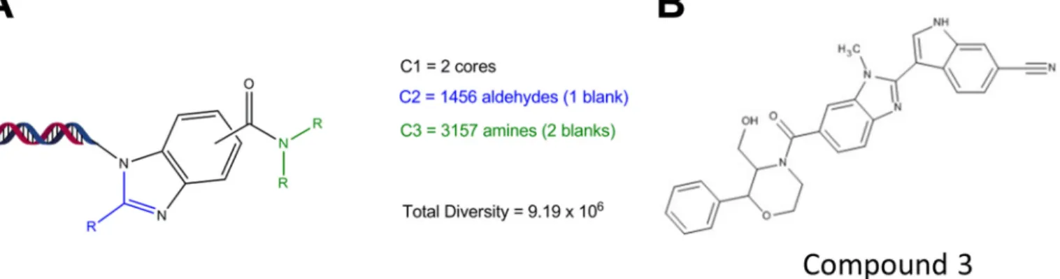
library (Fig. 4A) utilized two fluorinated aromatic acids in cycle
FIGURE 3.X-ray crystal structure of PDE12.A, ribbondiagram of the overall structure of PDE12 residues 161– 609 colored with a gradient frombluetored
beginning at the N terminus.B,surface representation of PDE12 colored by charge density.Blue,positive charge;red,negative charge;white,neutral.C,
comparison between the PDE12 (yellow) and CNOT6 (cyan) overall structure. The Mg2⫹ion common between the two structures is shown ingray. The Mg2⫹ ion unique to CNOT6 is shown ingreen.D,electron density around the Mg2⫹ion in the active site.
TABLE 2
Substrate kinetics and compound 3 inhibition
Substrate and inhibitor steady state kinetic studies were performed using the indicated enzyme and nucleic acid substrate. AMP formed was determined as a function of time from 20-min progress curves conducted in triplicate, and Equation 4 was fit to the initial rates to determinekcatandKmvalues, or Equation 5 was fit to determineKivalues
for compound3. The best fit parameter estimates are shown⫾the standard error of the fit.
2–5A OligoA-12
Compound 3,Ki
Km kcat kcat/Km Km kcat kcat/Km
M s⫺1
M⫺1s⫺1
M s⫺1
M⫺1s⫺1 n M
PDE12 103⫾4 27⫾0.5 2.6⫻105 280⫾100 11⫾3 0.39⫻105 17.5⫾0.8
chosen as the starting point for our hit optimization effort (Fig. 4B).
Structure-Activity Relationships for PDE12 Inhibitors—An
x-ray crystal structure of a complex of PDE12 with compound3
was determined using the 155– 609 construct (Fig. 5A, Protein
Data Bank code 4Z2B). Strong electron density for the com-pound could be seen in the active site, and we were able to fit the compound unambiguously by confirming the chirality of the
⬍4.4n⫽1.
TABLE3
Structure-activityrelationshipsforPDE12inhibitors
Compounddose-responseexperimentswereconductedandanalyzedasdescribedunder“ExperimentalProcedures.”PDE12,PDE12enzymeinhibition;CNOT6,CNOT6 enzymeinhibition;HeLa2–5A,IFN␣andpoly(IC)-stimulated2–5Alevelsincelllysate;EMCV,EMCVCPEinHeLaOhiocells;HRV16,inhibitionofHRV16infectionof SAECmeasuredbyimagingcapsidprotein.Forinhibitionassays,pXC50representsthenegativelogoftheIC50inmolar(pIC50).ForHeLa2–5A,pXC50representsnegative logoftheinflectionpointoftheincrease(pEC50).Thevaluespresentedarethemean共 S.D.fromthenumberofexperimentsshowninparentheses.Forsomeofthereplicate experimentaldeterminations,aparameterfitwasnotobtainedbecausetheinflectionpointwasoutsidethecompoundconcentrationrange.Thesevalueshadtobeexcluded fromthereportedmean:compound1,PDE12pIC50⬎9.1n ⫽ 1;HeLa2–5ApEC50⬎8.1n ⫽ 2.Compound2,HeLa2–5ApEC50 ⬍4.8n ⫽ 2;compound4,PDE12pIC50
1,followedby benzimidazoleformationwith1456 aldehydes
(cycle2),andthentheacidfunctionalitywascappedwith3157
amines(cycle3)toaffordanELTlibrarywith9.19million
enu-merated compounds.After selections, DNA sequencingwas
conducted,andthesequencesweretranslatedtoidentifythe
structures of the putative PDE12 binders. Several potential
activecompoundsweredesignedandsynthesized.Compound
bound species directly (Fig. 5B). Compound 3 shows a very optimized fit and displays several important ligand-protein
interactions (Fig. 5C). The 6-CN moiety on the indole forms a
favorable hydrogen bonding interaction (2.2 Å) with the back-bone NH of Thr-500. Any attempts to replace this nitrile with bioisosteres proved deleterious to potency (data not shown).
The carbonyl at 4-C of the benzimidazole has proximity to the bottom of the protein pocket making a strong interaction with
the nearby Mg2⫹ion, which is stabilized by interactions with
several water molecules in the region. Similar to the
apo-struc-ture, we observed only one Mg2⫹ion in the active site. The
primary alcohol at the 2-position on morpholine forms a crucial
FIGURE 4.Encoded library structure and initial PDE12 inhibitor.A,schematic of the ELT library used for PDE12 inhibitor structure. The helix represents the position of DNA tags used to identify protein-bound compounds following DNA sequencing.C1represents the variants in the core structure.C2represents the variants found at position R (blue).C3represents the variants found at position R (green).B,chemical structure of key active compound discovered by ELT screening, compound design, and synthesis (compound3). The biological activity for compound3is described in Table 3.
FIGURE 5.Co-crystal structure of compound 3 bound to PDE12.A,structure of compound3bound to the PDE12 catalytic domain construct, PDE12 (155– 609⌬206 –233).B,electron density of the inhibitor in the active site.C,schematic representation of the contacts between the inhibitor and PDE12.D,
compounds in this series did not inhibit CNOT6 even at the
highest concentration tested, 50M(Table 3).
Effect of PDE12 Inhibitors on Cellular 2–5A, RNase-L Activa-tion, and Virus Infection—PDE12gene inactivation resulted in
increased 2–5A induced by IFN and poly(I-C) (Fig. 2A). We
evaluated the effect of PDE12 inhibitors in HeLa cells to deter-mine whether inhibition of enzyme activity produced the same effect as gene inactivation. Compound 1 is one of our most
potent PDE12 inhibitors with a pIC50value for enzyme
inhibi-tion of 9.1. Compound 1 produced a maximum 4-fold increase in 2–5A levels, which is similar to what was seen with gene
inactivation. The pEC50value for this effect was 7.7 (Fig. 7A).
This 1.4 log decrease in potency from the pIC50value for PDE12
enzyme inhibition is consistent for all compounds tested in this series (Table 3). Compounds in the PDE12 inhibitor series did not affect CNOT6, the most closely related enzyme to PDE12. Nevertheless, it remains possible that they could produce bio-logical effects through unknown proteins. We evaluated the
effect of compound 1 in the HeLa⌬PDE12 cells. The
IFN-FIGURE 6.Kinetic characterization of PDE12 and CNOT6.Substrate and inhibitor steady state kinetic studies were determined using the indicated enzyme and nucleic acid substrate. AMP formed was determined as a func-tion of time from 20-min progress curves, and the initial rates were replotted as shown. Kinetic parameters determined from these experiments are sum-marized in Table 1.A,reaction rate as a function of 2–5A concentration.Blue,
PDE12;red,CNOT6.Linesrepresent fit of the data points to Equation 4.B,
reaction rate as a function of oligoA;blue,PDE12;red,CNOT6.Linerepresents fit of the data points to Equation 3.C,inhibition of PDE12 by compound3as a function of 2–5A concentration.Blue,no compound;red,10 nMcompound 3;green,30 nMcompound3;purple,90 nMcompound3;orange,180 nM
compound3.Linesrepresent a global fit of the data points to Equation 4 for 2–5A competitive inhibition.
H-bondinginteraction(1.4Å) withGlu-351.Anycapping of
thismoiety results in adramatic drop in potency(data not
shown). The structure also reveals a⫹-stacking interaction
betweentheneighboringphenylgroupandTyr-307.Removal
ofanyorbothofthephenylandhydroxylgroupsresultsina
drasticdropinpotency(compound4).Alkylationoftheindole
withethyl(compound2)improvesthepotencyduetoa
favor-ablehydrophobicinteraction of theethylatthe edgeof the
receptor. Preparation of the 5-bromobenzimidazole analog
(compound1)boostedpotencycomparedwithcompound2.
We hypothesized that substitutions at the 5-benzimidazole
positionimpart a steric effect causing the4-CO to adopt a
perpendicularorientation with respecttothebenzimidazole
ring maximizing the interaction between the CO and the
magnesium.
ModeofActionofPDE12Inhibitors—ThePDE12-compound
3co-crystalstructureindicatesthatthecompoundbindsinthe
activesitecleftthatwasidentifiedbymolecularmodeling(41).
AcomparisonbetweenthecrystalstructureofCNOT6witha
polyadenylate RNA substrate and our PDE12 compound 3
complexshowsthatourinhibitorisoccupyingnearlythe
equiv-alentvolumeandpositioninPDE12asthesubstrateoccupiesin
CNOT6(Fig.3D).Thisimpliesthatthecompoundmayinhibit
bycompetingwith2–5A.Toevaluatethishypothesisfurther,
we conducted steady state substrate kinetic experiments to
determinethemodeofinhibitionofcompound3.Theinitial
rateofAMPproducedwasanalyzedatfivefixedconcentrations
ofcompound3usingarangeof2–5Aassubstratefrom1to250
M(Fig.6C).The initialrateswere analyzedbyglobalcurve
fitting to equations representing competitive (Equation 5),
noncompetitive(Equation 6), oruncompetitive (Equation7)
modesofinhibition.Thecompetitivemodelprovidedthebest
fit (R2 ⫽ 0.997) compared with the noncompetitive model
(R2⫽0.976)andtheuncompetitivemodel(R2⫽0.940).The
competitiveKi valueforcompound1inhibitionwas17.5(共 8)
nM. Takentogether with thecrystal structureandmodeling
results,weconcludethatcompound3inhibitsPDE12by
com-petingwith2–5Asubstrate.
PDE12isamemberoftheEEPnucleasefamily.CNOT6isthe
mostsimilarenzymetoPDE12with31%aminoacidsequence
identity in the EEP nuclease domain (41).PDE12 has been
showntobeabletocleavetypical3⬘,5⬘-linkedoligoAsubstrates
(19,41,42),butCNOT6hasnotbeenshowntocleave2–5A
substrates. Wecompared thesteady state substratekinetics
using 2–5A (Fig. 6A) and oligoA (Fig. 6B) for PDE12 and
CNOT6.kcatandKm valuesweredeterminedbyfittingthe
ini-tialrateofAMPformationasafunctionofsubstrate
concen-trationtoEquation4.kcat/Kmvalueisgenerallyregardedasthe
keymeasureofsubstrateefficiency.PDE12andCNOT6cleave
bothsubstratesefficiently(kcat/Kmnear10
5
M⫺1s⫺1).PDE12
prefers2–5Awitha2-foldhigherkcat/Kmcomparedwith
oli-goA.CNOT6alsoprefers2–5A.Theseresultssuggestthatboth
enzymeshaveaslightpreferenceforcleaving2⬘,5⬘
-oligoadeny-late.However,chainlengthdifferencesbetween2–5A(3
nucle-otides)andoligoA(12nucleotides)mayalsocontributetothe
observedkineticparameters.TheIC50valuesforcompounds
poly(I-C)-induced level of 2–5A was increased compared with
the parent HeLa line as expected. Compound1did not increase
the 2–5A level further at any concentration (Fig. 7A). Thus, the
ability of compounds in this series to increase 2–5A is depen-dent upon the presence of a functional PDE12 enzyme.
IFN-poly(I-C) induces relatively high levels of cellular 2–5A. This allows for accurate determination of 2–5A but may differ from a viral infection. We evaluated the ability of PDE12
inhib-itors to affect 2–5A in HRV-infected HeLa cells (Fig. 7B). HRV
alone resulted in a 15% increase in 2–5A compared with back-ground levels observed in uninfected cells. Prior treatment with IFN resulted in a slightly greater increase. Addition of PDE12 inhibitors to HRV-infected cells resulted in a 4 – 6-fold increase above the background level. By comparison, 2–5A levels in the compound-treated, IFN-poly(I-C)-induced cells were typically
20-fold higher than background levels (Fig. 7A).
Activation of RNase-L is the primary mechanism for the anti-viral effect of 2–5A. High level activation of RNase-L results in cleavage of rRNA and reduced protein synthesis (2– 4). Cellular
rRNA was analyzed using an Agilent Bioanalyzer (Fig. 7C).
IFN-poly(I-C) treatment of HeLa cells for 2 h resulted in the appear-ance of a major cleavage product representing 10% of the
detectable 28S rRNA. Addition of compound 1resulted in a
further increase in the proportion of this cleavage product to 23% indicative of increased RNase-L activation in response to PDE12 inhibition.
PDE12 inhibitors were evaluated for their antiviral activity (Table 3). Compound 1 inhibited the EMCV-induced
cyto-pathic effect in HeLa Ohio cells with a pIC50of 6.7. The
com-pound inhibited HeLa Ohio cell proliferation in the absence of
EMCV infection with a pIC50of 5.7. BecausePDE12gene
inac-tivation is tolerated in the HeLa cell background, it is possible the effect on cell proliferation is due to a PDE12-independent
effect. We evaluated the effect of compound1on the
prolifer-ation of HeLa⌬PDE12 cells. Compound 1 inhibited the
prolif-eration of both HeLa and HeLa⌬PDE12 cells with a pIC50of 5.7.
Thus, the compounds in this series appear to have some anti-proliferative PDE12-independent activities at higher concen-trations. Typically, the antiviral activity of these compounds is 10 –30-fold more potent than the anti-proliferative activity (Table 3). The mechanism of the PDE12-independent anti-pro-liferative activity is unknown.
Our results suggest that PDE12 inhibitors exert an antiviral effect through modulation of the IFN-induced OAS/RNase-L effector pathway. This pathway may be altered in the HeLa cell background due to changes associated with oncogenesis. HRV replicates well in human small-airway epithelial cells so we used the HRV imaging assay to measure the effect of PDE12 inhibi-tors in a primary cell host. PDE12 inhibiinhibi-tors blocked the repli-cation of HRV in these cells (Table 3). For example, compound
1inhibited HRV infection with a pIC50of 6.9. In general, the
potency of compounds in this series for 2–5A modulation, EMCV inhibition, and HRV inhibition correlated with the potency of PDE12 enzyme inhibition.
Discussion
PDE12 is a widely expressed, abundant 2–5A-degrading enzyme that may have potential as an antiviral target. Two pre-vious lines of evidence support the antiviral role for PDE12, siRNA-mediated knockdown of protein expression and the effects of an inhibitor of PDE12 enzyme activity (17). The pro-posed mechanism for this activity is modulation of the IFN-induced OAS/2–5A/RNase-L pathway, although direct evi-dence supporting this has not been presented. In addition to PDE12, virus-encoded 2–5A nucleases and other host enzymes have been described that possess 2–5A nuclease activity, and modulation of these genes has been found to influence viral infectivity (12). Therefore, the role of PDE12 in regulation of host antiviral defense remains unclear.
We employed TALEN nuclease-mediated gene inactivation
to create a HeLa-derived cell line, HeLa⌬PDE12, with no
func-tional PDE12 expression. HeLa⌬PDE12 cells demonstrated
increased 2–5A compared with the parent HeLa cell line when treated with IFN and the double-stranded RNA mimic poly-(I-C). This confirms that PDE12 controls cellular 2–5A levels in
response to stimuli known to activate OAS.PDE12gene
inac-tivation also resulted in increased resistance to infection by EMCV, HRV, and RSV. The lack of PDE12 activity did not
pre-FIGURE 7.Cellular mechanism of PDE12 inhibitors.A,2–5A concentrations were determined following IFN and poly(I-C) treatment of the indicated cell line as a function of compound concentration. Blue, HeLa cells; red,
HeLa⌬PDE12 cells.B,2–5A concentrations were determined in HeLa cells infected with HRV (m.o.i.⫽30) after 2 days of infection in the presence or absence of IFN (100 units/ml) or PDE12 inhibitors (5M).C,rRNA from HeLa cells was extracted and analyzed using an Agilent Bioanalyzer.Lane 1,
untreated HeLa cells.Lane 2,HeLa cells treated for 2 h with IFN and poly(I-C).
Lane 3,HeLa cells treated for 2 h with IFN and poly(I-C) in the presence of 1M
Both ends of the active site appear to be more open in PDE12 relative to CNOT6. PDE12 contains a double glycine at residues 554 –555 compared with Phe-482–Asp-483 in cNOT6 at one end of the pocket. On the opposite end, CNOT6 contains an 11-residue insertion that provides a well defined end to the active site cleft. The absence of these residues in PDE12 opens up the cleft. The significance of these differences is unknown but may affect cleavage of longer substrates.
At the N terminus, PDE12 contains an immunoglobulin (Ig)-like domain that interacts with the surface of the catalytic domain. It is interesting to note that other EEP family enzymes do not appear to contain this domain. Although the function of the N-terminal Ig domain remains unknown, we speculate that it may function like other Ig domains as an interaction region to anchor the enzyme to another protein or lipid membrane. Alternatively, the basic patch on the surface of the Ig domain
(Fig. 3C) could interact with the phosphate backbone of longer
substrates.
PDE12 has been shown to be located in the mitochondrial
matrix and found to have the ability to cleave 3⬘,5⬘
-oligoadeny-late in addition to 2–5A (19, 41, 42). Moreover, overexpression of PDE12 reduced the levels of key mitochondrial proteins and rates of oxidative phosphorylation; thus, it appears that PDE12 functions as a mitochondrion-specific deadenylase controlling gene expression in a fashion similar to other EEP enzymes (41).
In contrast, we have found that PDE12 prefers 2–5A to 3⬘,5⬘
-oligoA as a substrate with a 6.5-fold higherkcat/Kmvalue. In
addition,PDE12gene inactivation or inhibition with small
mol-ecules results in higher cellular 2–5A in response to OAS acti-vation. Thus, PDE12 appears to be a biologically relevant regu-lator of the 2–5A-mediated IFN response as well as a reguregu-lator of mitochondrial gene expression. The localization of PDE12 to the mitochondrial matrix confounds our understanding of the subcellular compartmentalization of the OAS/2–5A/RNase-L pathway, which is typically thought of as being a cytoplasmic process. Perhaps a subpopulation of PDE12 is present in the cytoplasm. In contrast, OAS and RNase-L have been shown to be associated with mitochondria, so PDE12 may be part of a larger mitochondrion-associated innate defense response (45–
47). We have found thatPDE12gene inactivation and
inhibi-tors are generally well tolerated by HeLa cells. The effect of gene inactivation and inhibitors on mitochondrial biology has not yet been studied, but these effects will clearly be important in evaluating the potential of PDE12 inhibition for antiviral therapy.
As discussed in the Introduction, other host enzymes in the LigT-like group of 2H phosphodiesterases, including AKAP7 have been shown to degrade 2–5A (14). Moreover, EEP nucleases, including EPP1 (19) and CNOT6, have been shown to possess this activity. Thus, it seems that mammalian cells have evolved multiple mechanisms for down-regulating the OAS/2–5A/RNase-L pathway. The alternative subcellular localization of these enzymes or other levels of regulation may be important for preventing excessive RNA degradation and subsequent cellular damage. From a practical perspective, the redundancy within the host cell as well as virus-encoded 2–5A-cleaving enzymes may complicate the utility of future agents targeting these enzymes.
venttheexpressionofHRVorRSVstructuralproteinsinthe
earlystagesofHeLa⌬PDE12infection,butthespreadof
infec-tionobservedatlowerlevelsofviruswassignificantlyreduced
comparedwithinfectionoftheparentalHeLacells(Fig.2,C
andD).Thedelayedantiviralresponsemaybeattributedtothe
timerequiredforthehostcellstorespondtotheinitial
infec-tion,induceIFN,induceOAS,andthenaccumulate2–5Ato
activatetheantiviralproteinRNase-L.
Host targets like PDE12 mayprovide ameans for
break-throughdiscoveriesoftherapeuticagentswithbroadlyacting
antiviral potential. Phosphodiesterases like PDE4 and PDE5
haveproventobetractabledrugtargetsforavarietyof
indica-tions,includinginflammationanderectiledysfunction,
respec-tively(43).Despiteitsnamebaseduponanabilitytobreakthe
phosphodiester bonds linking the adenylate monomers of
2–5A,PDE12isnotamemberofthecyclicnucleotide
phos-phodiesterase superfamily that contains the enzymes PDE1
throughPDE11.PDE12isamemberoftheEEPsuperfamilyof
deadenylases basedupon aminoacid sequence homologyof
active sites.Other EEPenzymesthat include CNOT6,
Noc-turnin,andANGELcontrolarangeofbiologicalactivitiesby
regulatingtypical3⬘,5⬘-poly(A)taillengthandsubsequentRNA
stability(18).SmallmoleculeinhibitorsofenzymesintheEEP
nucleasefamilyhavenotbeenreported.Therefore,weelected
toinitiateourdrugdiscoveryeffortusingELT,anapproachthat
selects enzyme-binding small molecules from extremely
diversechemicallibraries(39).BaseduponananalysisofELT
compoundsthatboundPDE12,wegeneratedaseriesofsmall
moleculeinhibitorswithrepresentativesdescribedinTable3.
Treatmentofhostcellswiththesecompoundsmodulates
cel-lular2–5A,activatesRNase-L,andimpartsresistancetoviral
infection in a fashionthat mimicsPDE12 gene inactivation.
These moleculesmayrepresentattractive startingpoints for
host-directedantiviraldrugdiscovery.
Tofurtherourunderstandingoftheenzymeandaid
com-pound design,wedetermined thex-ray crystal structures of
PDE12intheapo-stateandwiththeenzymeboundto
com-pound3.ThestructuresofPDE12revealsimilaritieswithother
membersofthenucleasefamily.Nearlyalloftheresidues
deter-minedtoplayaroleinthecatalyticmechanismforAPE1(44)
areconservedinsimilarhydrogenbond networksand
struc-tural alignment in PDE12. These residues include Asp-561,
His-599,awatermolecule,theMg2⫹ion,Glu-351, Asn-301,
Asn-498,andAsp-496leadingustobelievethatthecatalytic
mechanismisthesameaswhatwaselucidatedforother
mem-bersofthisfamily.ComparisonofPDE12andCNOT6reveals
importantdifferencesthat mayaccountforsubstrate
prefer-enceandinhibitorbindingspecificitywithintheEEPsubfamily
ofnucleases.PDE12hasaninsertionoffourresiduestoextend
twoshort helices onone side of thesubstratepocket anda
numberofsingleresiduedifferencesatpositions316,500,555,
and 560. These changes result in overall differences in the
shapesofthePDE12andCNOT6substratepocketspresenting
unique surfaces involvedin substrateand inhibitor binding.
However,thepocketsofbothenzymesarelargeandopen
rel-ativetotheconservedgeometryofthecatalyticresiduesthat
may explain the observed flexibility in substrate selection