The handle
http://hdl.handle.net/1887/33100
holds various files of this Leiden University
dissertation
Author: Dane, Martijn
Structure and Function of the
Endothelial Glycocalyx in the
Microcirculation
Martijn Jacob Cornelis Dane, 2015
All rights are reserved. No part of this publication may be reproduced, stored, or transmitted in any form or by any means, without permission of the copyright owners.
ISBN: 978-94-6295-163-1
Cover: Martijn Dane
Layout: Martijn Dane
Printed by: Proefschriftmaken.nl || Uitgeverij BOXpress Published by: Uitgeverij BOXpress, ‘s Hertogenbosch
The research presented in this thesis was performed at the department of Nephrology of the Leiden University Medical Center.
The research decribed in this thesis was supported by a grant from the Dutch Kidney Foundation (C08.2265). Financial support by the Dutch Kidney Foundation and the Dutch Heart Foundation for the publication of this thesis is gratefully acknowledged.
Structure and Function of the
Endothelial Glycocalyx in the
Microcirculation
Proefschrift
ter verkrijging van
de graad van Doctor aan de Universiteit Leiden,
op gezag van Rector Magnificus prof. mr. C.J.J.M. Stolker,
volgens besluit van het College voor Promoties
te verdedigen op dinsdag 2 juni 2015
klokke 16.15 uur
door
Martijn Jacob Cornelis Dane
Promotor: Prof. Dr. A.J. Rabelink
Co-promotores: Dr. B.M. van den Berg
Dr. H Vink
Overige leden: Prof. Dr. V.W.M. van Hinsbergh
VU University Medical Centre,
Amsterdam, The Netherlands
Dr. J. van der Vlag
Radboud University Nijmegen Medical Centre, Nijmegen, the Netherlands
Prof. Dr. P. Reitsma
Prof. Dr. J.W. Jukema
Contents
Chapter 1 General Introduction 7
Chapter 2 A microscopic view on the renal endothelial glycocalyx 29
Adapted from: Am J Physiol Renal Physiol 00532 02014, 2015
Chapter 3 Prolonged shear stress modifies the composition of 49 the endothelial glycocalyx
In preparation
Chapter 4 Glomerular endothelial surface layer acts as a barrier 69 against albumin filtration
Am J Pathol 182: 1532-1540, 2013
Chapter 5 Association of kidney function with changes in 95 the endothelial surface layer
CJASN 9: 698-704, 2014
Chapter 6 Deeper penetration of erythrocytes into the 117
endothelial glycocalyx is associated with impaired microvascular perfusion
PloS One 9: e96477,2014
Chapter 7 General discussion and summary 137
Chapter 8 Nederlandse samenvatting 151
Introduction and outline
1
Published as part of: Am J Physiol Renal Physiol ajprenal 00532 02014, 2015.Martijn J.C. Dane1, Bernard M. van den Berg1, Dae Hyun Lee1, Margien G.S. Boels1,
Gesa L. Tiemeier1, M. Cristina Avramut2, Anton Jan van Zonneveld1, Johan van der
Vlag3, Hans Vink4, Ton J. Rabelink1
1Department of Nephrology, Einthoven laboratory for Vascular Medicine, LUMC, Leiden
University Medical Center, The Netherlands 2Department of Molecular Cell Biology, Section
Electron Microscopy LUMC, Leiden University Medical Center, The Netherlands 3Department
of Nephrology, Radboud Institute for Molecular Life Sciences, Radboud University Medical Center, Nijmegen, the Netherlands 4Department of Physiology, Maastricht University Medical
The endothelial glycocalyx
1
Figure 1: Schematic overview of the endothelial glycocalyx in a healthy and diseased condition.
Biochemical structure of the endothelial glycocalyx
The membrane bound part of the endothelial glycocalyx consists of proteoglycans, glycosaminoglycans, glycoproteins and glycolipids. Proteoglycans with their bound GAGs are the main contributors to the EG structure and function[6]. Of these GAGs, heparan sulfate and hyaluronan constitute up to 90% [7-9].
Heparan sulfate is a linear polysaccharide which consists of the repeating disaccharide β1– 4-linked D-glucuronic acid (GlcA) and α1–4-linked N-acetyl-D-glucosamine (GlcNAc). This polymer is covalently attached to a limited number of core proteins at cell surface, called heparan sulfate proteoglycans (HSPGs). The proteoglycans found on the luminal endothelial side are syndecans 1 and 4, glypican 1, versican and thrombomodulin. HS chains are processed in the Golgi apparatus were they undergo a series of modifications in which subsets of glucosamine residues can become N-deacetylated and N-sulfated and where glucuronic acids may undergo epimerization to L-iduronic acid (IdoA). In particular, C2 of uronic acid and C6 (and rarely C3) of glucosamine residues may become sulfated [10]. After intracellular processing, further extracellular modification can occur. Heparanase (HPSE1) and endosulfatases (SULF1,2) can cleave the HS chain or further modify the sulfate groups within HS, respectively [11]. (figure 2) Although they do attribute to a wide variety of specific HS binding sites for protein interactions that determine endothelial cell biology, regulation of these modifications is still not well understood. While the core proteins can function independently of the HS chains they carry, the HS chains predominantly dictate ligand-binding capability and therefore the biological roles of HSPG. Some examples are discussed in the next section. HS is structurally related to heparin, a highly sulfated GAG [10-12].
1
Figure 2
Figure 2: Schematic overview of production and modification of HA and HS. Top left: Hyaluronan
Function of the endothelial glycocalyx
Biomechanical properties
The endothelial glycocalyx is believed to be the primary molecular sieve for plasma proteins and, therefore, the origin of the oncotic forces that control transcapillary fluid exchange. This has led to a revised Starling principle in which the capillary filtration equilibrium is generated over the glycocalyx, rather than across the entire endothelial layer as had been widely assumed [14]. Within the glycocalyx, hyaluronan is a key determinant of many biomechanical and hydrodynamic properties such as its function in fluid exchange and shear sensing properties [15-19]. In particular, its physical properties in solution contribute to the function of hyaluronan [2,20,21]. Even small amounts of hyaluronan can bind large amounts of water molecules, forming a gel-like structure. Small molecules like water, electrolytes and nutrients can freely diffuse through the solvent. In contrast, large molecules, such as proteins, are excluded because of their hydrodynamic sizes in solution. In this way, hyaluronan contributes to the barrier function of the endothelial glycocalyx. The negative charge of the sulfated heparan groups in HS chains further adds to the barrier function of the glycocalyx, by repelling negatively charged circulating proteins like albumin.
An intact glycocalyx also serves as the primary sensor of shear stress on ECs. Hydrodynamic drag arising from flowing plasma through this layer transmits fluid shear stress through the thin sub-layer where proteoglycans, glycoproteins and glycolipids are directly linked to the endothelial cytoskeleton [22]. Both, the presence of hyaluronan, possibly by linking to the epithelial sodium channel (ENaC) in the endothelium, and HS appear to be critical for this function [23]. For example, enzymatic removal of hyaluronan or HS from the glycocalyx results in reduced nitric oxide (NO) release during shear [17]. Heparinase III, a bacterial enzyme that cleaves HS proteoglycans, has a dramatic effect on the ECs’ ability to produce NO to shear stress and transfer shear forces to the actin cytoskeleton [8,24]. Interestingly, the enzyme chondroitinase, employed to selectively degrade CS, did not affect shear-induced NO production, underpinning the importance of HS for the biomechanical properties of the endothelial glycocalyx [25].
Inflammation
1
NDST1 which results in expression of HS domains on glomerular endothelium that are associated with inflammation [29,30]. In agreement, we recently showed that endothelial deletion of NDST1 results in reduced leukocyte extravasation in experimental anti-GBM glomerulonephritis [30].
The migration of leukocytes across the endothelium occurs along chemoattractant gradients composed of chemokines that have bound to the endothelium [5]. Nearly all members of the chemokine family bind to HS by way of a positively charged C-terminal domain. Again, this binding may be modified by chemical editing of HS chains. For example, changing the domain structure of HS by inactivation of HS2ST (which causes a compensatory increase in glucosamine N-sulfation and 6-O-sulfation) results in enhanced binding of IL-8 to the endothelium and increases inflammation [31].
Endothelial HS has also been shown to be critical for the function of the complement system. The function of many of the components of the complement system, including C1, C1q, C1 inhibitor, C2, C4, C4b, FactorB, FactorD, FactorH, Properdin, and complement receptors CR3(CD11b/CD18) (24) and CR4 (CD11c/CD18) is dependent upon binding to specific glycosaminoglycan domains [32-34]. The clinical importance and the specificity of these interactions are illustrated by patients with mutations in the short consensus repeats 19 and 20 of the complement inhibitor, factor H. These mutations result in impaired binding of factor H to the glomerular endothelium, but not to other endothelial beds. Upon a trigger that activates the complement system this may result in glomerular endothelial injury and presents itself clinically as atypical hemolytic uremic syndrome [35].
Anticoagulation
Both the biomechanical properties as well as glycocalyx-protein interactions play a prominent role in preventing the blood from clotting. The gel-like properties of the glycocalyx prevent platelets from accessing the endothelium [40]. HS in the glycocalyx is required to activate antithrombin, a serine protease that inhibits thrombin and thus the conversion of fibrinogen to fibrin. By acting as a scaffold and simultaneously binding to both enzyme and substrate, HS greatly increases engagement of thrombin with antithrombin [41,42]. This interaction depends upon a specific pentasaccharide, an insight that led to the clinical development of fondaparinux. By binding the plasma protein Heparin cofactor II (HCII), heparan sulfate repeats in the glycocalyx further inhibit the pro-coagulant activity of thrombin. The latter pathway has been shown to counteract thrombus formation in the presence of endothelial injury [43].
1
Risk factors for endothelial glycocalyx degradationCurrent treatment options to stabilize the endothelial glycocalyx
The proposed role of the EG as target for cardiovascular risk factors implies that the EG could be an interesting target for therapies against the development of vasculature-born pathologies. First of all, as endothelial dysfunction seems to be closely associated with endothelial glycocalyx loss, current medication based on improving vascular or endothelial function will most likely also affect the endothelial glycocalyx. Stabilizing the endothelium, for example with angiopoietin 1 (Ang1), has been shown to enhance glycocalyx thickness in frog mesenteric cells and increase GAG production in human microvascular endothelium [60].
In contrast to these indirect effects, several options have been proposed to directly influence endothelial glycocalyx. This is done by GAG supplementation. One of the most well studied glycosaminoglycan supplements is sulodexide; a highly purified mixture of low molecular weight heparin (80%) and dermatan sulfate (20%). Although its function has been prescribed to different mechanisms over the past years, its anti-albuminuric effect has been demonstrated in several studies [61-63]. For example, in male participants with type 2 diabetes (T2D), sulodexide administration led to an increase in systemic glycocalyx thickness. It showed a reduction in plasma hyaluronidase, as well as a trend towards the normalization of systemic albumin clearance [64]. Although a recent study showed that sulodexide failed to demonstrate renoprotection in overt type 2 diabetic nephropathy patients [65], this can most likely be explained by the progression of type 2 diabetes. Restoring the EG has the most significant effects during the early stages of the disease, before irreversible morphological changes within the kidney occur. Indeed, animal studies showed that sulodexide ameliorates only early disease in models of radiation nephropathy and diabetic nephropathy in rats [66]. Nonetheless, the disadvantage of sulodexide is the heterogeneity of the composition, which might also explain the diversity in study results.
1
The endothelial glycocalyx in the glomerular filtration barrierThe kidney
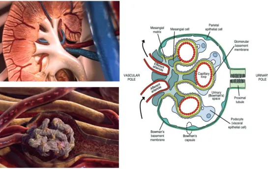
In addition to the general composition and function of the endothelial glycocalyx in the vasculature, we studied the role of the endothelial glycocalyx in renal filtration. The human kidney contains approximately 0.8-1.5*10^6 nephrons, which are the functional units of the kidney. The normal glomerular filtration rate ranges from 130-180 liters per day [72]. Circulating blood enters the glomerulus via the afferent arterioles. In the glomerulus ultrafiltration takes place (figure 3). Here, water and small solutes, like salts, can pass through the glomerular filtration barrier, while larger molecular like proteins and cells are retained within the circulation.
Figure 3: Structure and function of the kidney and glomerulus.One of the main functions of the
Barrier function in the glomerulus
Although still under some debate [73], most evidence points towards the glomerular filtration barrier (GFB) as the main site of filtration. Farquhar demonstrated, using electron microscopy, that in intact glomeruli only about 0.06% of plasma albumin gets filtrated [74,75]. Although tubular reabsorption may play a role in fine-tuning the leakage of albumin, the GFB is assumed to play the key role in filtration. This filtration function of the GFB is maintained by the three layers of the filtration barrier: endothelium, glomerular basement membrane (GBM) and podocytes. Together with the mesangium, these components form the glomerulus: a unique non-clogging filter that can endure the filtration for over a lifetime. Although all layers and its specific roles within the GFB are often studied separately, it should always be taken into account that all these layers depend on each other’s structure and function to form this highly specialized glomerular filtration barrier. For example, disturbed VEGF signaling from the podocytes affects endothelial health [76], and perturbed PDGF signaling from the endothelium affects the mesangium [77].
Until recently, it was reasoned that the endothelial layer could not contribute to the filtration barrier because of its large fenestrae. These fenestrae lack a diaphragm and can therefore be considered as holes of about 60-80 nanometer [78]. These holes support the passage of high volumes of water, but are also large enough to allow proteins like albumin to pass the endothelial layer. Consequently, most studies focused on the GBM and podocytes as main components of the filtration barrier. However, recently Weil et al. showed that changes in the endothelial layer were associated with glomerular filtration rate and albumin-creatinine ratio in T2D patients [79,80].
A role for the endothelium in the filtration barrier is further supported by the little variation in reflection coefficient between fenestrated and continuous capillary beds [81,82]. Ryan et al. demonstrated that under physiological conditions hardly any albumin is observed in the GBM or underlying podocytes [83]. This can be explained by the observation that endothelial fenestrae are filled with endothelial glycocalyx [84,85]. Consequently, the endothelial glycocalyx within the fenestrae was proposed to be the first barrier within the GFB that excludes proteins [86]. Glycosaminoglycan-degrading enzymes such as chondroitinase and heparinase have been shown to alter the charge selectivity of the glomerular filter [87,88]. In addition, even local displacement of only the non-covalently bound components of the renal glycocalyx has been demonstrated to result in a 12-fold increase in fractional albumin clearance [89]. This indicates that besides GAGs, the loosely bound plasma proteins are also essential for the structure of the endothelial glycocalyx and for the barrier function of the glomerulus.
1
the negatively charged albumin [90,91]. Although this is supported by data with non-metabolizable negatively charged probes that would support that electric charge would modify filtration of albumin [92,93], this concept has been challenged by invivo studies of the fractional clearance of negatively charged Ficoll as compared with Ficoll where negative charge selectivity did not exist [94,95].
Scope of this thesis
In the introduction we described a role for the EG in the onset of development in several vasculature-related pathologies like cardiovascular a disease, diabetes, but also renal failure. In this thesis the structure and function of the EG and its components in the vasculature in general and in the glomerular capillaries have been studied. In addition, methods to determine changes in the EG and the relation with known vascular damage markers in healthy and diseased subjects have been newly developed.
In Chapter 2, we mainly focus on the challenges in visualization and quantification
of the endothelial glycocalyx. Here, the different methods to study ESL thickness and composition, both in vitro and in vivo, are discussed.
Because it has been demonstrated that EG is absent in vitro, we hypothesized that mimicking the in vivo situation by subjecting the cells to prolonged shear stress would change the endothelial glycocalyx dimensions and composition. This will be further discussed in chapter 3.
The EG contributes to the permeability function of the endothelium throughout the whole vasculature. In the fenestrated endothelium of the kidney, the EG is also present within the fenestrae, suggesting a role for the EG in glomerular filtration. The main EG component that contributes to endothelial permeability has been proposed to be HA.
In chapter 4 we studied the role of the EG, and specifically HA, in the filtration barrier.
Therefore, we systemically removed HA with hyaluronidase and examined the effects on the function of the filtration barrier.
In chapter 5, we used a novel method to measure changes in the EG in end stage renal
disease patients. Using SDF imaging of the sublingual microvasculature, we looked into variations in the width of the RBC column as a measure for changes in the EG. In addition, we looked into endothelial activation and glycocalyx shedding to determine the association between renal function, endothelial dysfunction and glycocalyx shedding. Finally we studied the ability of the EG to recover after successful kidney transplantation.
In chapter 6 we studied the EG in a cohort of healthy but obese participants. Here
we demonstrated the relation between microvascular perfusion and EG thickness. Furthermore, these participants have a higher risk for the development of cardiovascular disease. Therefore this study functions as baseline measurement for future follow-up studies. Consequently the question whether early changes in EG predict cardiovascular events will be studied.
1
References1. Lennon FE, Singleton PA (2011) Hyaluronan regulation of vascular integrity. Am J Cardiovasc Dis 1: 200-213.
2. Laurent TC, Fraser JR (1992) Hyaluronan. FASEB J 6: 2397-2404.
3. Laurent T (1989) The biology of hyaluronan. Introduction. Ciba Found Symp 143: 1-20.
4. Rops AL, van der Vlag J, Lensen JF, Wijnhoven TJ, van den Heuvel LP, et al. (2004) Heparan sulfate proteoglycans in glomerular inflammation. Kidney Int 65: 768-785. 5. Massena S, Christoffersson G, Hjertstrom E, Zcharia E, Vlodavsky I, et al. (2010) A chemotactic gradient sequestered on endothelial heparan sulfate induces directional intraluminal crawling of neutrophils. Blood 116: 1924-1931.
6. Reitsma S, Slaaf DW, Vink H, van Zandvoort MA, oude Egbrink MG (2007) The endothelial glycocalyx: composition, functions, and visualization. Pflugers Arch 454: 345-359.
7. Oohira A, Wight TN, Bornstein P (1983) Sulfated proteoglycans synthesized by vascular endothelial cells in culture. J Biol Chem 258: 2014-2021.
8. Florian JA, Kosky JR, Ainslie K, Pang Z, Dull RO, et al. (2003) Heparan sulfate proteoglycan is a mechanosensor on endothelial cells. Circ Res 93: e136-142. 9. Rix DA, Douglas MS, Talbot D, Dark JH, Kirby JA (1996) Role of
glycosaminoglycans (GAGs) in regulation of the immunogenicity of human vascular endothelial cells. Clin Exp Immunol 104:60-65.
10. Esko JD, Lindahl U (2001) Molecular diversity of heparan sulfate. J Clin Invest 108: 169-173.
11. Esko JD, Selleck SB (2002) Order out of chaos: assembly of ligand binding sites in heparan sulfate. Annu Rev Biochem 71: 435-471.
12. Casu B, Lindahl U (2001) Structure and biological interactions of heparin and heparan sulfate. Adv Carbohydr Chem Biochem 57: 159-206.
13. Day AJ, Sheehan JK (2001) Hyaluronan: polysaccharide chaos to protein organisation. Curr Opin Struct Biol 11: 617-622.
14. Levick JR, Michel CC (2010) Microvascular fluid exchange and the revised Starling principle. Cardiovasc Res 87: 198-210.
15. van den Berg BM, Vink H, Spaan JA (2003) The endothelial glycocalyx protects against myocardial edema. Circ Res 92: 592-594.
16. van den Berg BM, Spaan JA, Vink H (2009) Impaired glycocalyx barrier properties contribute to enhanced intimal low-density lipoprotein accumulation at the carotid artery bifurcation in mice. Pflugers Arch 457: 1199-1206.
17. Mochizuki S, Vink H, Hiramatsu O, Kajita T, Shigeto F, et al. (2003) Role of hyaluronic acid glycosaminoglycans in shear-induced endothelium-derived nitric oxide release. Am J Physiol Heart Circ Physiol 285: H722-726.
19. Nagy N, Freudenberger T, Melchior-Becker A, Rock K, Ter Braak M, et al. (2010) Inhibition of hyaluronan synthesis accelerates murine atherosclerosis: novel insights into the role of hyaluronan synthesis. Circulation 122: 2313-2322.
20. Laurent TC, Laurent UB, Fraser JR (1995) Functions of hyaluronan. Ann Rheum Dis 54: 429-432.
21. Laurent TC, Laurent UB, Fraser JR (1996) The structure and function of hyaluronan: An overview. Immunol Cell Biol 74: A1-7.
22. Weinbaum S, Tarbell JM, Damiano ER (2007) The structure and function of the endothelial glycocalyx layer. Annu Rev Biomed Eng 9: 121-167.
23. Ramiro-Diaz J, Barajas-Espinosa A, Chi-Ahumada E, Perez-Aguilar S, Torres-Tirado D, et al. (2010) Luminal endothelial lectins with affinity for N-acetylglucosamine determine flow-induced cardiac and vascular paracrine-dependent responses. Am J Physiol Heart Circ Physiol 299: H743-751.
24. Thi MM, Tarbell JM, Weinbaum S, Spray DC (2004) The role of the glycocalyx in reorganization of the actin cytoskeleton under fluid shear stress: a “bumper-car” model. Proc Natl Acad Sci U S A 101: 16483-16488.
25. Pahakis MY, Kosky JR, Dull RO, Tarbell JM (2007) The role of endothelial glycocalyx components in mechanotransduction of fluid shear stress. Biochem Biophys Res Commun 355: 228-233.
26. Norgard-Sumnicht K, Varki A (1995) Endothelial heparan sulfate proteoglycans that bind to L-selectin have glucosamine residues with unsubstituted amino groups. J Biol Chem 270: 12012-12024.
27. Norgard-Sumnicht KE, Varki NM, Varki A (1993) Calcium-dependent heparin-like ligands for L-selectin in nonlymphoid endothelial cells. Science 261: 480-483. 28. Wang L, Fuster M, Sriramarao P, Esko JD (2005) Endothelial heparan sulfate
deficiency impairs L-selectin- and chemokine-mediated neutrophil trafficking during inflammatory responses. Nat Immunol 6: 902-910.
29. Rops AL, van den Hoven MJ, Baselmans MM, Lensen JF, Wijnhoven TJ, et al. (2008) Heparan sulfate domains on cultured activated glomerular endothelial cells mediate leukocyte trafficking. Kidney Int 73: 52-62.
30. Rops AL, Loeven MA, van Gemst JJ, Eversen I, Van Wijk XM, et al. (2014) Modulation of heparan sulfate in the glomerular endothelial glycocalyx decreases leukocyte influx during experimental glomerulonephritis. Kidney Int 86: 932-942 31. Axelsson J, Xu D, Kang BN, Nussbacher JK, Handel TM, et al. (2012) Inactivation
of heparan sulfate 2-O-sulfotransferase accentuates neutrophil infiltration during acute inflammation in mice. Blood 120: 1742-1751.
32. Wuillemin WA, te Velthuis H, Lubbers YT, de Ruig CP, Eldering E, et al. (1997) Potentiation of C1 inhibitor by glycosaminoglycans: dextran sulfate species are effective inhibitors of in vitro complement activation in plasma. J Immunol 159: 1953-1960.
1
34. Presanis JS, Hajela K, Ambrus G, Gal P, Sim RB (2004) Differential substrate andinhibitor profiles for human MASP-1 and MASP-2. Mol Immunol 40: 921-929. 35. Boels MG, Lee DH, van den Berg BM, Dane MJ, van der Vlag J, et al. (2013) The
endothelial glycocalyx as a potential modifier of the hemolytic uremic syndrome. Eur J Intern Med 24: 503-509.
36. Yung S, Thomas GJ, Davies M (2000) Induction of hyaluronan metabolism after mechanical injury of human peritoneal mesothelial cells in vitro. Kidney Int 58: 1953-1962.
37. Dyer DP, Thomson JM, Hermant A, Jowitt TA, Handel TM, et al. (2014) TSG-6 Inhibits Neutrophil Migration via Direct Interaction with the Chemokine CXCL8. J Immunol 192: 2177-2185.
38. Jiang D, Liang J, Noble PW (2011) Hyaluronan as an immune regulator in human diseases. Physiol Rev 91: 221-264.
39. Kim MY, Muto J, Gallo RL (2013) Hyaluronic acid oligosaccharides suppress TLR3- dependent cytokine expression in a TLR4-dependent manner. PLoS One 8: e72421. 40. Reitsma S, Oude Egbrink MG, Heijnen VV, Megens RT, Engels W, et al. (2011)
Endothelial glycocalyx thickness and platelet-vessel wall interactions during atherogenesis. Thromb Haemost 106: 939-946.
41. Olson ST, Bjork I (1991) Predominant contribution of surface approximation to the mechanism of heparin acceleration of the antithrombin-thrombin reaction. Elucidation from salt concentration effects. J Biol Chem 266: 6353-6364.
42. Li W, Johnson DJ, Esmon CT, Huntington JA (2004) Structure of the antithrombin- thrombin-heparin ternary complex reveals the antithrombotic mechanism of heparin. Nat Struct Mol Biol 11: 857-862.
43. He L, Vicente CP, Westrick RJ, Eitzman DT, Tollefsen DM (2002) Heparin cofactor II inhibits arterial thrombosis after endothelial injury. J Clin Invest 109: 213-219. 44. Parkinson JF, Vlahos CJ, Yan SC, Bang NU (1992) Recombinant human
thrombomodulin. Regulation of cofactor activity and anticoagulant function by a glycosaminoglycan side chain. Biochem J 283 ( Pt 1): 151-157.
45. Isermann B, Vinnikov IA, Madhusudhan T, Herzog S, Kashif M, et al. (2007) Activated protein C protects against diabetic nephropathy by inhibiting endothelial and podocyte apoptosis. Nat Med 13: 1349-1358.
46. Ye J, Esmon CT, Johnson AE (1993) The chondroitin sulfate moiety of
thrombomodulin binds a second molecule of thrombin. J Biol Chem 268: 2373-2379. 47. Lin JH, McLean K, Morser J, Young TA, Wydro RM, et al. (1994) Modulation
of glycosaminoglycan addition in naturally expressed and recombinant human thrombomodulin. J Biol Chem 269: 25021-25030.
48. Li W, Huntington JA (2008) The heparin binding site of protein C inhibitor is protease-dependent. J Biol Chem 283: 36039-36045.
50. Nieuwdorp M, Mooij HL, Kroon J, Atasever B, Spaan JA, et al. (2006) Endothelial glycocalyx damage coincides with microalbuminuria in type 1 diabetes. Diabetes 55: 1127-1132.
51. Han J, Woytowich AE, Mandal AK, Hiebert LM (2007) Heparanase upregulation in high glucose-treated endothelial cells is prevented by insulin and heparin. Exp Biol Med (Maywood) 232: 927-934.
52. Singh A, Friden V, Dasgupta I, Foster RR, Welsh GI, et al. (2011) High glucose causes dysfunction of the human glomerular endothelial glycocalyx. Am J Physiol Renal Physiol 300: F40-48.
53. van den Berg BM, Spaan JA, Rolf TM, Vink H (2006) Atherogenic region and diet diminish glycocalyx dimension and increase intima-to-media ratios at murine carotid artery bifurcation. Am J Physiol Heart Circ Physiol 290: H915-920.
54. Meuwese MC, Broekhuizen LN, Kuikhoven M, Heeneman S, Lutgens E, et al. (2010) Endothelial surface layer degradation by chronic hyaluronidase infusion induces proteinuria in apolipoprotein E-deficient mice. PLoS One 5: e14262.
55. Meuwese MC, Mooij HL, Nieuwdorp M, van Lith B, Marck R, et al. (2009) Partial recovery of the endothelial glycocalyx upon rosuvastatin therapy in patients with heterozygous familial hypercholesterolemia. J Lipid Res 50: 148-153.
56. Chappell D, Hofmann-Kiefer K, Jacob M, Rehm M, Briegel J, et al. (2009) TNF-alpha induced shedding of the endothelial glycocalyx is prevented by hydrocortisone and antithrombin. Basic Res Cardiol 104: 78-89.
57. Czarnowska E, Karwatowska-Prokopczuk E (1995) Ultrastructural demonstration of endothelial glycocalyx disruption in the reperfused rat heart. Involvement of oxygen free radicals. Basic Res Cardiol 90: 357-364.
58. Nieuwdorp M (2006) Loss of endothelial glycocalyx during acute hyperglycemia coincides with endothelial dysfunction and coagulation activation in vivo. Diabetes 55:480-486.
59. Girish KS, Kemparaju K (2007) The magic glue hyaluronan and its eraser hyaluronidase: a biological overview. Life Sci 80: 1921-1943.
60. Salmon AH, Neal CR, Sage LM, Glass CA, Harper SJ, et al. (2009) Angiopoietin-1 alters microvascular permeability coefficients in vivo via modification of endothelial glycocalyx. Cardiovasc Res 83: 24-33.
61. Gaddi AV, Cicero AF, Gambaro G (2010) Nephroprotective action of
glycosaminoglycans: why the pharmacological properties of sulodexide might be reconsidered. Int J Nephrol Renovasc Dis 3: 99-105.
62. Gambaro G, Kinalska I, Oksa A, Pont’uch P, Hertlová M, et al. (2002) Oral Sulodexide Reduces Albuminuria in Microalbuminuric and Macroalbuminuric Type 1 and Type 2 Diabetic Patients: The Di.N.A.S. Randomized Trial. Journal of the American Society of Nephrology 13: 1615-1625.
1
64. Broekhuizen LN, Lemkes BA, Mooij HL, Meuwese MC, Verberne H, et al. (2010)Effect of sulodexide on endothelial glycocalyx and vascular permeability in patients with type 2 diabetes mellitus. Diabetologia 53: 2646-2655.
65. Packham DK, Wolfe R, Reutens AT, Berl T, Heerspink HL, et al. (2012) Sulodexide Fails to Demonstrate Renoprotection in Overt Type 2 Diabetic Nephropathy. Journal of the American Society of Nephrology 23: 123-130.
66. Rossini M, Naito T, Yang H, Freeman M, Donnert E, et al. (2010) Sulodexide ameliorates early but not late kidney disease in models of radiation nephropathy and diabetic nephropathy. Nephrol Dial Transplant 25: 1803-1810.
67. Parish CR, Freeman C, Brown KJ, Francis DJ, Cowden WB (1999) Identification of sulfated oligosaccharide-based inhibitors of tumor growth and metastasis using novelin vitro assays for angiogenesis and heparanase activity. Cancer Res 59: 3433-3441.
68. van Kuppevelt TH, Jenniskens GJ, Veerkamp JH, ten Dam GB, Dennissen MA (2001) Phage display technology to obtain antiheparan sulfate antibodies. Methods Mol Biol 171: 519-534.
69. Ikeda Y, Charef S, Ouidja MO, Barbier-Chassefiere V, Sineriz F, et al. (2011) Synthesis and biological activities of a library of glycosaminoglycans mimetic oligosaccharides. Biomaterials 32: 769-776.
70. Lensen JF, Rops AL, Wijnhoven TJ, Hafmans T, Feitz WF, et al. (2005) Localization and functional characterization of glycosaminoglycan domains in the normal human kidney as revealed by phage display-derived single chain antibodies. J Am Soc Nephrol 16: 1279-1288.
71. Gil N, Goldberg R, Neuman T, Garsen M, Zcharia E, et al. (2012) Heparanase is essential for the development of diabetic nephropathy in mice. Diabetes 61: 208-216. 72. Silverthorn DU (2004) The kidneys. Human physiology. third edition ed: Pearson
education. pp. 598-624.
73. Comper WD, Haraldsson B, Deen WM (2008) Resolved: normal glomeruli filter nephrotic levels of albumin. J Am Soc Nephrol 19: 427-432.
74. Farquhar MG, Palade GE (1961) Glomerular permeability. II. Ferritin transfer across the glomerular capillary wall in nephrotic rats. JExpMed 114: 699-716.
75. Farquhar MG, Wissig SL, Palade GE (1961) Glomerular permeability. I. Ferritin transfer across the normal glomerular capillary wall. JExpMed 113: 47-66. 76. Eremina V, Sood M, Haigh J, Nagy A, Lajoie G, et al. (2003) Glomerular-specific
alterations of VEGF-A expression lead to distinct congenital and acquired renal diseases. JClinInvest 111: 707-716.
77. Schlondorff D, Banas B (2009) The mesangial cell revisited: no cell is an island. J Am Soc Nephrol 20: 1179-1187.
79. Weil EJ, Lemley KV, Mason CC, Yee B, Jones LI, et al. (2012) Podocyte detachment and reduced glomerular capillary endothelial fenestration promote kidney disease in type 2 diabetic nephropathy. Kidney Int 82: 1010-1017.
80. Satchell SC (2012) The glomerular endothelium emerges as a key player in diabetic nephropathy. Kidney Int 82: 949-951.
81. Levick JR, Smaje LH (1987) An analysis of the permeability of a fenestra. Microvasc Res 33: 233-256.
82. Michel CC, Curry FE (1999) Microvascular permeability. Physiol Rev 79: 703-761. 83. Ryan GB, Karnovsky MJ (1976) Distribution of endogenous albumin in the rat
glomerulus: role of hemodynamic factors in glomerular barrier function. Kidney Int 9:36-45.
84. Rostgaard J, Qvortrup K (1997) Electron microscopic demonstrations of filamentous molecular sieve plugs in capillary fenestrae. Microvasc Res 53: 1-13.
85. Avasthi PS, Koshy V (1988) The anionic matrix at the rat glomerular endothelial surface. Anat Rec 220: 258-266.
86. Salmon AH, Satchell SC (2012) Endothelial glycocalyx dysfunction in disease: albuminuria and increased microvascular permeability. J Pathol 226: 562-574. 87. Jeansson M, Haraldsson B (2003) Glomerular size and charge selectivity in the
mouse after exposure to glucosaminoglycan-degrading enzymes. J Am Soc Nephrol 14: 1756-1765.
88. Jeansson M, Haraldsson B (2006) Morphological and functional evidence for an important role of the endothelial cell glycocalyx in the glomerular barrier. Am J Physiol Renal Physiol 290: F111-116.
89. Friden V, Oveland E, Tenstad O, Ebefors K, Nystrom J, et al. (2011) The glomerular endothelial cell coat is essential for glomerular filtration. Kidney Int 79: 1322-1330. 90. Chang RL, Deen WM, Robertson CR, Brenner BM (1975) Permselectivity of the
glomerular capillary wall: III. Restricted transport of polyanions. Kidney Int 8: 212- 218.
91. Chang RL, Ueki IF, Troy JL, Deen WM, Robertson CR, et al. (1975) Permselectivity of the glomerular capillary wall to macromolecules. II. Experimental studies in rats using neutral dextran. Biophys J 15: 887-906.
92. Ohlson M, Sorensson J, Haraldsson B (2000) Glomerular size and charge selectivity in the rat as revealed by FITC-ficoll and albumin. Am J Physiol Renal Physiol 279: F84-91.
93. Bhalla G, Deen WM (2009) Effects of charge on osmotic reflection coefficients of macromolecules in fibrous membranes. Biophys J 97: 1595-1605.
94. Guimaraes MA, Nikolovski J, Pratt LM, Greive K, Comper WD (2003) Anomalous fractional clearance of negatively charged Ficoll relative to uncharged Ficoll. Am J Physiol Renal Physiol 285: F1118-1124.
1
96. Arkill KP, Knupp C, Michel CC, Neal CR, Qvortrup K, et al. (2011) Similarendothelial glycocalyx structures in microvessels from a range of mammalian tissues: evidence for a common filtering mechanism? Biophys J 101: 1046-1056. 97. Comper WD, Laurent TC (1978) Physiological function of connective tissue
polysaccharides. Physiol Rev 58: 255-315.
98. Weinbaum S, Zhang X, Han Y, Vink H, Cowin SC (2003) Mechanotransduction and flow across the endothelial glycocalyx. Proc Natl Acad Sci U S A 100: 7988-7995. 99. Adamson RH, Lenz JF, Zhang X, Adamson GN, Weinbaum S, et al. (2004) Oncotic
pressures opposing filtration across non-fenestrated rat microvessels. J Physiol 557: 889-907.
100. Lindstrom KE, Blom A, Johnsson E, Haraldsson B, Fries E (1997) High glomerular permeability of bikunin despite similarity in charge and hydrodynamic size to serum albumin. Kidney Int 51: 1053-1058.
101. Comper WD (2014) Albuminuria is controlled primarily by proximal tubules. Nat Rev Nephrol 10: 180.
A microscopic view on the renal endothelial
glycocalyx
2
Published as part of: Am J Physiol Renal Physiol ajprenal 00532 02014, 2015.Martijn J.C. Dane1, Bernard M. van den Berg1, Dae Hyun Lee1, Margien G.S. Boels1,
Gesa L. Tiemeier1, M. Cristina Avramut2, Anton Jan van Zonneveld1, Johan van der
Vlag3, Hans Vink4, Ton J. Rabelink1
1Department of Nephrology, Einthoven laboratory for Vascular Medicine, LUMC, Leiden
University Medical Center, The Netherlands 2Department of Molecular Cell Biology, Section
Electron Microscopy LUMC, Leiden University Medical Center, The Netherlands 3Department
of Nephrology, Radboud Institute for Molecular Life Sciences, Radboud University Medical Center, Nijmegen, the Netherlands 4Department of Physiology, Maastricht University Medical
Introduction
Endothelial cells perform key homeostatic functions such as regulating blood flow and permeability, preventing leukocyte activation, and aiding immune surveillance for pathogens. Endothelial activation therefore has been identified as an important effector mechanism in progression of renal disease as well as the associated development of cardiovascular disease. The primary interface between blood and the endothelium is the glycocalyx. This carbohydrate-rich gel-like structure mediates most of the regulatory functions of the endothelium. Because the endothelial glycocalyx is a highly dynamic and fragile structure ex-vivo, studying its dimensions and function has been proven to be a challenge. Tissue processing for staining and perfusion-fixation, usually will result in a partial or complete loss of the glycocalyx. Consequently, its functions and its potential as a therapeutic target have often been underappreciated. Here we will outline the different techniques to visualize structure function relationships in kidney and vasculature.
Imaging the endothelial glycocalyx
2
AB
C Figure 2
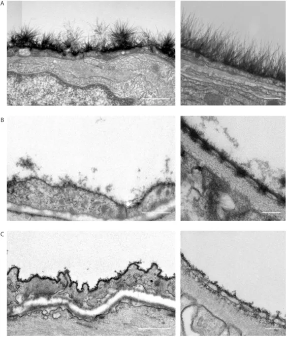
Figure 1: Three methods to stabilize and stain the endothelial glycocalyx for electron microscopy.
Electron microscopy
Preserving and staining the EG for EM
The high polysaccharide content of the EG interacts poorly with the commonly used post-fixation stains. As a result, the EG scatters few electrons and is indistinguishable from its environment in conventionally processed samples for TEM [6]. Thus, even if the pre-treatment of the sample maintains the structural integrity of the endothelial glycocalyx, an additional staining or preservation is required for visualization of the EG in TEM. This explains the absence of visible EG structures in almost all tissues that are conventionally processed for TEM. The first to succeed in staining the EG for electron microscopy was Luft in 1966, who used ruthenium red, or ammoniated ruthenium oxychloride, which stains the aldehyde fixed mucopolysaccharides in the EG and generates an electron dense stain in the presence of osmium tetroxide [7].
The presence of the luminal EG has subsequently been shown using TEM in combination with a variety of staining and preservation techniques such as ruthenium red, ferritin, and lanthanum. Most of these agents cationic dyes bind to the negative charge of the sulfated GAGs and form an electron dense contrast together with osmium tetroxide, to enable visualization [8-12]. Using alcian blue 8GX, van den Berg et al. were the first to be able to better stabilize the anionic carbohydrate structures in myocardial capillaries, thereby visualizing an impressive EG of up to 500 nm thick [13]. The saccharine nature of these visualized structures was confirmed by perfusion of gold-labelled lectins before the staining procedure. Recently, we were able to visualize the EG in the renal glomerulus using cupromeronic blue, a chemically more stable cationic dye resembling some alcian blue properties [14]. This revealed a staining of matrix polysaccharides on the luminal surface of the glomerular endothelial membranes and a dense staining of polysaccharide matrix within the fenestrae. Examples of the described staining procedures are shown
in figure 1.
High pressure freezing
2
3-Dimensional EM imaging
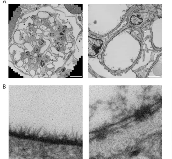
Over the recent years some interesting developments took the field of electron microscopy to a next level. These new methods might be used to better and more reliably study the EG. In regular EM imaging techniques, the highly detailed 2-dimensional EM images are prone to selection bias, as only a small area of interest is shown. To avoid this operator introduced selection bias, Faas et al. developed a method which provides high resolution and high detailed images while maintaining the lower resolution overview of the cellular context [15]. Examples of images in which virtual zooming was used on a stitch of a Cupromeronic blue stained glomerulus are shown in figure 3.
Interestingly, within one stitch of a mouse glomerulus different structural organizations of the EG can be observed, depending on its location (figure 3B non-fenestrated (top) vs fenestrated (bottom) endothelium). In addition 3D imaging can also be used to decrease bias and obtain more insight in the imaged structures. Based on earlier publications where bacterial flagellae were imaged [16], Arkill et al. succeeded to make a 3D reconstruction of the EG using 3D electron tomography. In electron tomography, an electron beam passes through the sample which is tilted after every image so that images can be taken at different angles. By reconstructing these images, a 3D image of the sample can be produced [17]. Although not used for EG imaging yet, some newer techniques, like SEM with a built-in microtome and focused ion beam scanning electron microscopy (FIB-SEM), will make the 3D analysis in electron microscopy even more detailed [18].
Figure 3
GBM
P EC
Glx
Figure 2: EG in a glomerular capillary of a high pressure frozen kidney section. EG was stained
A
B
Figure 3: Transmission electron microscopic images of a cupromeronic blue stained glomerulus.
2
Fluorescence microscopyStaining and imaging specific EG components
The carbohydrates within the EG have been imaged using specific lectins, mostly in combination with fluorescence microscopy. Lectins are carbohydrate binding proteins that recognize specific sugar moieties and 3D configurations. One of the most commonly used lectins is wheat germ agglutinin (WGA). Although lectin staining of the EG can be used to study changes in dimensions, they do not allow to determine changes in the specific composition of different GAGs. Consequently, antibodies are needed to more specifically determine compositional changes within the EG.
Over the recent years several antibodies have been obtained against specific carbohydrate domains within heparan sulfates (e.g. JM403, 10E4 and HEPSS1), hyaluronan and chondroitin sulfates or against the proteoglycan core proteins such as syndecan, glypican or perlecan [19-24]. Chondroitin sulfate and hyaluronan have been shown to be present on the endothelium, using an anti-CS antibody and by capitalizing on the protein binding properties of HA with a fluorescent hyaluronan binding protein (HABP), respectively [24-26]. Although all these techniques have shown the presence of glycocalyx components on the endothelial cells, a considerable part of these studies have been performed using epi-fluorescence microscopy, which has major limitations with respect to optical spatial resolution necessary for imaging the luminal endothelial glycocalyx.
LEA -CD31 -Syto41 LEA -WGA
A B
C WGA -Syto41 D
Figure 4: Endothelial glycocalyx imaged ex vivo with confocal- or multiphoton imaging
tech-niques. A) Mouse glomerulus visualised with anti-CD31 antibodies and TRITC-labeled conjugate
2
New labels for imaging the EG: Single chain antibodies
Measuring and imaging structural characteristics
Circulating markers of the glycocalyx
In response to reactive oxygen species (ROS) or other inflammatory mediators, both single GAGs and proteoglycans can be shed from the endothelium [29-31]. This has been suggested to be mediated by endoglycosidases such as heparanase and hyaluronidase, or proteases such as matrix metalloproteases [30,32,33]. Consequently, measuring shed glycocalyx components might be used as a marker for endothelial glycocalyx stability. For example, syndecan-1 and heparan sulfate are released from the tissue and can be detected in the circulating blood of patients with perioperative global or regional ischemia [34,35]. The glycocalyx core protein syndecan-1 acts as negative regulator of endothelial activation [36] and has been shown to be critical for processing and inactivation of heparanase on the cell surface [37]. In addition, release of the endothelium specific proteoglycan thrombomodulin has been shown to coincide with diabetes and diabetic nephropathy [38,39]. We demonstrated that patients with renal failure have increased circulating levels of syndecan-1 and thrombomodulin, which was reversed by kidney transplantation [40]. Furthermore, renal failure was shown to correlate with increased concentrations of shed HA [41]. Altogether, while several glycocalyx components are shedded upon endothelial activation, the exact location, mechanism and timing of shedding is complex and mostly unknown. Interpreting the results is still challenging.
Atomic Force microscopy
To determine the elastic properties of the EG, atomic force microscopy (AFM), has been shown to be a valuable tool. An AFM consists of a cantilever with a spherical tip which can scan the surface of the specimen. Reaching close contact with the specimen, forces between the tip and sample will result in deflection of this cantilever, that can be detected by changes in reflection of a laser spot. This technique has been previously used to measure mechanical stiffness in endothelial cells. However, by comparing endothelial cell layers with and without removal of the EG, the presence and stiffness of the EG could also be measured. This resulted in an estimated thickness of 400 nm of the EG on cultured endothelial cells, with a 50% EG thickness reduction after heparinase treatment [42]. Changes in the endothelial surface layer stiffness and thickness were associated with both endothelial activation and shedding of syndecan-1 and HA in renal failure patients and in animals [43].
Exclusion of macromolecules
2
before after
A B
40 kDa dextran (neutral) 40 kDa sulfated dextran
(anionic) Overlay
Figure 5
C
Figure 5: Exclusion properties of the endothelial surface layer in vivo. A) Intravital microscopic
Most of the techniques developed to estimate the changes in EG are based on this principle. Haraldsson et al. used Intralipid, a chylomicron-like suspension of purified soybean oil, egg-yolk phospholipids, glycerol, and water to use the physical exclusion properties of the EG for determining the EG thickness in TEM. After making electron micrographs of capillaries, the location of the lipid drop was identified as central or peripheral (being within 200 nm from the luminal endothelial surface) [46]. By comparing the distribution of these lipid particles before and after for example adriamycin treatment, changes in EG thickness could be estimated [47]. In vivo, a theoretically comparable method was used by Smith et al., who looked into the location of flowing microspheres. Using dual-flash epi-illumination, the velocity of infused fluorescently labelled microspheres was estimated in the near wall plasma-rich region of venules before and after light-dye treatment to degrade the EG. Using this technique, the theoretically suggested hydraulic resistivity of the surface layer was demonstrated [48]. Also, the EG thickness was estimated to be 0.33-0.44 um, which confirms the measurements by Vink and Duling [44].
Tracer dilution technique
Based on their in vivo observations in mice, the group of Vink et al. has mainly focused on the development of a method to measure changes in EG in patients. The tracer dilution technique was the first technique to estimate the glycocalyx volume. By comparing the circulating blood volume, using a glycocalyx impermeable tracer, and the total intravascular volume, using a glycocalyx permeable tracer, the total glycocalyx volume could be estimated. This concept was first applied when Nieuwdorp et al. showed that both acute hyperglycemia and type 1 diabetes mellitus coincided with a reduction of the estimated EG volume [49,50]. Unfortunately this technique has the disadvantage that it is very time-consuming and invasive to infuse labelled RBCs and Dex40.
RBC-EC gap and SDF imaging
Based on previous observations of the RBC exclusion zone in animal models, the presence of an RBC exclusion zone between the RBC column and the endothelium was demonstrated in trans-illuminated hamster cremaster muscle capillaries [51,52]. More recently we tested the hypothesis that the EG occupies this RBC-EC gap. When comparing the width of the RBC column with the internal anatomical vessel diameter as defined by the position of the fluorescent endothelium a decreased RBC-EC gap was observed after degradation of the EG with hyaluronidase [41].
2
Figure 5A
B
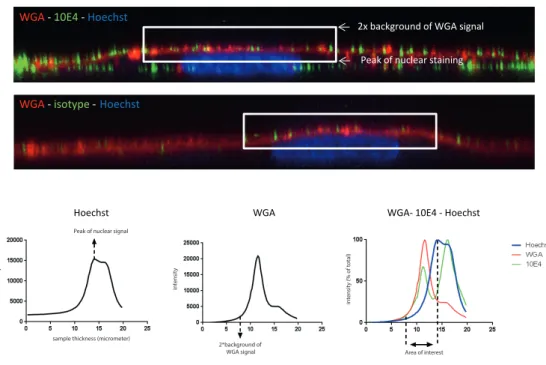
Figure 6: Sidestream darkfield (SDF) imaging to measure the perfused boundary region (PBR)
in the sublingual capillary bed.A) Recordings from the sublingual capillary bed made with the SDF
2
Summary and conclusionThe endothelial glycocalyx is proposed to mediate almost all of the endothelial functions and structural or compositional perturbation of this glycocalyx associates with a variety of vasculature related pathologies. However, monitoring the EG is challenging, since the stability of the layer highly depends on its environment (e.g. blood pressure, loosely bound plasma proteins and water). Nevertheless, several imaging methods have been developed to study the endothelial glycocalyx over the last decades. Using electron microscopy, the presence of the endothelial glycocalyx has been observed in vascular tissue. An important remaining future challenge for imaging with electron microscopy (but also for other visualization techniques) is the preservation of the EG without using stabilizing perfusion fixation techniques. This would allow staining and dimensional quantification of the EG in conventionally processed patient biopsies.
References
1. Rabelink TJ, de Boer HC, van Zonneveld AJ (2010) Endothelial activation and circulating markers of endothelial activation in kidney disease. Nat Rev Nephrol 6: 404-414.
2. Deanfield JE, Halcox JP, Rabelink TJ (2007) Endothelial function and dysfunction: testing and clinical relevance. Circulation 115: 1285-1295.
3. Ikegami-Kawai M, Suzuki A, Karita I, Takahashi T (2003) Increased hyaluronidase activity in the kidney of streptozotocin-induced diabetic rats. J Biochem 134: 875-880. 4. Wijnhoven TJ, van den Hoven MJ, Ding H, van Kuppevelt TH, van der Vlag J, et
al. (2008) Heparanase induces a differential loss of heparan sulphate domains in overt diabetic nephropathy. Diabetologia 51: 372-382.
5. Cohen-Mazor M, Sela S, Mazor R, Ilan N, Vlodavsky I, et al. (2008) Are
primed polymorphonuclear leukocytes contributors to the high heparanase levels in hemodialysis patients? Am J Physiol Heart Circ Physiol 294: H651-658.
6. Erlandsen SL, Kristich CJ, Dunny GM, Wells CL (2004) High-resolution visualization of the microbial glycocalyx with low-voltage scanning electron microscopy: dependence on cationic dyes. J Histochem Cytochem 52: 1427-1435. 7. Luft JH (1966) Fine structures of capillary and endocapillary layer as revealed by
ruthenium red. Fed Proc 25: 1773-1783.
8. Bruegger D, Jacob M, Rehm M, Loetsch M, Welsch U, et al. (2005) Atrial natriuretic peptide induces shedding of endothelial glycocalyx in coronary vascular bed of guinea pig hearts. Am J Physiol Heart Circ Physiol 289: H1993-1999.
9. Jacob M, Bruegger D, Rehm M, Welsch U, Conzen P, et al. (2006) Contrasting effects of colloid and crystalloid resuscitation fluids on cardiac vascular permeability. Anesthesiology 104: 1223-1231.
10. Chappell D, Jacob M, Paul O, Rehm M, Welsch U, et al. (2009) The glycocalyx of the human umbilical vein endothelial cell: an impressive structure ex vivo but not in culture. Circ Res 104: 1313-1317.
11. Becker BF, Chappell D, Jacob M (2010) Endothelial glycocalyx and coronary vascular permeability: the fringe benefit. Basic Res Cardiol 105: 687-701.
12. Vogel J, Sperandio M, Pries AR, Linderkamp O, Gaehtgens P, et al. (2000) Influence of the endothelial glycocalyx on cerebral blood flow in mice. J Cereb Blood Flow Metab 20: 1571-1578.
13. van den Berg BM, Vink H, Spaan JA (2003) The endothelial glycocalyx protects against myocardial edema. Circ Res 92: 592-594.
14. Dane MJ, van den Berg BM, Avramut MC, Faas FG, van der Vlag J, et al. (2013) Glomerular endothelial surface layer acts as a barrier against albumin filtration. Am J Pathol 182: 1532-1540.
2
16. Pigino G, Geimer S, Lanzavecchia S, Paccagnini E, Cantele F, et al. (2009) Electron-tomographic analysis of intraflagellar transport particle trains in situ. J Cell Biol 187: 135-148.
17. Arkill KP, Neal CR, Mantell JM, Michel CC, Qvortrup K, et al. (2012) 3D reconstruction of the glycocalyx structure in mammalian capillaries using electron tomography. Microcirculation 19: 343-351.
18. Kizilyaprak C, Bittermann AG, Daraspe J, Humbel BM (2014) FIB-SEM Tomography in Biology. Methods Mol Biol 1117: 541-558.
19. Zeng Y, Waters M, Andrews A, Honarmandi P, Ebong EE, et al. (2013) Fluid shear stress induces the clustering of heparan sulfate via mobility of glypican-1 in lipid rafts. Am J Physiol Heart Circ Physiol 305: H811-820.
20. Giantsos-Adams KM, Koo AJ, Song S, Sakai J, Sankaran J, et al. (2013)
Heparan Sulfate Regrowth Profiles Under Laminar Shear Flow Following Enzymatic Degradation. Cell Mol Bioeng 6: 160-174.
21. Singh A, Ramnath RD, Foster RR, Wylie EC, Friden V, et al. (2013) Reactive oxygen species modulate the barrier function of the human glomerular endothelial glycocalyx. PLoS One 8: e55852.
22. Singh A, Friden V, Dasgupta I, Foster RR, Welsh GI, et al. (2011) High glucose causes dysfunction of the human glomerular endothelial glycocalyx. Am J Physiol Renal Physiol 300: F40-48.
23. Singh A, Satchell SC, Neal CR, McKenzie EA, Tooke JE, et al. (2007) Glomerular endothelial glycocalyx constitutes a barrier to protein permeability. J Am Soc Nephrol 18: 2885-2893.
24. Zeng Y, Ebong EE, Fu BM, Tarbell JM (2012) The structural stability of the endothelial glycocalyx after enzymatic removal of glycosaminoglycans. PLoS One 7: e43168.
25. Lopez-Quintero SV, Cancel LM, Pierides A, Antonetti D, Spray DC, et al. (2013) High glucose attenuates shear-induced changes in endothelial hydraulic conductivity by degrading the glycocalyx. PLoS One 8: e78954.
26. Foster RR, Armstrong L, Baker S, Wong DW, Wylie EC, et al. (2013) Glycosaminoglycan Regulation by VEGFA and VEGFC of the Glomerular Microvascular Endothelial Cell Glycocalyx in Vitro. Am J Pathol 183: 604-616. 27. van den Hoven MJ, Wijnhoven TJ, Li JP, Zcharia E, Dijkman HB, et al. (2008)
Reduction of anionic sites in the glomerular basement membrane by heparanase does not lead to proteinuria. Kidney Int 73: 278-287.
28. Rops AL, van den Hoven MJ, Baselmans MM, Lensen JF, Wijnhoven TJ, et al. (2008) Heparan sulfate domains on cultured activated glomerular endothelial cells mediate leukocyte trafficking. Kidney Int 73: 52-62.
29. Mulivor AW, Lipowsky HH (2004) Inflammation- and ischemia-induced shedding of venular glycocalyx. Am J Physiol Heart Circ Physiol 286: H1672-1680.
31. Kliment CR, Tobolewski JM, Manni ML, Tan RJ, Enghild J, et al. (2008)
Extracellular superoxide dismutase protects against matrix degradation of heparan sulfate in the lung. Antioxid Redox Signal 10: 261-268.
32. Lipowsky HH, Gao L, Lescanic A (2011) Shedding of the endothelial glycocalyx in arterioles, capillaries, and venules and its effect on capillary hemodynamics during inflammation. Am J Physiol Heart Circ Physiol 301: H2235-2245.
33. Mulivor AW, Lipowsky HH (2009) Inhibition of glycan shedding
and leukocyte-endothelial adhesion in postcapillary venules by suppression of matrixmetalloprotease activity with doxycycline. Microcirculation 16: 657-666. 34. Rehm M, Bruegger D, Christ F, Conzen P, Thiel M, et al. (2007) Shedding of the
endothelial glycocalyx in patients undergoing major vascular surgery with global and regional ischemia. Circulation 116: 1896-1906.
35. Snoeijs MG, Vink H, Voesten N, Christiaans MH, Daemen JW, et al. (2010) Acute ischemic injury to the renal microvasculature in human kidney transplantation. Am J Physiol Renal Physiol 299: F1134-1140.
36. Voyvodic PL, Min D, Liu R, Williams E, Chitalia V, et al. (2014) Loss of syndecan-1 induces a pro-inflammatory phenotype in endothelial cells with a dysregulated response to atheroprotective flow. J Biol Chem 289: 9547-9559.
37. Shteingauz A, Ilan N, Vlodavsky I (2014) Processing of heparanase is mediated by syndecan-1 cytoplasmic domain and involves syntenin and alpha-actinin. Cell Mol Life Sci.
38. Iwashima Y, Sato T, Watanabe K, Ooshima E, Hiraishi S, et al. (1990) Elevation of plasma thrombomodulin level in diabetic patients with early diabetic nephropathy. Diabetes 39: 983-988.
39. Oida K, Takai H, Maeda H, Takahashi S, Tamai T, et al. (1990) Plasma
thrombomodulin concentration in diabetes mellitus. Diabetes Res Clin Pract 10: 193-196.
40. Vlahu CA, Lemkes BA, Struijk DG, Koopman MG, Krediet RT, et al. (2012) Damage of the endothelial glycocalyx in dialysis patients. J Am Soc Nephrol 23: 1900-1908. 41. Dane MJ, Khairoun M, Lee DH, van den Berg BM, Eskens BJ, et al. (2014)
Association of kidney function with changes in the endothelial surface layer. Clin J Am Soc Nephrol 9: 698-704.
42. Oberleithner H, Peters W, Kusche-Vihrog K, Korte S, Schillers H, et al. (2011) Salt overload damages the glycocalyx sodium barrier of vascular endothelium. Pflugers Arch 462: 519-528.
43. Padberg J-S, Wiesinger A, Marco GSd, Reuter S, Grabner A, et al. (2014) Damage of the endothelial glycocalyx in chronic kidney disease. Atherosclerosis in press. 44. Vink H, and Duling BR. (2014) Identification of distinct luminal domains for
macromolecules, erythrocytes, and leukocytes within mammalian capillaries. Circ Res 79: 581-589
2
46. Hjalmarsson C, Johansson BR, Haraldsson B (2004) Electron microscopic evaluationof the endothelial surface layer of glomerular capillaries. Microvasc Res 67: 9-17. 47. Jeansson M, Bjorck K, Tenstad O, Haraldsson B (2009) Adriamycin alters glomerular
endothelium to induce proteinuria. J Am Soc Nephrol 20: 114-122.
48. Smith ML, Long DS, Damiano ER, Ley K (2003) Near-wall micro-PIV reveals a hydrodynamically relevant endothelial surface layer in venules in vivo. Biophys J 85: 637-645.
49. Nieuwdorp M, Mooij HL, Kroon J, Atasever B, Spaan JA, et al. (2006) Endothelial glycocalyx damage coincides with microalbuminuria in type 1 diabetes. Diabetes 55: 1127-1132.
50. Nieuwdorp M, van Haeften TW, Gouverneur MC, Mooij HL, van Lieshout MH, et al. (2006) Loss of endothelial glycocalyx during acute hyperglycemia coincides with endothelial dysfunction and coagulation activation in vivo. Diabetes 55: 480-486. 51. Vink H, Constantinescu AA, Spaan JA (2000) Oxidized lipoproteins degrade
the endothelial surface layer : implications for platelet-endothelial cell adhesion. Circulation 101: 1500-1502.
52. Constantinescu AA, Vink H, Spaan JA (2001) Elevated capillary tube hematocrit reflects degradation of endothelial cell glycocalyx by oxidized LDL. Am J Physiol Heart Circ Physiol 280: H1051-1057.
53. Han Y, Weinbaum S, Spaan JA, Vink H (2006) Large-deformation analysis of the elastic recoil of fibre layers in a Brinkman medium with application to the endothelial glycocalyx. J Fluid Mech 554: 217-235.
54. Nieuwdorp M, Meuwese MC, Mooij HL, Ince C, Broekhuizen LN, et al. (2008) Measuring endothelial glycocalyx dimensions in humans: a potential novel tool to monitor vascular vulnerability. J Appl Physiol (1985) 104: 845-852.
55. Groner W, Winkelman JW, Harris AG, Ince C, Bouma GJ, et al. (1999) Orthogonal polarization spectral imaging: a new method for study of the microcirculation. Nat Med 5: 1209-1212.
56. Donati A, Damiani E, Domizi R, Romano R, Adrario E, et al. (2013) Alteration of the sublingual microvascular glycocalyx in critically ill patients. Microvasc Res.
57. Mulders TA, Nieuwdorp M, Stroes ES, Vink H, Pinto-Sietsma SJ (2013) Non-invasive assessment of microvascular dysfunction in families with premature coronary artery disease. Int J Cardiol.
58. Lee DH, Dane MJ, van den Berg BM, Boels MG, van Teeffelen JW, et al. (2014) Deeper penetration of erythrocytes into the endothelial glycocalyx is associated with impaired microvascular perfusion. PLoS One 9: e96477.
59. Broekhuizen LN, Lemkes BA, Mooij HL, Meuwese MC, Verberne H, et al. (2010) Effect of sulodexide on endothelial glycocalyx and vascular permeability in patients with type 2 diabetes mellitus. Diabetologia 53: 2646-2655.
Prolonged shear stress modifies the composition
of the endothelial glycocalyx
3
In preparationMartijn JC Dane1 & Dae Hyun Lee1, Bernard M. van den Berg1, Margien GS Boels1,
Johan van der Vlag2, Anton Jan van Zonneveld1, Ton. J. Rabelink1
1Department of Nephrology, Einthoven laboratory for Vascular Medicine, LUMC,
Leiden University Medical Centre, The Netherlands 2Department of Nephrology, Radboud
Abstract
3
IntroductionThroughout the vasculature, the endothelium is covered by a membrane-bound glycocalyx with adsorbed plasma proteins [1,2]. The membrane bound part of the glycocalyx contains glycoproteins, glycosaminoglycans and proteoglycans (PG), i.e. core proteins with long side-branches that consist of repeated disaccharide glycosaminoglycans (GAGs). Examples of GAGs present in the endothelial glycocalyx are heparan sulfate (HS), hyaluronan (HA) and chondroitin sulfate (CS) [2].
Heparan sulfate is attached to membrane bound core-proteins such as syndecans 1 and -4, glypican-1 and versican, forming proteoglycans. Starting with glycosylation at serine residues on a core protein, HS and CS GAGs are produced in the Golgi apparatus. Here, especially HS is extensively modified. N-Deacetylase/N-Sulfotransferase (NDST) modifies the N-acetylglucosamine by N-deacetylation or subsequent N-sulfation, while some glucuronic acids are epimerized to L-iduronic acid (IdoA). Hereafter, the sulfation pattern of the HS chain is modified by several sulfotransferases, which sulfates C2 of the uronic acids and C6 (and rarely C3) of the glucosamine residues. After modification, the HSPG is transported to the membrane where it can be modified again by endosulfatases (SULF1 and SULF2) and heparanases. In contrast to heparan sulfates, hyaluronan is not directly attached to the membrane via a core-protein, but can be bound to hyaluronan synthase, CD44, or hyaluronidase. Hyaluronan can also be cross-linked by various hyaluronan binding proteins, such as versican, aggrecan, neurocan and tumor necrosis factor-stimulated gene 6 (TSG-6), some of which are also able to bind HS [3,4]. This interconnected matrix of carbohydrates has been proposed to be involved in almost all functions of the endothelial layer, such as inflammation, coagulation, permeability, shear sensing and regulation of perfusion [2,5,6]. Perturbation of this layer has been associated with cardiovascular disease, sepsis, renal failure and diabetes [5].
In vitro, the endothelial surface layer lacks most of its barrier function in cultured HUVECs when compared to the in vivo situation [15,16]. In addition, the endothelial glycocalyx dimensions observed in vitro are reduced [17]. One of the explanations for these differences between in vivo and in vitro is lack of a proper endothelial environment during culture [18-20]. Especially, shear stress has been shown to be involved in glycocalyx production and function. For example, shear stress has been shown to result in incorporation of hyaluronan within the endothelial glycocalyx [21]. In vivo, perturbation of the endothelial glycocalyx was observed in areas of disturbed flow, areas that are also vulnerable for development of atherosclerosis [22-24]. These findings have been also demonstrated in cultured endothelial cells through the exposure to either atheroprone or atheroprotective waveforms during prolonged culture and suggest that uniform laminar shear stress is beneficial for presence of a healthy endothelial glycocalyx [25].
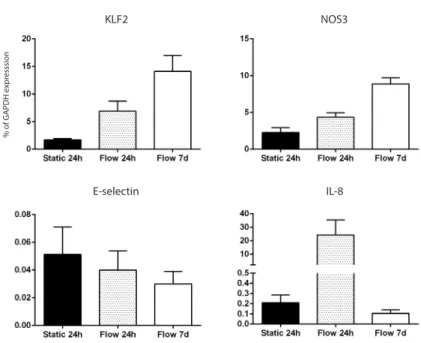
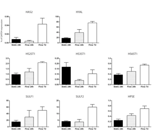
In this study we hypothesized that prolonged laminar shear stress leads to compositional, dimensional and consequently also functional changes within the endothelial glycocalyx. To study this, primary isolated human umbilical vein endothelial cells (HUVECs) were cultured under continuous laminar flow (shear stress is 10 dyne/cm2) for 7 days. Wheat
3
Material and methodsCells
HUVECs were isolated from umbilical cords by perfusion, and 20 minutes incubation, with trypsin at 37°C. Freshly isolated HUVECs were cultured on 0.5% gelatin coated 75 cm2 flasks (Greiner Bio-one) in EGM2 medium (Lonza, Basel, Switzerland) supplemented
with antibiotics/antimycotics (Life technology, Carlsbad, California, USA) and used for experiments at passage 1-3.
Flow experiments
Flow experiments were performed using an ibidi flow system (Ibidi, Martinsried, Germany). HUVECs were seeded into closed perfusion chambers (ibiTreat 0.4 µ-Slide I or VI, Luer ) at a concentration of 1.5x10⁶ cells per mL. Cells were allowed to adhere for 3
hours. Hereafter, the chamber was connected to a computer-controlled air pressure pump and a fluidic unit with a two-way switching valve. The pump setup allowed pumping of 16 mL cell culture medium from two reservoirs in a unidirectional way through the flow channel over the monolayer of endothelial cells at a constant shear stress of 10 dyne/ cm2. Medium was refreshed after 1 and 4 days of culture. The chamber and the reservoirs
containing the medium were kept in an incubator at 37°C and 5% CO2. RNA was isolated
from cells subjected to shear stress in a 0.4 µ-Slide I Luer flow chamber, while the 6 lanes of a 0.4 µ-Slide VI Luer were used for immunofluorescent staining experiments.
RNA Isolation and qRT-PCR
Total RNA was isolated from HUVECs cultured in a µ-Slide using Trizol reagent (Life Technologies, Carlsbad, California, USA) and isolation kit (Qiagen, Hilden, Germany). Reverse transcription was performed using a 5 minute 65°C incubation of 500 ng total RNA with deoxyribonucleotide triphosphates (Life Technologies) and oligo(dT) (Life Technologies), cDNA was synthesized using M-MLV First-Strand Synthesis (Life Technologies), and detection was carried out using SYBR Green Master Mix (Life technologies). Levels of expression were determined by normalizing to GAPDH levels.
Cell culture and confocal immunofluorescence microscopy