Address for correspondence Dr. Shagufta Anwar,
Department of Dermatology, Bahawal-Victoria Hospital,
Quaid-e-Azam Medical College, Bahawalpur. Email: [email protected]
Original Article
Frequency of cutaneous manifestations in
patients of hepatitis C infection
Introduction
The hepatitis C virus (HCV) is an RNA virus that belongs to the family flaviviridae.1 HCV
replicates in the cytoplasm of hepatocytes, but is not directly cytopathic. Persistent infection appears to rely on rapid production of virus and continuous cell-to-cell spread, along with a lack of vigorous T-cell immune response to HCV antigens. The HCV turnover rate can be quite high with replication ranging between 1010 to 1012 virions per day and a predicted viral half-life of 2 to 3 hours.2 The rapid viral
replication and lack of error proofreading by the viral RNA polymerase are reasons why the HCV RNA genome mutates frequently.3There
are six known genotypes (numbered 1 through 6) and more than 50 subtypes (e.g., 1a, 1b, 2a...).4 Frequent HCV mutations and numerous
subtypes have made the search for an HCV vaccine challenging. Chronic hepatitis C is the most common cause of chronic liver disease and cirrhosis, and the most common indication for liver transplantation in the United States (U.S.), Australia, and most of Europe.5,6
Approximately 170 million people are affected with HCV worldwide, comprising ~3% of the global population.4 Hepatitis C virus (HCV) is
the most common chronic blood borne infection in the U.S., and is involved in 40% of chronic liver disease.4,5
Shagufta Anwar, Muhammad Khalid, Jamil Ahmad Shaheen
Department of Dermatology, Quaid-e-Azam Medical College, Bahawal-Victoria Hospital, Bahawalpur
Abstract
Objective To determine the frequency of cutaneous manifestations in patients suffering from hepatitis C infection.Methods In this cross-sectional study, one hundred diagnosed patients of hepatitis C, admitted in medical units of Bahawal-Victoria Hospital, Bahawalpur, Quaid-e-Azam Medical College, Bahawalpur were registered over a period of six months. Cutaneous manifestations in these patients were recorded and analyzed.
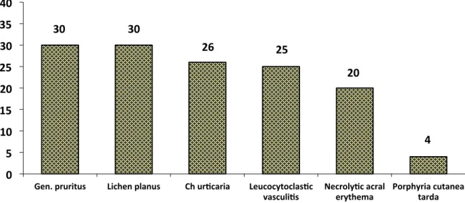
Results Out of 100 patients, 51 (51%) were males and 49 (49%) were females. Majority of the patients (73%) were 20 to 59 years old. Most of the patients had more than one cutaneous manifestation. These included generalized pruritus 30%, lichen planus 30%, urticaria 26%, leukocytoclastic vasculitis 25%, necrolytic acral erythema 20% and porphyria cutanea tarda 4%.
Conclusion Cutaneous manifestations of hepatitis are not uncommon. These may be the first clinical sign of chronic hepatitis C infection. Generalized pruritus, lichen planus, urticaria, leukocytoclastic vasculitis, necrolytic acral erythema and porphyria cutanea tarda were the important cutaneous manifestations recorded. Screening such patients on the basis of these dermatoses and investigating accordingly may help in early diagnosis and prevention of complications of this grave disease.
Key words
Hepatitis C virus infection is one of the commonest chronic viral infections in the world, with about 300 million people chronicallyinfected worldwide. Chronic HCV infection leads to cirrhosis of liver if not treated properly.8 Physicians know hepatic
cirrhosis and its complication since the time of Hippocrates. W.H.O. has estimated that cirrhosis is responsible for 1.1% of all deaths worldwide. About 175 million people in the world have cirrhosis of liver. Cirrhosis comprises 10th most common cause of death in USA. About 30% patients of cirrhosis die in hepatic coma.
Hepatitis C infection is very common in this southern area of Punjab. It is associated with many cutaneous manifestations. These skin manifestations may lead to screening and early diagnosis of this chronic disease. To determine the frequency of these skin changes among hepatitis C patients was the objective of this study.
Methods
Patients of both genders having positive anti-HCV antibodies on the basis of BIOTEC Latex Kit method® and presence of HCV RNA by polymerase chain reaction (PCR) (Qualitative), were included in the study. Patients having age less than 15 years, known alcoholics, patients of primary biliary cirrhosis and patients with HBsAg-positive test were excluded from the study.
Cases of hepatitis C with positive HCV evidence, according to inclusion criteria, admitted in medical units of Bahawal-Victoria Hospital, Bahawalpur were considered. One hundred cases of positive HCV were registered in the study. Informed consent was taken from the patients and all the information was collected on pre-designed proforma, with two parts, part-I comprising sociodemographic details like age, sex, occupation and educational status while part-II consisting study variables. The cutaneous manifestations
were observed in each patient and dermatological diagnosis was re-confirmed by senior consultant dermatologist (MK and JAS) and investigated where needed. The patients who had anti-HCV antibodies in their serum were subjected to HCV RNA by PCR (Qualitative). Cryoglobulins and the levels of complement were analyzed in patients who had positive serologic tests for rheumatoid factor (RF). Patients with co-existent liver diseases (co-infection with hepatitis B virus), alcoholic liver disease and primary biliary cirrhosis were excluded.
All the information collected on the proforma was analyzed using statistical package for social sciences (SPSS) version 10.0. Frequencies for individual cutaneous manifestations and their percentages were calculated in hepatitis C patients in general, as well as, with respect to sex and age. Account was also taken of the cutaneous features with or without history of antiviral therapy. Mean and standard deviation was calculated for age.
Results
Figure 1 Frequency of different cutaneous manifestations in 100 HCV patients.
Figure 1 shows the frequency of different cutaneous manifestations seen in the study population. Out of 100 patients, generalized pruritus was seen in 30% (18 male and 12 female), lichen planus in 30% (17 male and 13 female), urticaria in 26% (13 male and 13 female), leukocytoclastic vasculitis in 25% (14 male and 11 female), necrolytic acral erythema in 20% (12 male and 8 female) and porphyria cutanea tarda in 40% (3 male and 1 female),
Generalized pruritus was seen in 30 (30%) cases. On examination, 6 had dry skin, and 2 excoriated papules, the skin in the remaining wasnormal. In 5 of 30 patients with pruritus, a moderate cholestasis was present.
Cutaneous and mucosal lichen planus (LP), confirmed by histopathological examination, were noted in 30 (30%) patients, 17 males and 13 females. These patients presented with cutaneous lesions of various sized pruritic papules and plaques mostly over the extremities. 14 patients had cutaneous lesions only and 4 patients had cutaneous, as well as, oral lesions and oral lichen planus alone was present in 12 patients. In some cases there were whitish streaks over the oral mucosa, while in others painful erosive lesions were seen over the tongue. The LP lasted more than one year.
25 patients of leukocytoclastic vasculitis, presented with palpable purpura, erythematous plaques, erosions and ulcers over the feet and lower legs. Histopathology revealed a cutaneous leukocytoclastic vasculitis. In 5 of these, RF was positive, the complement levels were low and cryoglobulinemia was detected.
Necrolytic acral erythema was reported in 20% patients as erythematous, scaly plaques on hands and feet. Histopathology was suggestive of the disease. In 4 patients of PCT, there was history of photosensitivity and blistering on face and hands, hyperpigmentation, hypertrichosis and scarring but the biochemical diagnosis could not be confirmed due to unavailability of laboratory tests.
The serum levels of ALT and AST were normal in 22 of the 100 chronic HCV infected patients (22%). Fifty-five patients (55%) showed mild to severe elevations of the serum transaminases. RF was positive (>20 IU/ml) in 44 of 100 patients (44%). In 5 serum samples from the RF positive patients, cryoglobulinemia and altered complement levels were detected. Forty patients (40%) had received or were on antiviral therapy, which was a combination of interferon and ribavirin. None of the patients were on interferon therapy alone or on ribavirin therapy only. All
30 30
26 25
20
4
0 5 10 15 20 25 30 35 40
Gen. pruritus Lichen planus Ch ur9caria Leucocytoclas9c
the patients were on supportive/symptomatic therapy.
Discussion
In the present study, total one hundred patients were included. 51 (51%) were male and 49 (49%) were female. The male predominance has been observed in various studies conducted in Pakistan, as well as, internationally previously, so is the case in this study. This male to female difference may be due to delayed consultation by female patients and gender inequality in utilization of health care facilities in Pakistan. The other factor may be that, as compared to females, males are relatively more exposed to the risk factor for the transmission of HCV i.e. transmission through barbers and intravenous drug abuse. Fifty seven percent patients were illiterate.
Epidemiological studies have revealed that HCV infection is uncommon in age groups younger than 20 years and prevalent in persons older than 40 years.5Our results show only 8 patients of less than 20 years with a frequency of 8%, hence an almost similar scenario but we found the infection also common in the age range of 40-49 years. This may indicate that in our region younger persons are becoming a victim to the disease.
Pruritus was found more often in patients with severe fibrosis and cirrhosis. Pruritus with non-specific excoriations was a common finding with a frequency of 30%. Several etiologies can be considered. Pruritus could be a direct effect of HCV infection or related to IFN therapy. Cholestasis alone could be another cause.10 The prevalence of pruritus in
HCV infected patients varies from one country to another, and the epidemiology of HCV differs substantially between countries. It is, therefore, difficult to compare the results. For example, the HCV rate in patients with pruritus was 0.7% in a study from France11 while in another French study, pruritus was found in 15% of HCV positive patients.12
The relationship between LP and HCV is debatable and several studies have been conducted. A retrospective study by Beaird et al.13 reported 70% frequency of HCV in patients of LP. Another case-control study on 340 LP patients revealed 55% frequency.14 Epidemiological study by Tameez-ud-Deen et al.15 on patients of LP have reported an association of 32.7% while Mahboob et al.16 reported a frequency of 23.5%. All these studies were conducted on patients of LP while in our study HCV positive patients were examined for features of LP. We found a frequency of 30%. This difference in frequency could be due to our detection of LP in HCV patients rather than HCV detection in LP patients.
In several studies, a possible link between urticaria and HCV infection was mentioned. A Japanese study by Kanazawa et al.17 in 1996,
showed a statistically significant association between urticaria and hepatitis C. A study, in Pakistan, on patients of chronic urticaria by Ahmed et al.18 showed a frequency of 13.16%
cases positive for anti-HCV antibodies. The demographic data revealed an almost equal gender distribution. A study carried out by Umar et al.19 in Pakistan showed a similar
male to female ratio.
Cutaneous vasculitis has been associated with HCV infection. Karlsberg et al.20 did a
systematic dermatological evaluation of 408 patients with hepatitis C and vasculitis was found in 10 (3%) patients. In a comparative study on essential mixed cryoglobulinemia in HCV infected vs. noninfected patients, 21% of HCV infected patients presented with cutaneous features of palpable purpura.21Our
type II and type III mixed cryoglobulinemia and HCV infection. The initial observation was by Pascual et al.21 in 1990 who found
anti-HCV antibodies in patients with type II cryoglobulinemia.Porphyria cutanea tarda was seen in 4 (4%) of our cases and it is frequently associated with HCV infection.22
Chronic HCV is a leading cause of cirrhosis in Bahawalpur. As there is no vaccine yet available against hepatitis C virus and it is the commonest cause of cirrhosis in this part of world hence needs more meticulous approach to prevent its transmission, through avoidance of risk factors and early detection, if a patient presents with cutaneous manifestation. Even if the cirrhosis develops, early detection and prompt treatment of these viral infections improve the overall outcome of the patients and prevent from development of hepatocellular carcinoma. Once the cirrhotic process has begun, the incidence of hepatocellular carcinoma ranges from 1% to 4%. Hepatitis C is reaching epidemic proportions and is a significant cause of morbidity worldwide. Timely intervention can stabilize the disease and positively impact morbidity and mortality. This underscores the importance of detecting individuals infected with HCV. Since dermatologic manifestations may be the only and most apparent sign of chronic HCV, it is important that health care professionals be aware of these dermatologic manifestations. The cutaneous features are not only themselves a cause of morbidity, but they can also provide an indirect clue for the underlying disease. Such an observation leads to early detection and initiation of therapy. Accurate and timely diagnosis of HCV is critical to prevent the life threatening complications. Antiviral therapy for HCV may also be effective in curing the cutaneous disease for example, cryoglobulinemia. Moreover, such identification can help to prevent further transmission of the disease.
Conclusion
Cutaneous manifestations may be the first clinical sign of chronic HCV infection. Screening for HCV infection in certain dermatological conditions may lead to antiviral treatment being effective in curing cutaneous diseases. Moreover, such identification will help prevent further transmission of HCV.
References
1. Lauer GM, Walker BD. Hepatitis C virus infection. N Engl J Med. 2001;345:41-52. 2. Neumann AU, Lam NP, Dahari H et al.
Hepatitis C viral dynamics in vivo and the antiviral efficacy of interferon-alpha therapy. Science. 1998;282:103-7. 3. Bukh J, Miller RH, Purcell RH. Genetic
heterogeneity of hepatitis C virus: Quasispecies and genotypes. Semin Liver Dis. 1995;15:41-63.
4. National Institutes of Health Consensus Development Conference Statement: Management of hepatitis C 2002 (June 10-12, 2002). Gastrenterology. 2002;123:2082-99.
5. Wasley A, Alter MJ. Epidemiology of hepatitis C: geographic differences and temporal trends. Semin Liver Dis. 2000;20:1-16.
6. Centers for Disease Control and Prevention. Recommendations for prevention and control of hepatitis C virus (HCV) infection and HCV-related chronic disease. MMWR Recomm Rep. 1998;47 :1-39.
7. Charlton M. Hepatitis C infection in liver transplantation. Am J Transplant. 2001;1:197-203.
8. Shah NH, Shabbier G. A review of published literature on hepatitis C and B virus prevalence in Pakistan. J Coll Physicians Surg Pak. 2002;12:368-71.
9. Cribier B, Santinelli F, Schmitt C et al.
Should patients with pruritus be testedfor
hepatitis C virus infection? A
case-controlled study. Br J Dermatol.
2000;142:1234-64.
10. Dega H, Frances C, Dupin N et al. Pruritus and the hepatitis C virus. Ann Dermatol Venereol. 1998;125:9-12. 11. Khokhar N, Gill ML, Malik GJ. General
12. Dervis E, Serez K. The prevalence of dermatologic manifestations related to chronic hepatitis C virus infection in a study from a single center in Turkey. Acta Dermatovenerol Alp Panonica Adriat. 2005;14:93-8.
13. Beaird LM, Kabloon N, Franco J, Fairley JA. Screening of HCV in lichen planus patients. J Am Acad Dermatol. 2001;44:311-2.
14. Chuang TY, Stitle L, Brashear R, Lewis C. Hepatitis C virus and lichen planus: A case control study of 340 patients. J Am Acad Dermatol. 1999;41:787-9.
15. Tameez-ud-Deen, Naqqash S, Butt AQ. Lichen planus and hepatitis C virus infection: An epidemiologic study. J Pak Assoc Dermatol. 2003;13:127-9.
16. Mahboob A, Haroon TS, Iqbal Z et al. Frequency of anti-HCV antibodies in patients with lichen planus. J Coll Physicians Surg Pak. 2003;13:248-52. 17. Paoletti V, Mammarella A, Basili S et al.
Prevalence and clinical features of skin disease in chronic hepatitis C infections. A prospective study in 96 patients. Panminerva Med. 2002;44:349-52. 18. Ahmed I, Wahid Z, Ahmed Z. Chronic
urticaria: frequency of anti-HCV antibodies. J Pak Assoc Dermatol. 2003;13:179-83.
19. Umar M, Bushra HT, Shuaib A et al. Spectrum of chronic liver disease due to hepatitis C virus infection. J Coll Physicians Surg Pak. 2000;10:380-3.
20. Karlisberg PL, Le WM, Casey DL et al. Cutaneous vasculitis and rheumatoid factor positivity as presenting signs of hepatitis C virus induced mixed cryoglobulinemia. Arch Dermatol. 1995;131:1119-23.
21. Ito A, Kazama T, Ito K et al. Purpura with cold urticaria in a patient with hepatitis C virus infection-associated mixed cryoglobulinemia type III: Successful treatment with interferon beta. J Dermatol. 2003;30:321-5.
22. 22Chuang TY, Stitle L, Brashear R, Lewis C. Porphyria cutanea tarda and hepatitis C virus: A case control study and meta-analysis of the literature. J Am Acad Dermatol. 1999;41:31-6.
23. Gisbert JP, Garcia-Buey L, Pajares JM, Moreno-Otero R. Prevalence of hepatitis C virus infection in porphyria cutanea tarda: systematic review and meta-analysis. J Hepatol. 2003; 39: 620-627 24. Poynard T, Yuen MF, Ratziu V, Lai CL.
Viral hepatitis C. Lancet. 2003;362 :2095-2100.