Address for correspondence Dr. Shehla Shaukat, Dr. Ghazala Butt Assistant Professors,
Department of Dermatology, Unit-I, King Edward Medical University/ Mayo Hospital, Lahore.
Email: [email protected] [email protected]
Review Article
Cutaneous manifestations of COVID-19
Introduction
The
severe
acute
respiratory
syndrome
coronavirus 2 (SARS
‐
CoV
‐
2), a single-stranded
RNA virus of Coronaviridae family, was first
isolated from Wuhan China in December, 2019
and due to rapid spread of disease across the
globe, coronavirus disease 2019 (COVID
‐
19)
was declared as pandemic condition by WHO in
March, 2020. Till date, more than 6,573,540
cases of COVID-19, with 388,041 deaths and
3,170,505 recoveries have been reported
worldwide.
1The virus enters the respiratory system and
attaches to angiotensin converting enzyme 2
ACE2 receptors on the cell surfaces.
2The
presenting symptoms of COVID-19 are flu-like
including fever, dry cough, dyspnea, anorexia,
ageusia and anosmia which later on progresses
to pneumonia and acute respiratory distress
syndrome in some patients. In most of the cases
it is asymptomatic.
2During the viremic stage of disease, the virus
particles reach the integument also and
subsequent to direct viral exposure and indirect
immune dysregulation, skin is expected to be
involved in COVID-19, like other common
systemic
viral
infections.
Scanty
data,
international and national, exist about the
cutaneous manifestations of this new disease;
however, a variety of dermatological findings
have been reported (discussed later) and this
spectrum is likely to expand in future. The
frequency varies from 0.2% from China to 20%
from Italy.
Shehla Shaukat, Ghazala Butt, Ijaz Hussain
Department of Dermatology Unit-I, King Edward Medical University/ Mayo Hospital, Lahore.
Abstract
The severe acute respiratory syndrome coronavirus 2 (SARS‐CoV‐2) and the disease it causes,termed coronavirus disease 2019 (COVID‐19), has rapidly swept across the world since its first known human manifestation on December 8, 2019. Confirmed cases are present in over 160 countries. Patients of COVID-19 infection present with diverse cutaneous manifestations seen in 0.2% to 20% of patients. This article summarizes different dermatological manifestations in patients with COVID-19 through literature review using Google Scholar, Sci Hub, PubMed and other online review articles. Case reports, case series and other studies which mentioned cutaneous manifestations in the patients with COVID-19 infection were added. The most common cutaneous manifestation of COVID-19 was found to be maculopapular (morbilliform) exanthem. Papulovesicular rash, urticaria, painful acral red purple papules (COVID toes), livedo reticularis and petechiae were other presentations. Majority of lesions were localized on the trunk; however, involvement of the hands and feet was also noted. Cutaneous involvement usually followed the respiratory symptoms; nonetheless, in a minority, it preceded systemic features. Majority of the studies failed to report any correlation between COVID-19 severity and skin lesions. Cutaneous manifestations may help in early diagnosis of disease and prompt treatment of COVID-19.
Key words
As this a novel virus so new signs and
symptoms are also discovered with the passage
of time. Many cutaneous findings have also been
reported
in
international
literature.
The
cutaneous manifestations of COVID -19 include
erythematous
lesions,
petechiae,
urticaria,
vesicular eruption and chilblain of toes.
3Pathogenesis
[2,4,5]SARS-2 virus can affect skin by direct and
indirect pathways (
Figure 1
).
1.
Virus particles reach endothelial cells of
cutaneous vasculature by hematogenous
route directly or carried by inflammatory
cells.
2.
SARS-CoV-2, like other viral infections is
suggested to induce autoimmunity; different
underlying mechanisms may be molecular
mimicry, release of self-antigens and epitope
spreading.
3.
Hypoxic injury to skin enhances the
anaerobic metabolism and subsequent lactic
acid accumulation further reduces cutaneous
blood flow.
4.
Virus uses ACE2 receptors, expressed in
cutaneous tissues, to enter the host cell,
followed by down regulation of ACE2
receptors. ACE-2 is a negative regulator of
the
renin-angiotensin
system
(RAS).
Interference
of
ACE2-RAS
underlies
different vascular pathologies.
Hypercoagulable state is a feature of sever
COVID-19 infection. Different underlying
mechanisms may be activation of cogulation
pathway
and
suppression
of
fibrinolytic
pathways by IL-6 and other cytokines; direct
activation of cogulation system by virus
particles; and production of antiphospholipid
antibodies.
Cutaneous manifestations
Cutaneous manifestations can be grouped into
three types depending on the pathogenesis.
6,71.
Dermatoses due to immune response of the
body.
2.
Dermatoses due to systemic involvement.
3.
Dermatoses due to complications
of
treatment or personal protective equipment
(PPE).
Dermatosis due to immune response of the
body
1.
Morbilliform Rash/Maculopapular Rash
Morbilliform rash consists of macular lesions
that are red and usually 2-10mm in diameter but
may be confluent in places.
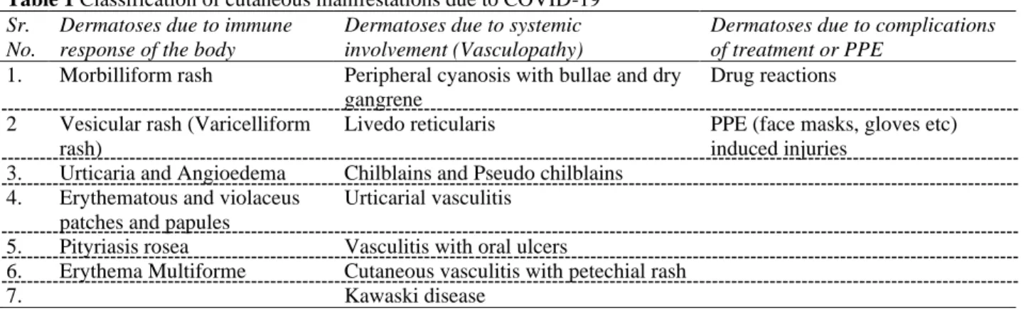
Table 1 Classification of cutaneous manifestations due to COVID-19 Sr.
No.
Dermatoses due to immune response of the body
Dermatoses due to systemic involvement (Vasculopathy)
Dermatoses due to complications of treatment or PPE
1. Morbilliform rash Peripheral cyanosis with bullae and dry gangrene
Drug reactions 2 Vesicular rash (Varicelliform
rash)
Livedo reticularis PPE (face masks, gloves etc) induced injuries
3. Urticaria and Angioedema Chilblains and Pseudo chilblains 4. Erythematous and violaceus
patches and papules
Urticarial vasculitis 5. Pityriasis rosea Vasculitis with oral ulcers
6. Erythema Multiforme Cutaneous vasculitis with petechial rash
7. Kawaski disease
It has been observed that a generalized macular
or maculopapular exanthem (morbilliform)
appeared to be the most common cutaneous
manifestation in COVID-19, with 36.1% of
patients presenting such lesions on their skin.
A 71-year-old Caucasian woman presented with
fever, productive cough and worsening shortness
of breath which started 10 days before in the
emergency department in Milan. Over the
following days a maculopapular itchy rash
appeared on the trunk resembling a Grover
disease. In another similar case, a 77-year-old
Caucasian
woman
presented
with
neck
lymphonodal enlargement, fever, cough and
diffuse maculopapular exanthem (morbilliform)
on the trunk in Milan hospital.
4Recalcati reported 14 patients of COVID-19
with maculopapular erythematous rash.
8Hunt
reported diffuse, morbilliform, maculopapular,
and nonpruritic rash on the trunk and extremities
sparing the face along with fever in the patients
of COVID-19 in New York.
9Mahe reported
erythematous macular rash which first appeared
on the antecubital fossa and later spread to the
trunk and axillary folds in COVID-19 patient in
France. Skin rash appeared four days after
fever.
10Najarian reported erythematous macules
with islands of normal appearing skin between
them, arranged in a morbilliform pattern on the
legs, thighs, forearms, arms, shoulders, back,
chest and abdomen 1 day after onset of
respiratory symptoms in the patients of
COVID-19.
112.
Vesicular Rash (Varicelliform Rash)
A papulovesicular rash (vesicles) was seen in
34.7%
of
COVID-19
patients in
Italy.
Monomorphic disseminated vesicular lesions
and acral vesiculo-pustular lesions are quite
specific. A 72-year-old Caucasian woman,
otherwise healthy, presented to the Emergency
Department in Milan with headache, arthralgia,
myalgia and fever. Four days later, a
papular-vesicular, pruritic eruption appeared on
sub-mammary folds, trunk and hips.
4Vesicular eruption has also been seen as an early
presentation of COVID-19, in 15% of cases
before other symptoms. Marzano reported
Varicella-like itchy papulovesicular exanthem
on the trunk and limbs in 22 COVID-19
patients.
12Recalcati reported one patient with
chickenpox-like vesicles on the trunk with little
to no itching. They healed within a few days No
correlation was seen with disease severity.
83.
Urticaria
Urticarial lesions healed without medication.
8Fernandez reported one female COVID-19
patient with urticarial rash 6 days after the onset
of symptoms in Spain.
13Zhang in Wuhan, China
reported
two
COVID-19
patients
with
Urticaria.
14Henry reported one female patient of COVID-19
with pruritic urticarial eruption on the face,
hands and feet (acral involvement) 48 hours
before onset of respiratory symptoms in
France.
15Urticarial exanthem has also been seen as an
early diagnostic clue for COVID-19 infection. A
61-year-old Spanish medical doctor presented
with progressive cutaneous rash for the last 4
days. He was treating patients with coronavirus
infection for three weeks. On presentation, his
temperature was 37.3ºC. He did not complain of
respiratory symptoms (as cough or dyspnoea),
headache, malaise, sore throat or nasal
congestion. Physical examination showed an
urticarial
rash
consisting
of
confluent,
edematous and erythematous papules on his
thighs, arms, and forearms. Palms and soles
were spared. Cutaneous lesions were mildly
itchy.
164.
Erythematous and Violaceous patches and
papules
Kolivras found violaceous, infiltrated plaques on
an erythematous background on the dorsal
aspect of toes and lateral sides of feet 3 days
after onset of respiratory symptoms. These were
painful also.
17Mazzotta (Italy) reported one
COVID-19 patient with erythemato-violet,
rounded lesions of 5-15 mm in diameter, with
blurred limits on the plantar surface of 1st right
toe and dorsal surface of the 2nd toe on both
feet. It was associated with intense itching and
burning.
18Alramthan (Qatar) reported two cases
with acral ischemic lesions presenting as red
purple papules on the dorsal aspect of fingers.
195.
Pityriasis Rosea
Ehsani
et al
reported pityriasis rosea in a patient
with COVID-19. A scaly annular eruption over
arms and trunk was observed in the 27-year old
man. The lesions were pruritic and disseminated
over 5 days. The lesions resolved after topical
steroids and oral antihistamine therapy.
206.
Erythema multiforme
It is an immune mediated skin reaction to
viruses or drugs. It appears as macule, papule
and typical target-like lesion on acral parts.
Histologically apoptotic individual keratinocytes
in epidermis and perivascular lymphohistiocytic
infilteration is seen in dermis. In a study by
Janah
et al
, two cases have been reported with
erythema multiforme in patients with COVID-19
who were on treatment and the lesions resolved
despite continuation of treatment.
21Dermatoses due to systemic involvement
(Vasculopathy)
1.
Peripheral cyanosis with bullae and dry
gangrene
Recalcati
et al
reported cases with associated
cutaneous peripheral cyanosis with bullae and
dry gangrene.
8Zhang
et al
reported seven cases
with acral cyanosis, bullae and dry gangrene.
14Ma
et al
also reported acral dry gangrene in
patients with COVID-19.
22Young
et al
and
Suerez
et al
also reported cases with acral
purpura, cyanosis and acral ischemia in their
study.
23,24A network pattern of erythematous to dusky
cutaneous
vessels
due
to
microvascular
occlusion or injury is commonly seen on lower
legs around the malleoli. This pattern has been
reported in patients with COVID-19. In the
study by Magro
et al
significant deposition of
terminal complement components C5b-9, C4d
mannose binding lectin (MBL) associated serine
protease (MASP)2 was observed in the
microvasculature of skin biopsy samples of the
COVID-19 patients with livedo reticularis and
purpuric lesions. This study suggests that
microvascular injury is mediated by complement
activation and associated procoagulant state.
5De
Masson
et al
also reported a case of livedo in
their study.
3Manalo
et al
studied two cases with
unilateral livedo which was transient in nature in
patients with COVID-19.
253.
Chilblains and Pseudo chilblains
Acral eruption of erythematous or dusky papules
and macules associated with swelling of the
digits is called Perniosis or chilblains. These
lesions have observed in the patients with
COVID-19 in Italy and other countries.
22,3Pseudo chilblain lesions have also been reported.
Recalcati
et al
reported cases with Perniosis like
lesions in their patients with COVID-19. The
patient had acral eruption of
erthemato-violaceous papules and macules with digital
swelling.
26Similar findings were seen in the
study by Alramthan and Aldarajii.
19De Masson
et al
in the retrospective study in France
reported acral lesions as the most common
finding with suspected or confirmed cases of
COVID-19 (142/277 cases). Out of these 142
cases 106 patients (75%) had chilblain-like
lesions.
34.
Urticarial Vasculitis
Estebanez reported a case presenting with
erythematous yellowish papules on heels
bilaterally
which
became
confluent
and
developed into pruritic erythematous patches
resembling urticarial vasculitis.
27,65.
Vasculitis with Oral ulcers
Chaux- Bodard
et al
reported a case of
45-year-old COVID-19 positive female patient with an
irregular ulcer on the dorsal side of the tongue
along with vasculitic lesions on the acral part.
286.
Kawasaki disease
It is an acute febrile illness characterized by
vasculitis
of
the
medium-sized
arteries,
including
coronary
arteries.
An
atypical
Kawasaki disease has been reported in many
countries including France (25 cases), United
Kingdom (more than 12 cases) and United States
of America (15 cases). Patients present with
erythematous rash, conjunctivitis, glossitis and
high grade fever. Systemic complications
include abdominal pain and gastrointestinal
symptoms and cardiac inflammation.
29,307.
Cutaneous vasculitis with petechial rash:
Bouaziz and Castelnovo in their studies reported
cases presenting with signs of vasculitis and
associated petechial rash along with acral
ischemia and Raynaud’s phenomenon.
31,32Dermatoses due to treatment
Figure 2 Papulo-vesicular Eruption in a COVID -19 patient
Figure 3 Urticarial Eruption in COVID -19 patient
Figure 4 Cutaneous small vessel vasculitis involving legs, feet and arms of a COVID-19 patient
exanthematous pustulosis, acneiform eruption,
Petechiae, leucocytoclastic vasculitis, hair loss
and photosensitivity.
6Sernicola
et al
reported
incidents of toxic erythema and eosinophilia in
patients being treated with tocilizumab.
33Robustelli
et al
reported cases with acute
Figure 5 Scabies and Vasculitic lesions both seen in the same patient of COVID-19
Dermatoses due to PPE
Self protection of medical staff by wearing
protective clothing, masks, goggles and gloves is
mandatory in COVID wards. Prolong wearing of
PPE is associated with multiple skin problems
which can lead to break in the skin barrier
function. The problems include pruritus,
erythema, scratches, blisters, rhagades, papules,
edema, exudation/crust and lichenification.
These findings were observed in the study by
Pei
et al
in China.
35Pruritus ranged from mild to
severe. Other cutaneous lesions were reported in
73.1% of the participants with 38.8% noticed
erythema, scratch (22.9%), blister (13.8%),
rahagades (13.6%), papule/edema (12.8%),
exudation/crust
(6.8%)
and
lichenification
(5.6%). Majority of the lesions were reported on
the face. Singh
et al
reported various dermatoses
associated with donning of PPE. The most
common was irritant contact dermatitis seen in
39.5% cases followed by friction dermatitis in
25.5% cases. The injuries were caused by
goggles (51.9%), N95 masks (30.77%) and face
shields (17.31%), pruritus and erythema were
common symptoms. Nasal bridge, cheeks and
chin were the common sites of lesions.
36Reports
of injuries have been reported in other studies as
well.
37-40COVID-19 patients in our local population
In Pakistan COVID-19 infection is also
spreading exponentially. As of 3
rdJune, 2020,
there have been about 80,500 confirmed cases
with 28,900 recoveries and 1,690 deaths in the
country. Cutaneous manifestations have also
been found in patients infected with this virus.
Cutaneous presentations in few of the
COVID-19 infected patients from Pakistani population
are shown below
(Figure 2-5)
.
Acknowledgement
We wish to acknowledge and express our
appreciation to Dr. Yousuf A. Mallick (FCPS),
Consultant Dermatologist, The Indus Hospital
Karachi, for his contribution for sharing
COVID-19 patients photographs to be added in
this review article.
References
1. https://www.worldometers.info/coronavirus/ 2. Sachdeva M, Gianotti R, Shah M, Lucia B, Tosi D, Veraldi S, et al. Cutaneous manifestations of COVID-19: Report of three cases and a review of literature [published online ahead of print, 2020 Apr 29]. J Dermatol Sci.2020;S0923-1811(20)30149-3.
doi:10.1016/j.jdermsci.2020.04.011
3. de Masson A, Bouaziz J-D, Sulimovic L, Cassius C, Jachiet M, Ionescu M-A, Rybojad M, Bagot M, Duong T-A, on behalf of the SNDV (French Union of Dermatologists-Venereologists), Chilblains are a common cutaneous finding during the COVID-19 pandemic: a retrospective nationwide study from France J Am Acad
Dermatol (2020), doi:
https://doi.org/10.1016/j.jaad.2020.04.161. 4. Gianotti R, Zerbi P, Dodiuk-Gad R.
Histopathological study of skin dermatoses in patients affected by COVID-19 infection in the Northern part of Italy, J Cosmet Dermatol. Sci. Appl. (2020) In press. 5. Magro C, Mulvey J, Berlin D, Nuovo G,
thrombosis in the pathogenesis of severe COVID-19infection: a report of five cases.
Transl Res
2020.https://doi.org/10.1016/j.trsl.2020.04.0 07.
6. Suchonwanit P, Leerunyakul K, Kositkuljorn C, Cutaneous manifestations in COVID-19: Lessons learned from current evidence, J Am Acad Dermatol (2020), doi: https://doi.org/10.1016/j.jaad.2020.04.094 7. Galvan Casas c, Catala A, Carretero
Hernandez G, Rodriguez-Jimenez P, Fernandez Nieto D, Rodriguez-Villa Lario A et al. Classification of the cutaneous manifestations of COVID‐19: a rapid prospective nationwide consensus study in Spain with 375 cases. Br J Dermatol. 2020. https://doi.org/10.1111/bjd.19163
8. Recalcati S, Cutaneous manifestations in COVID-19: a first perspective, J Eur Acad Dermatol Venereol. (March 26) (2020), doi:http://dx.doi.org/10.1111/ jdv.16387 [Epub ahead of print].
9. M. Hunt, C. Koziatek, A case of COVID-19 pneumonia in a young male with full body rash as a presenting symptom, Clin Pract
Cases Emerg Med. 2020.
doi:http://dx.doi.org/10.5811/cpcem.2020.3. 47349
10. A. Mahé, E. Birckel, S. Krieger, C. Merklen, L. Bottlaender, A distinctive skin rash associated with Coronavirus Disease 2019? J Eur Acad Dermatol Venereol. 2020. 11. D.J. Najarian, Morbilliform exanthem
associated with COVID-19, JAAD Case
Rep. (2020),
doi:http://dx.doi.org/10.1016/j.jdcr.2020.04. 015.
12. A.V. Marzano, G. Genovese, G. Fabbrocini, P. Pigatto, G. Monfrecola, B.M. Piraccini, et al., Varicella- like exanthem as a specific COVID-19-associated skin manifestation: multicenter case series of 22 patients, J. Am. Acad. Dermatol. (April 16) (2020), doi:http://dx.doi.org/10.1016/j.jaad.2020.04. 044 pii: S0190- 9622(20)30657-5.
13. Fernandez-Nieto D, Ortega-Quijano D, Segurado-Miravalles G, Pindado-Ortega C, Prieto-Barrios M, Jimenez-Cauhe J, Comment on: cutaneous manifestations in COVID-19: a first perspective. Safety concerns of clinical images and skin biopsies, J Eur Acad Dermatol Venereol. 2020,
doi:http://dx.doi.org/10.1111/jdv.16470 [Epub ahead of print].
14. Zhang J, Dong X, Cao Y, Yuan y, Yang Y, Yan Y, et al. Clinical characteristics of 140 patients infected with SARS-CoV-2 in Wuhan, China, Allergy (February 19) (2020),
doi:http://dx.doi.org/10.1111/all.14238 [Epub ahead of print].
15. Henry D, Ackerman M, Sancelme E, Finon A, Esteve E, Urticarial eruption in COVID-19 infection, J Eur Acad Dermatol Venereol.
2020. doi:
http://dx.doi.org/10.1111/jdv.16472 [Epub ahead of print].
16. Quintana-Castanedo L, Feito-Rodríguez M, Valero-López I, ChiloechesFernández C, Sendagorta-Cudós E, Herranz-Pinto P, Urticarial exanthem as early diagnostic clue for COVID-19 infection. J Am Acad Dermatol. Case Reports (2020), doi: https://doi.org/10.1016/j.jdcr.2020.04.026. 17. Kolivras, F. Dehavay, D. Delplace, F. Feoli,
I. Meiers, L. Milone, et al., Coronavirus (COVID-19) infection- induced chilblains: a case report with histopathological findings, J Am Acad Dermatol Case Rep. 2020. doi:http://dx.doi.
org/10.1016/j.jdcr.2020.04.011.
18. Mazzotta F, Troccoli T, Acute Acro-Ischemia in the Child at the time of COVID19. Dermatologia Pediatrica, Bari, (2020) In press.
19. Alramthan A, Aldaraji W, A case of COVID-19 presenting in clinical picture resembling chilblains disease. First report from the Middle East, Clin Exp Dermatol.
2020. doi:
https://doi.org/10.1111/ced.14243. [Epub ahead of print]
20. Ehsani AH, Nasimi M, Bigdelo Z. Pitryasis rosea as a cutaneous manifestation of COVID-19 infection. J Eur Acad Dermatol Venereol. 2020;10.1111/jdv.16579. [Epub ahead of print]
21. Janah H, Zinebi A, Elbenave J. Atypical erythema multiforme palmar plaques lesions due to Sars-Cov-2. J Eur Dermatol venereal. 2020. https://doi.org/10.1111/jdv.16623. [Epub ahead of print]
22. Ma J, Xia P, Zhou Y, Liu Z, Zhou X, Wang J et al. Potential effect of blood purification therapy in reducing cytokine storm as a late complication of critically ill COVID-19.
Clin Immunol. 2020;214:
23. Young S, Fernandez AP. Skin manifestations of COVID-19. Clev Clin J Med. 2020. doi: 10.3949/ccjm.87a.ccc031. 24. Suarez-Valle A, Fernandez-Nieto D,
Diaz-Guimaraens B, Dominguez-Santas M, Carretero I, Perez-Garcia B. Acro‐ischemia in hospitalized COVID‐19 patients. J Eur
Acad Dermatol Venereol.
2020;10.1111/jdv.16592. [Epub ahead of print]
25. Manalo IF, Smith MK, Cheeley J, Jacobs R. A dermatologic manifestation of COVID-19: Transient livedo reticularis. J Am Acad
Dermatol. 2020.
https://doi.org/10.1016/j.jaad.2020.04.018. 26. Recalcati S, Barbagallo T, Frasin LA,
Prestinari F, Cogliardi A, Provero MC et al. Acral cutaneous lesions in the time of COVID-19. J Eur Dermatol Venereol. 2020. Doi: 10.1111/jdv.16533. [Epub ahead of print]
27. Estebanez A, Perez-Santiago L, Silva E, Guillen-Climent S, Garcia-Vazquez A, Ramon MD. Cutaneous manifestations in COVID-19: a new contribution. J Eur Acad 28. Chaux- Bodard AG, Deneuve S, Desoutter
A. Oral manifestation of Covid-19 as an inaugural symptom? J Oral Med oral Surg. 2020;26(2):18.
https://doi.org/10.1051/mbcb/2020011 29. Graeme M. COVID toes and Kawasaki rash:
5 cutaneous signs in COVID-19. Available at:
https://www.medscape.com/viewarticle/930 180
30. Pediatric Intensive Care Society. PICS Statement: Increased number of reported cases of novel presentation of multi-system inflammatory disease. April 27, 2020.
Available at
https://picsociety.uk/wpcontent/uploads/202 0/04/PICS-statement-re-novel-KD-C19-presentation-v2-27042020.pdfKawasaki disease
31. Bouaziz JD, Doung T, Jachiet M, Velter C, Lestang P, Cassius C et al. Vascular skin symptoms in COVID-19: a French observational study. J Eur Acad Dermatol Venereol. 2020;10.1111/jdv.16544. [Epub ahead of print]
32. Castelnovo L, Capelli F, Tamburello A, Faggioli PM, Mazzone A. Symmetric cutaneous vasculitis in COVID‐19 pneumonia. J Eur Acad Dermatol Venereol. 2020; doi: 10.1111/jdv.16589. [Epub ahead of print]
33. Sernicola A, Carnicelli G, Fraia MD, Chello C, Furhan C, Muharremi R et al. Toxic erythema and eosinophilia associated to tocilizumab therapy in COVID-19 patient. J Eur Acad Dermatol Venereol. 2020;10.1111/jdv.16620. [Epub ahead of print]
34. Robustelli Test E, Vezzoli P, Caugno A, Rapuno F, Gianatti A, Rongioletti F et al. Acute Generalized Exanthematous Pustulosis with Erythema Multiforme‐Like lesions in a COVID‐19 woman. J Eur Acad
Dermatol Venereol.
2020;10.1111/jdv.16613. [Epub ahead of print]
35. Pei S, Xue Y, Zhao S, Alexander N, Mohamad G, Chen X et al. Occupational skin conditions on the frontline: A survey among 484 Chinese healthcare professionals caring for Covid-19 patients. J Eur Acad Dermatol Venereol. 2020. https://doi.org/10.1111/jdv.16570. [Epub ahead of print]
36. Singh M, Pawar M, Bothra A, Maheshwari A, Dubey V, Tiwari A et al. Personal protective equipment induced facial dermatoses in healthcare workers managing COVID-19 cases. J Eur Acad Dermatol Venereol. 2020;10.1111/jdv.16628. [Epub ahead of print]
37. Lan J, Song Z, Miao X, et al. Skin damage among healthcare workers managing coronavirus disease-2019 [published online ahead of print, 2020 Mar 18]. J Am Acad Dermatol. 2020;S0190-9622(20)30392-3. doi:10.1016/j.jaad.2020.03.014
38. Yin Z. Covid-19: Countermeasure for N95 mask-induced pressure sore [published online ahead of print, 2020 Apr 17]. J Eur
Acad Dermatol Venereol.
2020;10.1111/jdv.16490. doi:10.1111/jdv.16490
39. Campbell V, Middleton D, Donnelly J, Hunter H. Localised mid-face Miliaria as a consequence of filtering face piece (FFP) respirator use during the COVID-19 pandemic. J Eur Acad Dermatol Venereol. 2020;10.1111/jdv.16624. [Epub ahead of print]
![Figure 1 Pathogenesis of cutaneous manifestations in COVID-19 (Adopted and modified from Wu et al [])](https://thumb-us.123doks.com/thumbv2/123dok_us/7871115.2098527/2.918.94.807.827.1035/figure-pathogenesis-cutaneous-manifestations-covid-adopted-modified-wu.webp)