Minimizing BK Virus Renal Allograft Loss Through
Immunosuppression Regimen Modification
Alyson E. Meyer, PharmD Candidate, Class of 2016 Dr. Jennifer Deyo, PharmD, Primary Investigator
Dr. Robert Dupuis, PharmD, Faculty Advisor
Honors Essay
UNC Eshelman School of Pharmacy University of North Carolina at Chapel Hill
April 1, 2016
Minimizing BK Virus Renal Allograft Loss Through
Immunosuppression Regimen Modification
Alyson E. Meyer, PharmD Candidate1; Jennifer Deyo, PharmD2; Tomasz Kozlowski, MD3; Ruth
Ann Lee, PharmD4; Timothy J. Bruflat, PharmD1; Robert Dupuis, PharmD5;
Author’s Affiliations:
1UNC Eshelman School of Pharmacy, Chapel Hill, NC, 27514
2University of North Carolina Memorial Hospital Department of Pharmacy, Chapel Hill, NC, 27514 3UNC School of Medicine – Department of Surgery, Chapel Hill, NC, 275143
4UNC Healthcare, Chapel Hill, NC, 27514
5UNC Eshelman School of Pharmacy – Department of Pharmacotherapy, Chapel Hill, NC, 27514
ABSTRACT
Purpose: The primary objective of this study was to examine graft loss rate following institution specific immunosuppression (IS) modulation in the treatment of BK virus re-‐activation post-‐ renal transplant.
Methods: A retrospective chart analysis identified 212 patients over 18 years of age receiving a single renal allograft at UNC Hospitals between 2005 and 2013 with reactivated BK virus as confirmed by the presence of decoy cells on urine cytology through follow-‐up until August 2014.
Results: Of the 212 renal allografts with reactivated BK, 33 (15.6%) had progressed to BKVAN (BK virus-‐associated nephropathy). Renal biopsy identified 15 patients with Stage A BKVAN, 16 with Stage B and 2 with Stage C. IS regimen changes included reduction in tacrolimus trough goals (60.6%), reduction in mycophenolic acid (24.2%), discontinuation of mycophenolic acid (81.8%), the addition of leflunomide (57.5%) or azathioprine (18%) and re-‐initiation of corticosteroids (65%). IS changes preserved graft function in 97% of patients, with only one graft lost (3%) following confirmed diagnosis with BKVAN.
Conclusion: UNC Hospitals reports a graft loss rate of only 3% among all diagnosed stages of BKVN. In contrast to current guidelines, UNC follows a protocol with more aggressive screening, a lower threshold for acquiring blood PCRs and IS adjustment, along with the addition of leflunomide and prednisone therapy.
INTRODUCTION
BK Virus Background
BK virus is a member of the polyoma virus family that resides as a latent infection, remaining dormant within the kidneys in 80-‐90% of the general population1, 2. Despite the large
percentage of the population carrying this virus, BK virus manifests as clinical disease in immunocompromised patients. Bone marrow transplant recipients suffer from hemorrhagic cystitis while renal transplant patients risk graft loss through BK virus nephropathy (BKVAN)1, 2, 3. BK virus is an opportunistic infection that, once reactivated, is a progressive, lytic infection
that is spread through cell-‐to-‐cell contact1, 2.
Cases of diagnosed BKVAN following other solid organ transplants have thus far been limited. Joseph, A, et al. have recently reported the diagnosis of BKVAN in 2 patient cases following heart transplantation where both patients were receiving tacrolimus, MMF and prednisone as immunosuppressive therapy after surgery. Decreased renal function has been seen in cardiac transplant patients, defined as an eGFR<30 mL/min/1.73m2, in 10.9% of the
These recent reports provide evidence that other solid organ transplants could be at risk of BK viral reactivation, aside from renal transplants.
BK Virus Risk Factors
Since BK virus resides within the urogenital tract, renal transplant patients are more susceptible to reactivation of this infection. Reactivation can be due to direct and indirect causes. Direct causes include damage to the organ prior to transplant, cold ischemic time or improper/rough handling of the kidney1. All of these factors can weaken the kidney and thus cause it to be more susceptible to infection post-‐transplant. Indirect causes associated with
reactivation are the potent immunosuppression regimens that are required of patients after transplantation. The emergence of more potent agents, including tacrolimus, mycophenolic acid and cyclosporine has been accompanied with an increased incidence of BK viral reactivation in the renal transplant patient population1, 6.
Additional patient demographics can place certain patient populations at an increased risk of BK viral reactivation post-‐transplant. These include the seropositive status of donors/recipients, deceased donors, male gender, older age, Caucasian race, concurrent diagnosis with diabetes mellitus and any episodes of acute rejection7. Any concurrent disease states or patient characteristics that result in weakened kidneys pose additional risks2.
BK Virus Pathophysiology, Detection and Diagnosis
It is recommended that screening kidney transplant recipients for BK virus replication via decoy cells on urine cytology should be done every other week for the first three months, then monthly until month 6, then every 3 months until 2 years post-‐transplant. If urine cytology is positive, a monthly BK PCR (plasma screening) is done for the first 6 months and then every 3 months until 2 years post-‐transplant1. If plasma levels remain constant, or increase, a renal biopsy is required for definitive BKVAN diagnosis.
BK viral reactivation typically lacks viral infection symptoms and will initially manifest as renal dysfunction, seen as an elevation in a patient’s serum creatinine. This increase ranges from minimal to significant depending on severity of infection. The initial detection of BK virus reactivation is indicated by the presence of decoy cells on urine cytology, known as BK viruria. Decoy cells are characterized by an abnormally large nucleus, indicative of a viral inclusion and actively replicating BK virus2. Progression of infection moves from the urine to the blood, BK
viremia, and can be confirmed by a blood PCR with positive detection of BK virus copies. BK viremia is seen in approximately 13% of renal transplant patients8, 9. Further progression of the
infection occurs with infection of the graft itself, as BKVAN. A definitive diagnosis of BKVAN can only be completed via renal biopsy. Approximately 8% of renal transplant patients will progress to BKVAN, resulting in graft loss in 10 to >90% of patients11. The BK virus can progress from the
urine, to the blood and ultimately to the kidney, with no other clinical manifestations other than impaired renal function and an elevated SCr. 1-‐3,6.
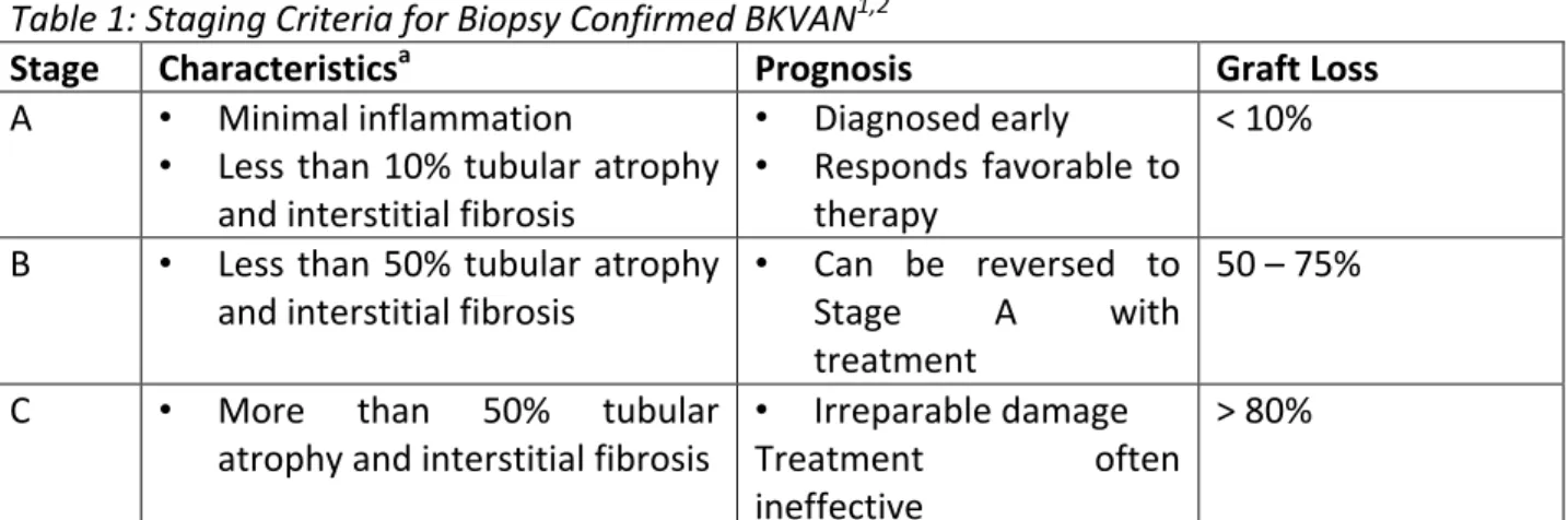
Stages and Prognosis of BKVAN
Table 1: Staging Criteria for Biopsy Confirmed BKVAN1,2
Stage Characteristicsa Prognosis Graft Loss
A • Minimal inflammation
• Less than 10% tubular atrophy and interstitial fibrosis
• Diagnosed early
• Responds favorable to therapy
< 10%
B • Less than 50% tubular atrophy and interstitial fibrosis
• Can be reversed to
Stage A with
treatment
50 – 75%
C • More than 50% tubular
atrophy and interstitial fibrosis
• Irreparable damage
Treatment often
ineffective
> 80%
a Percentage of tubular atrophy and interstitial fibrosis is based on the biopsy sample that is taken during diagnosis.
Currently, there is no widely accepted protocol to follow for patients presenting with BK virus reactivation. This study aims to evaluate the use of a more intense post-‐transplant monitoring and lower threshold for immunosuppression regimen changes which are currently
used at UNC Hospitals.
The mainstays of therapy in BKVAN treatment rely heavily on reduction in immunosuppression agents. It is generally accepted to reduce one agent at a time until a delayed progression of BK virus replication is seen. Should a reduction not suffice, additional agents have been tested as adjuncts to therapy, including, but not limited to leflunomide, IVIG, fluoroquinolones, brincidofovir and cidofovir1,8. However, there is no conclusive evidence as to
the benefit of these agents thus far.
METHODS Study Design
The study was conducted as a retrospective chart analysis to assess outcomes in renal transplant patients; specific to the infection of BK virus, viral progression, treatment and potential graft loss. Patients were included that had received a renal transplant at UNC Hospitals during 2005 and 2013 and tested positive for BK virus at any point after transplant during their follow-‐up through August 2014.
Primary Objective
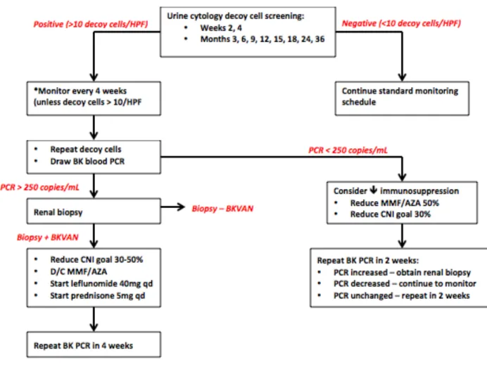
The primary objective of this study was to examine the graft loss rate due to BK virus following the UNC-‐specific IS modulation post-‐renal transplant, as seen in Figure 1. In assessing the effectiveness of the institution-‐specific protocol, the information gathered would potentially assist in the development of a standardized protocol in the prevention and treatment of BK virus in renal transplant patients nationwide to increase primary graft survival.
Figure 1: UNC Hospitals Protocol
Participants
A list of all patients receiving a renal transplant at UNC Hospitals between 2005 and 2013 was compiled. This list was further limited to only include transplant recipients that tested positive for BK virus based on urine cytology, at any time post-‐transplantation during follow-‐up.
Inclusion Criteria
All adult patients receiving a renal transplant at UNC Hospitals between January 2005 and December 2013 were considered for inclusion, with documented follow-‐up through August 2014. The included date range was chosen based on the time when the EMR TransChart, an EMR with specific functionality for transplant patients and better documentation, was introduced at UNC Hospitals. Additionally, it was the use of TransChart that prompted a more consistent following of the UNC BK Virus Protocol that required more significant monitoring. As it was observed that the protocol was followed more closely, these dates allow us to analyze the specific treatment changes used at UNC.
Exclusion Criteria
Patients were excluded if they were less than 18 years old, received a combined organ transplant (liver/kidney, liver/pancreas, heart/kidney, etc.). Combined transplant patients were excluded due to differing immunosuppression regimens employed to prevent multiple graft rejection. This exclusion was chosen to prevent skewing the data for the incidence of BKVAN in renal transplant patients specifically.
Data Collection
Data collection was performed using multiple EMRs that had been used at UNC Hospitals by the transplant treatment team during the study inclusion period, including WebCis, TransChart and EPIC. Patient demographics that were gathered included age at time of transplant, ethnicity, gender, reason for transplant, donor type (alive vs. deceased), induction antibodies, time to first decoy cell (+) on urine cytology, time to first blood BK PCR (+), time to first BKVAN (+) and stage of BKVAN as confirmed by renal biopsy, medications and drug levels in the month prior to BKVAN (including tacrolimus, cyclosporine, mycophenolic acid and steroids), and rejection data on BKVAN patients including type of rejection (acute and chronic), time before/after BKVAN diagnosis as well as medications for treatment.
Statistical Analysis
All statistical analysis was completed by the transplant department at UNC Hospitals using GraphPad™ software. The patient population was subdivided into two groups, no-‐BKVAN and BKVAN. Results were considered significant with a T-‐test value of p ≤ 0.05.
RESULTS
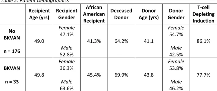
Table 2: Patient Demographicsa
Recipient
Age (yrs) Recipient Gender
African American Recipient
Deceased
Donor Age (yrs) Donor Gender Donor
T-‐cell Depleting Induction No
BKVAN n = 176
49.0
Female 47.1%
Male 52.8%
41.3% 64.2% 41.1
Female 54.7%
Male 42.5%
86.1%
BKVAN n = 33
49.8
Female 36.3%
Male 63.6%
45.4% 69.9% 43.8
Female 53.8%
Male 46.2%
77.7%
a In each group, there were some transplant patient’s charts that did not have the specific donor information reported. As such, calculations of patient demographics were completed using the information that was reported. In the No BKVAN group, donor age was only reported for 147 donors, gender for 141 donors and T-‐cell depleting induction for 94 donors. In the BKVAN cohort, donor age was only reported for 28 donors, gender for 28 donors and induction information for 27 donors.
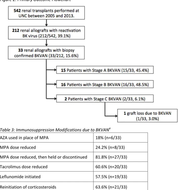
Primary Outcome
A total of 542 patients received a single renal transplant at UNC Hospitals between 2005 and 2013. Of these, 212 renal allografts had reactivated BK and 33 (15.6%) had progressed to BKVN. Renal biopsy identified 15 patients with Stage A BKVAN, 16 with Stage B and 2 with Stage C. The immunosuppression changes made, as seen in Table 3, preserved graft function in 97% of patients, with only one graft lost (1/33, 3%) following confirmed diagnosis with BKVAN. The one graft that was lost had flourid disease, stage C BKVAN.
Figure 2: Primary Outcome Flowchart
Table 3: Immunosuppression Modifications due to BKVANa
AZA used in place of MPA 18% (n=6/33)
MPA dose reduced 24.2% (n=8/33)
MPA dose reduced, then held or discontinued 81.8% (n=27/33)
Tacrolimus dose reduced 60.6% (n=20/33)
Leflunomide initiated 57.5% (n=19/33)
Reinitiation of corticosteroids 63.6% (n=21/33)
Immunosuppressives Doses at 1, 3, 6, and 12-‐months post-‐transplant
Table 4: Average prednisone doses 1, 3, 6 and 12-‐months post-‐transplant
Prednisone (mg/day) Month post-‐
transplant 1 3 6 12
No BKVAN
(n) Mean 6.77 (33) 7.04 (35) 7.38 (38) 6.73 (42)
BKVAN
(n) 10.56 (9) 10.17 (15) 8.13 (8) 6.72 (16)
p-‐value <0.01 Not significant Not significant Not significant
Table 5: Average mycophenolic acid doses 1, 3, 6 and 12-‐months post-‐transplant
Mycophenolic Aid (MPA, mg/day) Month post-‐
transplant 1 3 6 12
No BKVAN
(n) Mean 1332.66 (177) 1228.34 (163) 1102.47 (158) 1085.33 (150) BKVAN
(n) 1154.54 (33) 1038.33 (24) 1004.71 (17) 948.89 (9)
p-‐value <0.01 <0.02 Not significant Not significant
Table 6: Average tacrolimus doses 1, 3, 6 and 12-‐months post-‐transplant
Tacrolimus (mg/day) Month post-‐
transplant 1 3 6 12
No BKVAN
(n) Mean 8.56 (175) (173) 7.70 (168) 7.38 (156) 7.01
BKVAN
(n) 9.03 (33) 7.52 (31) 7.04 (29) 5.59 (26)
p-‐value Not significant Not significant Not significant <0.01
DISCUSSION
The emergence of BK virus infection in renal transplant patients poses a serious risk to graft survival in this population. Since the virus progresses without the typical symptoms of viral disease, including fever, malaise, myalgias, leukopenia, anemia, it can often go unnoticed until it has infected the graft itself2. The published rates of graft loss for stages A, B and C BKVAN are
<10%, ~50% and <80%, respectively1,10. The key to preserve the graft and graft function is to prevent infection or to prevent the progression of infection.
Weiss, AS et al. compared two strategies for BKVAN management, immunosuppression withdrawal vs. immunosuppression reduction. The results found that the early elimination of one immunosuppressant is significantly more effective in preserving graft function in renal transplant patients with a 1-‐year graft survival rate of 87.8% with immunosuppression withdrawal vs. 56.2% with immunosuppression reduction (p-‐value 0.03)11.
The American Society of Transplantation infectious diseases community of practice supports treatment via immunosuppression reduction of BK virus infection after a positive blood BK PCR test and suggest 2 potential strategies by reducing the calcineurin inhibitor 25-‐ 50%, followed by reduction of the anti-‐proliferative by 50% and then discontinuation of the anti-‐proliferative, or by first reducing the anti-‐proliferative by 50%, then reduce calcineurin inhibitor 25-‐50% and then discontinue the anti-‐proliferative. However, there was no reported efficacy on using either of these strategies on graft function or loss1. Additional strategies, including the addition of fluoroquinolones as a means of preventing BK viruria have been considered post-‐renal transplant. Knoll, GA, et al. sought to determine if levofloxacin could be used to prevent BK viruria and found a 29% incidence of BK viruria in the group treated with levofloxacin vs. 33.3% in the placebo group (p=0.58)12. Additionally, there was an increased risk of infection resistant to fluoroquinolones seen in the group treated with levofloxacin (58.3% vs. 33.3% in the placebo group, risk ratio 1.75)12.
Schaub, S, et al. assessed the mean time to viremia clearance after reduction of target tacrolimus trough levels, followed by a reduction of MMF dose. These changes resulted in a mean clearance of BK virus plasma levels within 4 months of initiation. These results support the strategy of using immunosuppression reduction. However, there was limited follow-‐up and the endpoints of the study did not look at rates of BK nephropathy or graft loss after changes were made in immunosuppressive therapy13.
The protocol used by UNC Hospitals (see Figure 1) follows a similar methodology as enumerated in the published literature. However, the UNC protocol deviates by employing more aggressive urine screening post-‐transplant to attempt to identify patients with viral replication early on in BK virus reactivation to prevent the spread of infection or potential graft loss. Additionally, a lower threshold for BK blood PCR levels is used when making the decision to decrease a patient’s immunosuppression regimen. This protocol has proven to be effective in minimizing graft loss in renal transplant patients secondary to BKVAN, with a 3% graft loss during the study period and patient follow-‐up through August 2014.
be seen upon following the BKVAN protocol used at UNC Hospitals, which first decreases mycophenolic acid and tacrolimus doses as initial therapy.
The addition of steroids upon after biopsy-‐confirmed BKVAN diagnosis is not a universally used practice, however it has been implemented as protocol at UNC Hospitals as adjunct therapy. Mean doses of prednisone after transplant were recorded, as seen in Table 4 and found that there was a slight higher average dose in the BKVAN group 1-‐month post-‐ transplant vs. no-‐BKVAN. Further analysis of doses and prednisone use was investigated in a further study assess overall use within this patient population.
As an addition to published treatment protocols, UNC Hospitals uses an antiproliferative agent with the addition of leflunomide to replace mycophenolic acid derivatives, upon diagnosis with BKVAN. Leflunomide is an antiviral that has an active metabolite, which inhibits BKV replication, hypothesized to be through nonspecific pyrimidine depletion. The addition is done in combination with total reduction of immunosuppression continuing the CNI and adding corticosteroid therapy. There is a lack of prospective randomized trials to confirm the efficacy of additive antivirals within this patient population14. However, UNC protocol dictates the addition upon confirmatory diagnosis of BKVAN and has only seen 1 graft loss over the study period further supporting this therapeutic addition.
Urine cytology is a non-‐invasive, less expensive means of assessing for BK virus replication. Performing these screens more frequently will allow providers to catch earlier stages of BK virus replication in the hopes of preventing the spread of infection. While blood BK PCR tests are more costly, Smith, F, et al. found significant cost savings with the reduction in immunosuppression in patients with significant viremia15. It is proposed that these savings would be enough to cover the more costly BK blood PCR tests required for monitoring.
Study Limitations
A limitation of this study was the required use of multiple EMRs in this retrospective chart analysis. The date range of data collection was chosen, as there was more consistent use of TransChart for transplant patients, however there was inconsistency in some of the data that was reported, and in the case of donor information there was some information omitted from the records entirely. Additionally, the switch to a new EMR during the data collection proved to be difficult for gathering any follow-‐up data.
While average doses of tacrolimus were recorded for each patient at specified time-‐ points post-‐transplant, there was no data recorded on the measured serum levels of tacrolimus. As the trough level of tacrolimus is the measure by which doses are changed, it would be important to see if the doses were successful in getting patients to their optimal level to improve graft survival.
The time at which the data was collected resulted in a variable about of follow-‐up for each patient, depending on his or her transplantation date. This time could have been too short to see any true reactivation or progression to nephropathy in some patients, which would warrant a longer follow-‐up to see additional outcomes.
time of 5 years is optimal in determining graft loss with BKVAN and was not necessarily the amount of follow-‐up for some patients included in this trial.
Future Directions
The results of this study demonstrate the success of the post-‐renal transplant immunosuppression regimen used at UNC Hospitals in minimizing the potential graft loss due to BK virus. However, some of the patients included in the date range of data collection had a relatively short follow-‐up period. Future directions for this research would include completing the data collection for 3-‐year graft survival follow-‐up as a better indicator of graft survival and completing data collection for rejection episodes and treatment prior to BK virus diagnosis. Additionally, having information regarding donor specific antibodies collected prior to transplant would allow practitioners to be aware of patients that may be at a higher risk for BK virus reactivation and may require alternative immunosuppression therapy goals.
CONCLUSIONS
UNC Hospitals center specific results include a graft loss rate of 3% among all diagnosed stages of BKVN. In contrast to current guidelines, this center follows a protocol with more aggressive post-‐transplant urine cytology screening, a lower threshold for acquiring blood PCRs and immunosuppression adjustment, along with the addition of leflunomide and prednisone therapy and discontinuation of antiproliferatives with biopsy-‐proven BKVAN. The results of this study support the continued use of our current center BK protocol to minimize renal allograft loss secondary to BKVAN.
REFERENCES
1. Hirsch, HH, Randhawa, P, et. al. BK Polyomavirus in Solid Organ Transplantation. Am J Transplantation. 2013; 13: 179-‐188.
2. Bonvoisin, C, Weekers, L, Xhignesse, P, Grosch, S, Milicevic, M, Krzesinski, JM. Polyomavirus in Renal Transplantation: A Hot Problem. Transplantation. 2008; 85 (7s): S42-‐S48.
3. Hariharan, S. BK virus nephritis after renal transplantation. Kidney International. 2006; 69: 655-‐662.
4. Joseph, A, et al. BK Virus Nephropathy in Heart Transplant Recipients. Am J Kidney Dis.2015; 65 (6): 949-‐955.
5. Ducharme-‐Smith, A, at al. Prevalence of BK Polyomavirus Infection and Association with Renal Dysfunction in Pediatric Heart Transplant Recipients. J Heart Lung Transplant. 2015; 34(2):222-‐226.
6. Nickeleit, V, Hirsch, HH, Binet, IF, Gudat, F, Prince, O, Dalquen, P, Thiel, G, Mihatsch, MJ. Polyomavirus Infection of Renal Allograft Recipients: From Latent Infection to Manifest Disease. J Am Soc Nephrol. 1999; 10: 1080-‐1089.
7. Huang, G, et al. Risk Factors for BK Virus Infection and BK Virus-‐Associated Nephropathy Under the Impact of Intensive Monitoring and Pre-‐emptive Immunosuppression Reduction. Transplant Proc. 2014; 46 (10):3448-‐54.
9. Schachtner, T, Muller, K, Stein, M, Diezemann, C, Sefrin, A, Babel, N,Reinke, P. BK Virus-‐ Specific Immunity Kinetics: A Predictor of Recovery from Polyomavirus BK-‐Associated Nephropathy. Am Jour of Transplantation. 2011; 11: 2443-‐2452.
10. Egli, A, et al. Cytomegalovirus and polyomavirus BK posttransplant. Nephrol Dial Transplant; 2007: (8) viii72-‐viii82
11. Weiss, AS, et al. Aggressive Immunosuppression Minimization Reduces Graft Loss Following Diagnosis of BK Virus-‐Associated Nephropathy: A Comparison of Two Reduction Strategies. Clin Jour Am Soc Nephrol. 2008; 3: 1812-‐1819.
12. Knoll, GA, et al. Levofloxacin for BK Virus Prophylaxis Following Kidney Transplantation: A Randomized Clinical Trial. JAMA. 2014; 312 (20): 2106 – 2114.
13. Schaub S, et al. Reducing immunosuppression preserves allograft function in presumptive and definitive polyoma-‐virus-‐associated nephropathy. Am Jour Transplantation. 2010; 10: 2615–2623.
14. Kuypers, DRJ. Management of polyomavirus-‐associated nephropathy in renal transplant recipients. Nat Rev Nephrol. 2012; 8: 390-‐402.
15. Smith, F, et al. Screening to Prevent Polyoma Virus Nephropathy in Kidney Transplantation: A Cost Analysis. Am Jour Transplantation. 2009; 9: 2177-‐2179.