1 Abstract
The effect of nitric oxide (NO)-release kinetics on the antibacterial activity of NO-releasing silica nanoparticles against the nosocomial pathogens Pseudomonas aeruginosa and Staphylococcus aureus is demonstrated.Silica nanoparticles were synthesized and modified to release NO via secondary amine modification with N-diazeniumdiolate NO donors. Planktonic Pseudomonas aeruginosa and Staphylococcus aureus were exposed toa series of NO-releasing silica particles with similar 2 h NO totals but different release kinetics (half-lives). Enhanced antibacterial efficacy was observed against Pseudomonas aeruginosa at pH 7.4 for nanoparticles with faster NO-release kinetics. At pH 7.4, minimal bactericidal efficacy was observed against Staphylococcus aureus,even for silica nanoparticles storing greater NO payloads. However, antibacterial activity was enhanced by exposing Staphylococcus aureus to NO-releasing silica particles at a lower pH (6.4). This was attributed to faster NO-release kinetics at lower pH due to more rapid proton-initiated decomposition of N-diazeniumdiolate NO donors.Collectively, these results demonstrate the enhanced NO-mediated bactericidal efficacy of rapid NO-release kinetics against nosocomial pathogens, providing further insight into the design of NO-releasing
2 Introduction
Despite advances in treating bacterial infections, the incidence of nosocomial infections increases annually, resulting in nearly two million cases of infection per year in the United States.1 In hospitals, biofilms composed of the nosocomial pathogens Pseudomonas aeruginosa and Staphylococcus aureus cause systemic infection in burn wounds, surgical sites, and the lungs.2 In fact, P. aeruginosa and S. aureus together account for nearly a third of all hospital-acquired lung infections.2 These infections are currently treated using conventional antibiotics; however, this method is becoming ineffective as the prevalence of antibiotic-resistant bacteria increases.1 The ramifications of resistance are severe, and it has been estimated that nearly 70% of lethal nosocomial infections are caused by antibiotic-resistant strains of these pathogens.3 Thus, the lack of suitable current drugs paired with the severity of hospital-acquired infections warrants research into alternative therapeutics.
Nitric oxide (NO), a highly reactive gas endogenously produced during the immune response to invading pathogens, may represent an alternative strategy to treating hospital-acquired infections. The broad-spectrum antibacterial activity of NO stems from its production of peroxynitrite (ONOO-) and dinitrogen trioxide (N2O3), reactive byproducts which exert
3
Due to its gaseous and highly reactive nature, NO must be contained in a scaffold to harness its antimicrobial properties. The Schoenfisch lab works on developing NO donor-modified macromolecular scaffolds that store and deliver tunable amounts of NO at
physiological conditions for antibacterial applications.6,7Silica has attracted attention as an NO delivery vehicle due to its low cytotoxicity and easily tunable physicochemical characteristics.3 Hetrick et al. first reported the utility of NO-releasing silica nanoparticles as antibacterial agents against the nosocomial pathogens S. aureus and P. aeruginosa.3,8 In these studies, Hetrick suggested that certain chemical properties (e.g., NO flux and release kinetics) of the NO-releasing silica nanoparticles influenced NO-mediated antibacterial action.3,8 Subsequent studies focused on varying these parameters to maximize bactericidal efficacy against pathogenic bacteria. The effect of release kinetics (i.e., half-life) on bactericidal efficacy of NO-releasing silica nanoparticles has been systematically studied against dental pathogens.10 By synthesizing nanoparticles with similar sizes and NO totals but different half-lives, Backlund et al. reported that the antibacterial activity of NO-releasing silica nanoparticles against oral pathogens depends on NO-release kinetics.10 Indeed, rapid NO-release kinetics enhanced antibacterial activity against Gram-positive oral pathogens, whereas Gram-negative
periodontopathogens were more susceptible to particles with extended NO-release kinetics.10 While these studies demonstrate the importance of NO-release rates on bactericidal efficacy, the effect of NO-release kinetics on the antibacterial activity of NO-releasing silica particles against nosocomial pathogens has yet to be systematically evaluated.
4
to yield similar 2h NO totals, but different release kinetics (half-lives). The 2 h minimum bactericidal concentrations (MBC2h) were determined against P. aeruginosa and S. aureus to
evaluate the role of NO-release kinetics on antibacterial activity independent of total NO release. This study provides insight into the design of future NO-releasing materials to combat
nosocomial infections.
Methods
I. Synthesis of nitric oxide-releasing silica particles
The synthesis of NO-releasing silica particles has been reported previously.10,11 Briefly, hybrid silica particles were synthesized using a Stöber method, involving the co-condensation of the aminosilanes N-(2-aminoethyl)-3-aminopropyltrimethoxysilane (AEAP3), N
-methylaminopropyltrimethoxysilane (MAP3), and N
-(6-aminohexyl)aminopropyl-trimethoxysilane (AHAP3) with a tetramethylorthosilicate (TMOS) alkoxysilane backbone.12 Silanes (TMOS with either AEAP3, MAP3, or AHAP3) were combined and injected in a bolus into a reaction mixture containing water (27.84 mL), ammonium hydroxide (9.8 mL) and EtOH to a total volume of 100 mL. After 2 h, the reaction was poured off and collected via
centrifugation. The particles were centrifuged and the supernatant decanted twice more to
remove unreacted silanes and residual solvent. After the third centrifugation, the supernatant was decanted once more and the particles were dried under vacuum overnight. The next day, the resultant secondary amine-modified particles (50 mol% MAP3/TMOS, 60 mol%
5
The hybrid silica nanoparticle secondary amines were then modified with N
-diazeniumdiolate NO donors, which spontaneously release NO under physiological conditions.11 A portion (30 mg) of the particles was suspended in a solution of 9:1 N,N-dimethylformamide (DMF) to methanol (MeOH) with 25 μL of sodium methoxide. The solution was then placed in a Parr reaction container connected to an in-house NO reactor and flushed with argon six times to remove oxygen in solution. The solution was then held under 10 atm of NO for three days with constant stirring. After three days, the unreacted NO was removed from the solution by purging the chamber with argon six times once more. The particles were collected via centrifugation and subsequent decanting of the supernatant. The pellet was centrifuged and the supernatant decanted twice more to remove residual solvent and NaOMe. The resulting N-diazeniumdiolate-modified silica nanoparticles were dried for 2 h and stored in a -20°C freezer in a vacuum-sealed bag until use.
II. Hybrid particle characterization
Dynamic light scattering (DLS) was utilized to measure particle size (i.e., hydrated diameter) and monodispersity.10 Samples were suspended in water at either 0.1 or 0.2 mg/mL, and sonicated for 20 min prior to analysis at room temperature. Particle morphologies and sizes were also evaluated using Scanning Electron Microscopy.
2.4 Characterization of nitric oxide release
6
mL/min to carry liberated NO to the analyzer. Analysis was terminated when NO levels decreased to <10 ppb/mg.
2.5 Bactericidal efficacy of nitric oxide-releasing silica nanoparticles
7 Results and Discussion
I. Nitric oxide-releasing silica nanoparticle characterization
The Stöber method was utilized to synthesize hybrid alkoxysilane/aminosilane silica nanoparticles (50 mol% MAP3/TMOS, 60 mol% AHAP3/TMOS, and 80 mol%
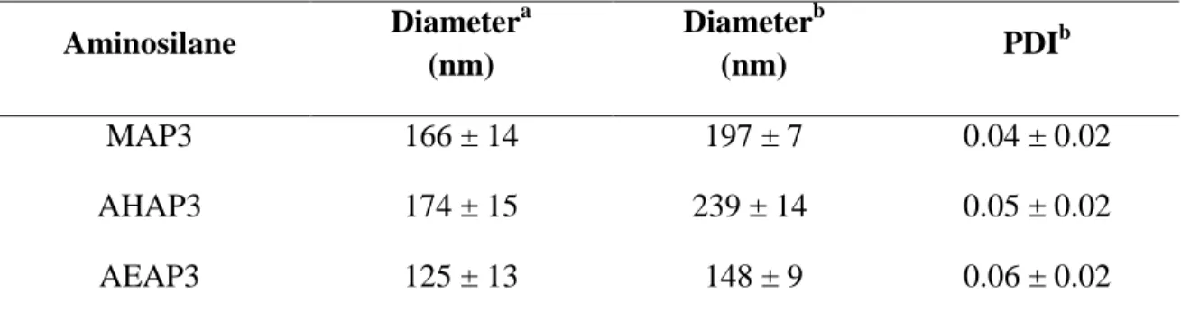
AEAP3/TMOS).12 For clarity, the particles will hereafter be referred to as MAP3, AHAP3, and AEAP3. Synthetic parameters were tuned to produce particles of similar sizes (~150 nm) and a high degree of monodispersity, as indicated by low polydispersity index (PDI) values (≤0.06; Table 1). As previous reports have demonstrated that particle size influences bactericidal efficacy, maintaining a constant particle size was crucial to this study.13,14 Thus, by tuning the identity of the aminosilane precursors while maintaining constant nanoparticle diameters, we were able to evaluate NO-mediated bactericidal efficacy independent of particle size.
Table 1. Silica nanoparticle characterization.Results presented as mean ± standard deviation for n = 3 or more pooled experiments. aGeometric diameter estimated using scanning electron microscopy. bHydrodynamic diameter and particle PDI measured in water using DLS.
Aminosilane Diameter
a
(nm)
Diameterb
(nm) PDI
b
MAP3 166 ± 14 197 ± 7 0.04 ± 0.02
AHAP3 174 ± 15 239 ± 14 0.05 ± 0.02
8
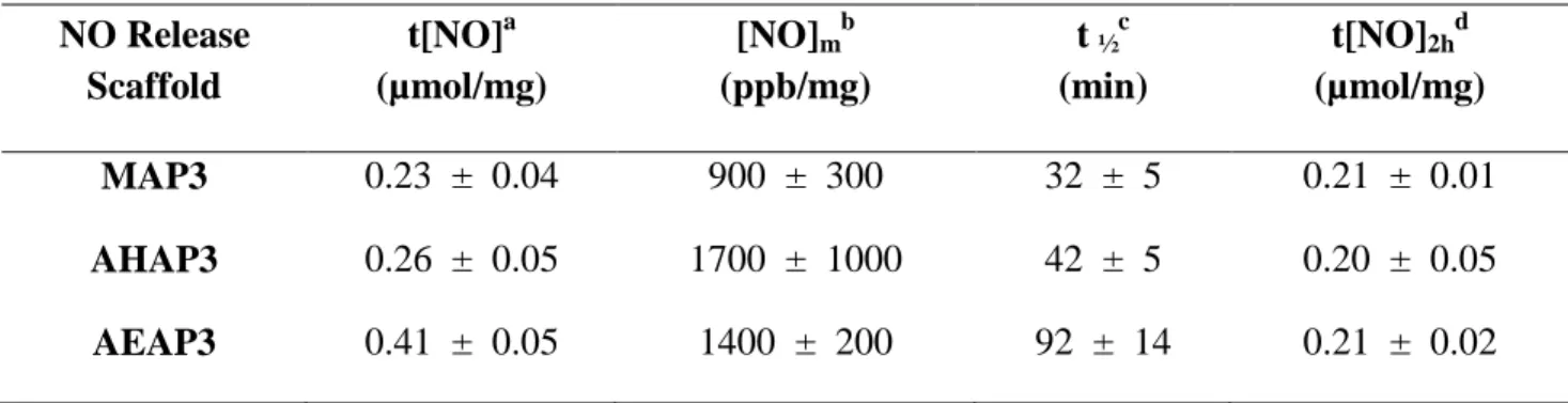
Due to the proton-initiated NO release from N-diazeniumdiolate NO donors, NO-release kinetics depend on aminosilane structure.11 As such, NO release from MAP3, AHAP3, and AEAP3 silica nanoparticles were quantified using an NOA to obtain release totals, 2 h NO-release totals, and NO-NO-release kinetics (half-lives).10 The total NO release for the MAP3, AHAP3, and AEAP3 particles was 0.23, 0.26, and 0.41 µmol/mg, respectively (Table 2). More importantly, 2 h NO-release totals were similar (~0.20 µmol/mg) across all three particle
systems, corresponding to the NO dose delivered during the 2 h MBC assays. The NO-release half-lives were significantly longer for AEAP3 (91.8 min) compared to MAP3 (31.6 min) and AHAP3 (42.2 min). The extended NO-release kinetics characteristic of AEAP3 were attributed to intramolecular hydrogen bonding between neighboring cationic amines on the aminosilane scaffold stabilizing the N-diazeniumdiolate NO donor and inhibiting rapid NO donor
decomposition.10,15 The bactericidal efficacies of MAP3, AHAP3, and AEAP3 were thereby compared as a function of NO-release kinetics, independent of particle size and 2 h NO-release totals.
Table 2. Characterization of NO-releasing silica particles in PBS (pH = 7.4 at 37 oC) by means of a chemiluminescent nitric oxide analyzer. Results shown for 50 mol% MAP3, 60 mol% AHAP3, and 80 mol% AEAP3 NO-releasing silica particles are presented as mean ± standard deviation for n = 3 or more pooled experiments. aTotal amount of NO released. bMaximum NO flux achieved. cTime to release half of total NO payload. dTotal amount of NO released after 2 h.
NO Release Scaffold
t[NO]a (µmol/mg)
[NO]mb
(ppb/mg)
t ½c
(min)
t[NO]2hd
(µmol/mg)
MAP3 0.23 ± 0.04 900 ± 300 32 ± 5 0.21 ± 0.01
9
II. Kinetic-dependent killing of planktonic Pseudomonas aeruginosa
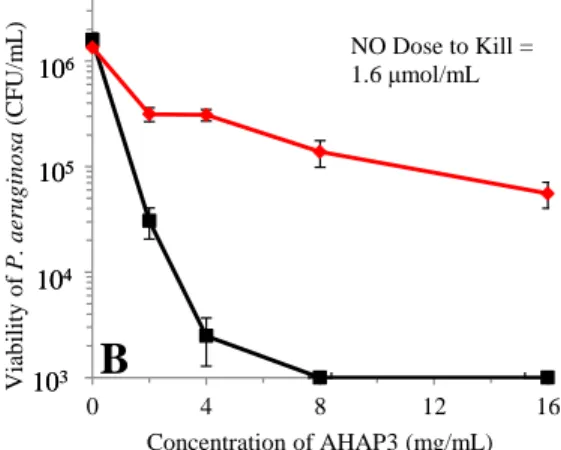
To study the role of NO-release kinetics on bactericidal efficacy, 2 h bacteria killing assays were conducted against nosocomial pathogens in Tris-PBS (pH = 7.4; 37 oC). P. aeruginosa was readily susceptible to NO, with bactericidal NO doses < 2 µmol/mL (Figure 1). Furthermore, bactericidal efficacy was found to be dependent on NO-release kinetics (Figure 1). The most effective killing of P. aeruginosa was achieved using NO-releasing MAP3 particles with the most rapid NO-release kinetics. The minimum bactericidal concentration (MBC2h) for
NO-releasing MAP3 was 4 mg/mL, corresponding to a bactericidal NO dose of 0.8 µmol/mL. This bactericidal NO dose is lower that of both NO-releasing AHAP3 and AEAP3, which required NO doses of 1.6 and 3.2 µmol/mL, respectively, to kill P. aeruginosa. We hypothesize that the rapid release kinetics characteristic of MAP3 (31.6 minutes) compared to AHAP3 (42.2 min) and AEAP3 (91.8) facilitates enhanced killing. More rapid NO-release kinetics also account for the enhanced bactericidal efficacy of AHAP3 compared to AEAP3.
10
and elicit NO-mediated killing.10,16 Conversely, Gram-negative bacteria that do not possess this intracellular mechanism (e.g., A. actinomycetemcomitans and P. gingivalis) benefit from a slower build-up of NO.10 The differential sensitivity to NO-release kinetics between P. aeruginosa and other Gram-negative pathogens may enhance the selective targeting of future NO-based therapeutics against different types of bacterial infections.
0 4 8 12 16
Viab ilit y o f P . a eru g in o sa (C FU/m L )
Concentration of AHAP3 (mg/mL) NO Dose to Kill = 1.6 μmol/mL 106 105 104 103 106 105 104
103
B
0 2 4 6 8
Viab ilit y o f P . a eru g in o sa (C FU/m L )
Concentration of MAP3 (mg/mL) 106
105
104
103
A
NO Dose to Kill = 0.8 μmol/mL
0 4 8 12 16
Viab ilit y o f P . a eru g in o sa (C FU/m L )
Concentration of AEAP3 (mg/mL) 106
105
104
103
NO Dose to Kill = 3.2 μmol/mL
C
11
III. Efficacy of nitric oxide-releasing silica particles against nosocomial pathogens
Previous reports have shown that Gram-positive species demonstrate reduced susceptibility to NO treatment compared to Gram-negative pathogens.9,17 For example, Backlund et al.
12
Table 3. Characterization of NO-releasing silica particles in PBS (pH = 7.4 at 37 oC) by means of a chemiluminescent nitric oxide analyzer. Results shown for 50 mol% and 70 mol% MAP3 NO-releasing silica particles and presented as mean ± standard deviation for n = 3 or more pooled experiments. aTotal amount of NO released. bMaximum NO flux achieved. cTime to release half of total NO payload. dTotal amount of NO released after 2 h.
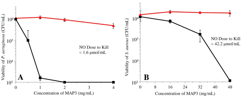
Figure 2. Bactericidal efficacy of 70 mol% MAP3 particles in Tris-PBS (pH = 7.4) against (A) Pseudomonas aeruginosa and (B) Staphylococcus aureus after 2 h exposure. NO-releasing particles denoted by rectangles (■) and non-NO-releasing controls denoted by triangles (▲). Error bars signify standard deviation of the mean bacterial viability (CFU/mL). For all measurements n = 3 or more pooled experiments.
NO Release Scaffold
t[NO]a (µmol/mg)
[NO]mb
(ppb/mg)
t ½c
(min)
t[NO]2hd
(µmol/mg) 50 mol% MAP3 0.22 ± 0.04 900 ± 300 31.6 ± 4.8 0.21 ± 0.01 70 mol% MAP3 0.84 ± 0.14 9200 ± 4300 19.8 ± 3.3 0.81 ± 0.15
0 1 2 3 4
Viab ilit y o f P . a eru g in o sa (C FU/m L )
Concentration of MAP3 (mg/mL)
106
105
104
103
NO Dose to Kill = 1.6 μmol/mL
A
0 8 16 24 32 40 48
Viab ilit y o f S . a u reu s (C FU/m L )
Concentration of MAP3 (mg/mL)
NO Dose to Kill = 42.2 μmol/mL
B
105104
103
13
The greater susceptibility of Gram-negative pathogens to treatment by NO lends further support toward the future use of NO as an antibacterial therapeutic. Conventional antibiotics are more effective at eradicating Gram-positive species, causing Gram-negative infections to spread and commonly remain untreated.1 Furthermore, Gram-negative species are more likely to develop antibiotic resistance compared to Gram-positive pathogens, reducing the availability of effective treatment strategies for Gram-negative infections.1 Thus, the greater antibacterial activity of NO-releasing therapeutics against Gram-negative species demonstrates the promise NO holds as an alternative strategy for treating hospital-acquired Gram-negative bacterial infections.
IV. Kinetic-dependent killing of planktonic Staphylococcus aureus
Backlund et al. previously observed that more rapid NO-release kinetics at lower pH
14
Table 4. Characterization of MAP3 NO-releasing silica particles in PBS (37 oC) at different pH values (7.4 and 6.4) using a chemiluminescent nitric oxide analyzer.Results shown for 70 mol% MAP3 NO-releasing particles and presented as mean ± standard deviation for n = 3 or more pooled experiments. aTotal amount of NO released. bMaximum NO flux achieved. cTime to release half of total NO payload. dTotal amount of NO released after 2 h.
While the bactericidal concentration of NO-releasing 70 mol% MAP3 was 48 mg/mL against S. aureus at pH 7.4, faster NO-release resulted in enhanced killing (3-log) of S. aureus at pH 6.4 at a reduced concentration of 32 mg/mL MAP3 particles (Figure 3). Thus, killing was enhanced at pH 6.4 (bactericidal NO dose 27.2 μmol/mL) compared to pH 7.4 (bactericidal NO dose 42.2 μmol/mL) due to the enhanced N-diazeniumdiolate NO donor decomposition and faster NO-release kinetics. The benefit of fast NO-release kinetics for more effective killing of both P. aeruginosa and S. aureus may influence the design of future NO-releasing therapeutics for treatment of hospital-acquired infections implicating both of these nosocomial pathogens.
pH t[NO]
a
(µmol/mg)
[NO]mb
(ppb/mg)
t ½c
(min)
t[NO]2hd
15
Figure 3. Bactericidal efficacy of 70 mol% MAP3 particles against S. aureus in (A) Tris-PBS (pH = 7.4) and (B) PBS (pH = 6.4) after 2 h exposure. NO-releasing material denoted by squares (■) and non-NO-releasing controls denoted by triangles (▲). Error bars signify standard deviation of the mean bacterial viability (CFU/mL). For all measurements n = 3 or more pooled experiments.
0 8 16 24 32 40 48
Viab ilit y o f S . a u reu s (C FU/m L )
Concentration of MAP3 (mg/mL) NO Dose to Kill =
42.2 μmol/mL
A
105 104 103 1060 8 16 24 32 40 48
Viab ilit y o f S . a u reu s (C FU/m L )
Concentration of MAP3 (mg/mL) NO Dose to Kill =
16 Conclusions
17 References
[1] Antibiotic resistance threats in the United States, 2013. Centers for Disease Control and Prevention Department of Health and Human Services 2013.
[2] Magill SS, Edwards JR, Bamberg W, Beldavs ZG, Dumyati G, Kainer MA, Lynfield R, Maloney M, McAllister-Hollod L, Nadle J, Ray SM, Thompson DL, Wilson LE, Fridkin SK. Multistate point-prevalence survey of health care-associated infections. N Engl J Med
2014;370:1198-208.
[3] Hetrick EM, Shin JH, Stasko NA, Johnson CB, Wespe DA, Holmuhamedov E, et al. Bactericidal efficacy of nitric oxide-releasing silica nanoparticles. ACS Nano 2008;2:235-46.
[4] Schairer DO, Chouake JS, Nosanchuk JD, Friedman AJ. The potential of nitric oxide releasing therapies as antimicrobial agents. Virulence 2012;3:271-9.
[5] Privett BJ, Broadnax AD, Bauman SJ, Riccio DA, Schoenfisch MH. Examination of bacterial resistance to exogenous nitric oxide. Nitric Oxide 2012;26:169-73.
[6] Riccio DA, Schoenfisch MH. Nitric oxide release: Part I. Macromolecular scaffolds. Chem Soc Rev 2012;41:3731-41.
[7] Carpenter AW, Schoenfisch MH. Nitric oxide release: Part II. Therapeutic applications. Chem Soc Rev 2012;41:3742-3752
[8] Hetrick EM, Shin JH, Paul HS, Schoenfisch MH. Anti-biofilm efficacy of nitric oxide-releasing silica nanoparticles. Biomaterials 2009;30:2782-9.
[9] Lu Y, Slomberg DL, Sun B, Schoenfisch MH. Shape‐and nitric oxide flux‐dependent bactericidal activity of nitric oxide‐releasing silica nanorods. Small 2013;9:2189-98.
[10] Backlund CJ, Worley BV, Sergesketter AR, Schoenfisch MH. Kinetic-dependent bactericidal efficacy of nitric oxide-releasing silica particles against oral pathogens. Acta Biomaterialia. In preparation.
18
[12] Stöber W, Fink A, Bohn E. Controlled growth of monodisperse silica spheres in the micron size range. J Colloid Interface Sci 1968;26:62-9.
[13] Carpenter AW, Slomberg DL, Rao KS, Schoenfisch MH. Influence of scaffold size on bactericidal activity of nitric oxide-releasing silica nanoparticles. ACS Nano 2011;5:7235-44.
[14] Slomberg DL, Lu Y, Broadnax AD, Hunter RA, Carpenter AW, Schoenfisch MH. Role of size and shape on biofilm eradication for nitric oxide-releasing silica nanoparticles. ACS Appl Mater Interfaces 2013;5:9322-9.
[15] Lu Y, Sun B, Li C, Schoenfisch MH. Structurally diverse nitric oxide-releasing poly (propylene imine) dendrimers. Chem Mater 2011;23:4227-33.
[16] Kakishima K, Shiratsuchi A, Taoka A, Nakanishi Y, Fukumori Y. Participation of nitric oxide reductase in survival of Pseudomonasaeruginosa in LPS-activated macrophages. Biochem Biophys Res Commun 2007;355:587-91.