RESEARCH ARTICLE
HIV-1 infection depletes human CD34
+
CD38
-hematopoietic progenitor cells via
pDC-dependent mechanisms
Guangming Li1☯, Juanjuan Zhao2☯, Liang Cheng1, Qi Jiang1, Sheng Kan2, Enqiang Qin3, Bo Tu3, Xin Zhang3, Liguo Zhang4‡, Lishan Su1,4‡, Zheng Zhang1,2☯‡*
1 The Lineberger Comprehensive Cancer Center, University of North Carolina, Chapel Hill, NC, United States of America, 2 Research Center for Clinical & Translational Medicine, Beijing 302 Hospital, Beijing, China, 3 Treatment and Research Center for Infectious Diseases, Beijing 302 Hospital, Beijing, China, 4 Key laboratory of Infection and Immunity, Institute of Biophysics, Chinese Academy of Science, Beijing, China
☯These authors contributed equally to this work. ‡ These authors are joint senior authors on this work.
Abstract
Chronic human immunodeficiency virus-1 (HIV-1) infection in patients leads to multi-lineage hematopoietic abnormalities or pancytopenia. The deficiency in hematopoietic pro-genitor cells (HPCs) induced by HIV-1 infection has been proposed, but the relevant mecha-nisms are poorly understood. We report here that both human CD34+CD38-early and CD34+CD38+intermediate HPCs were maintained in the bone marrow (BM) of humanized mice. Chronic HIV-1 infection preferentially depleted CD34+CD38-early HPCs in the BM and reduced their proliferation potential in vivo in both HIV-1-infected patients and human-ized mice, while CD34+CD38+intermediate HSCs were relatively unaffected. Strikingly, depletion of plasmacytoid dendritic cells (pDCs) prevented human CD34+CD38-early HPCs from HIV-1 infection-induced depletion and functional impairment and restored the gene expression profile of purified CD34+HPCs in humanized mice. These findings suggest that pDCs contribute to the early hematopoietic suppression induced by chronic HIV-1 infection and provide a novel therapeutic target for the hematopoiesis suppression in HIV-1 patients.
Author summary
Multi-lineage hematopoietic abnormalities generally occur during chronic infection which results in a disorder of human leukocyte development and differentiation, contrib-uting to human immunodeficiency virus-1 (HIV-1)-infection induced immune-patho-genesis in AIDS patients. Although successful antiretroviral therapy can reduce plasma viral loads to undetectable levels and ameliorate HIV-1-associated hemato-suppression, immune cell development is only partially restored. The mechanism for the abnormal hematopoiesis occurring during chronic HIV-1 infection remains unclear. HIV-1 infec-tion may directly or indirectly funcinfec-tionally impair hematopoietic progenitors by either viral products or induction of persistent inflammatory responses, leading to hematopoiesis
a1111111111 a1111111111 a1111111111 a1111111111 a1111111111 OPEN ACCESS
Citation: Li G, Zhao J, Cheng L, Jiang Q, Kan S, Qin E, et al. (2017) HIV-1 infection depletes human CD34+CD38-hematopoietic progenitor cells via
pDC-dependent mechanisms. PLoS Pathog 13(7): e1006505.https://doi.org/10.1371/journal. ppat.1006505
Editor: Guido Silvestri, Emory University, UNITED STATES
Received: February 17, 2017
Accepted: July 2, 2017
Published: July 31, 2017
Copyright:©2017 Li et al. This is an open access article distributed under the terms of theCreative Commons Attribution License, which permits unrestricted use, distribution, and reproduction in any medium, provided the original author and source are credited.
Data Availability Statement: All relevant data are within the paper and its Supporting Information files.
obstacles. Here, we show that HIV-1 infection significantly depleted and functionally impaired human hematopoietic progenitors in the bone marrow of both HIV-1-infected patients and humanized mice through a plasmacytoid dendritic cell (pDC)-dependent mechanism, as depletion of pDCs significantly recovered cell numbers and functions and gene expression profiles of hematopoietic progenitor cells in humanized micein vivo. Our study clarifies a novel mechanism underlying hemato-suppression induced by chronic HIV-1 infection and provides a novel strategy to halt HIV-1 disease.
Introduction
Human immunodeficiency virus-1 (HIV-1) infection in patients leads to multi-lineage hema-topoietic abnormalities, including anemia, granulocytopenia and thrombocytopenia [1,2]. Abnormalities in fetal hematopoiesis have also been reported in aborted fetuses from HIV-1 seropositive women [3]. The defect in hematopoietic progenitor cells (HPCs) or hematopoiesis induced by HIV-1 has been proposed [1,4,5]. In addition, the degree of the hematopoietic pathology correlates with the stage of disease progression [6], and end-stage disease is charac-terized by pancytopenia [1]. Long-term bone marrow (BM) cultures from HIV-1-infected patients exhibit low CD34+progenitor cell growth and differentiation [7,8], indicating func-tional impairment of early hematopoietic progenitors. Although the successful highly active antiretroviral therapy (HAART) clearly ameliorates HIV-1-associated hemato-suppression, it does not completely restore blood cell development [9]. These observations indicate that hematopoietic failure is an important aspect of HIV-1 infection-induced pathogenesis [10].
HPCs are comprised of diverse populations, including both early and intermediate progeni-tors. Each subpopulation expresses distinct sets of cell surface antigens, although they all express the cell surface antigen CD34 [11,12]. Early and intermediate populations can be dis-tinguished by the expression of CD38, with the former being negative for CD38 and the latter being positive for this antigen. Functionally, intermediate progenitors include common mye-loid progenitors that can give rise to all myemye-loid, erythroid and megakaryocyte lineages. Due to limited access to the BM in humans, properties of human HPC subsets and their alterations in healthy and HIV-1 disease states have been difficult to characterize.
The mechanisms underlying abnormal hematopoiesis in HIV-1 infection remains unclear due to the paucity of robust animal models that mimic human hemato-suppressionin vivo. Although previous studies failed to detect HIV-1 infection of HPCs, recent reports indicated that HIV-1 could directly infect HPC subsets and lead to their impairment [13–16]. In addi-tion, HIV-1 proteins such as Nef [17] and prolonged treatment with antiretroviral drugs could also compromise hematopoietic progenitors [18]. Although these studies investigated a litany of direct and indirect causes of HIV-1-associated hemato-suppression, how HIV-1 affects hematopoiesisin vivoremains unclear.
There is emerging evidence that certain cytokines induced during inflammation have sig-nificant effects on HPCs in the BM. Type I and II interferon (IFN) [19–23], tumor necrosis fac-tor (TNF) [24–26] and lipopolysaccharide (LPS) [27,28] directly stimulate HPC proliferation and differentiation, thereby increasing the short-term output of mature effector leukocytes. However, chronic inflammatory cytokine signaling can lead to functional exhaustion of HPCs [19,22,28]. Our previous study demonstrated that plasmacytoid dendritic cells (pDCs), the major type I interferon (IFN-I)-producing cells during acute or chronic HIV-1 infection, could inhibit viral replication while significantly contributing to HIV-1 infection-induced immune-pathogenesis, including increased immune cell death and reduced immune
AI095097 to LS), from the Science and Technology Innovation Fund of Shenzhen (JCYJ
20170412151650600 to ZZ). The funders had no role in study design, data collection and analysis, decision to publish, or preparation of the manuscript.
reconstitution of human CD45+cells in humanized micein vivo[29]. These findings suggest that pDCs play a pivotal role in the hemato-suppression induced by chronic HIV-1 infection.
In this study, we sought to understand the role of pDCs in HIV-1-associated hemato-sup-pression in a humanized mouse modelin vivo. We discovered that HIV-1 infection depleted CD34+CD38-early HPCs and functionally impaired human CD34+HPCs in the BM of patients and humanized mice with HIV-1 infection. This phenomenon was further found to be dependent on pDCs, as depletion of pDCs significantly recovered HPC cell numbers and multi-lineage colony-forming functions. Our present study therefore reveals a novel mecha-nism for hematopoiesis suppression induced by chronic HIV-1 infection and provides a new strategy to rescue HPC function and halt HIV-1 disease progression.
Results
Development and maintenance of functional human HPCs in humanized
mice
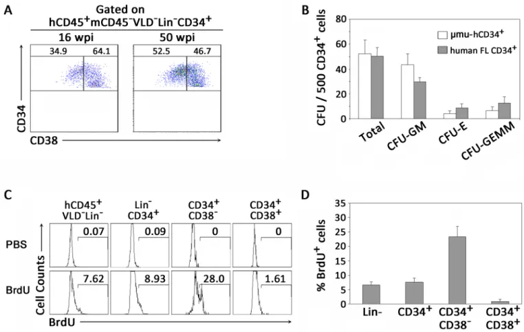
By gating on live human CD45+cells (VLD-mCD45-) with a lymphoid morphology that lacked common markers (Lineage−) for T cells (CD3), B cells (CD19 and CD20) or NK cells (CD56 and CD16), we identified human BM-derived HPCs as CD34+cells, which included early CD34+CD38-and intermediate CD34+CD38+subpopulations (S1 Fig). We then analyzed the human HPCs from the BM of humanized mice at various time points after human CD34+cell transplantation. The early and intermediate HPCs could be detected significantly at both 16 weeks and 50 weeks after CD34+cell transplantation with relatively stable levels (Fig 1A).
To determine if human HPCs derived from humanized mice were functional, human CD34+cells were isolated from the BM of humanized mice and human fetal livers, and col-ony-forming unit (CFU) assays were performed by culturing CD34+HPCs in a complete methylcellulose medium system. Two weeks later, HPCs derived from the BM of humanized mice produced 50 colonies on average for every 500 CD34+cells plated, similar to that of human fetal liver-derived CD34+cells (Fig 1B). All hematopoietic lineages were generated in cultures from the BM of humanized mice. In addition, HPCs from humanized mice could pro-liferate and differentiate into various blood lineage cellsin vitroat a similar frequency to that of CD34+cells from human fetal livers, including colony-forming unit-granulocyte and mac-rophage (CFU-GM), colony-forming unit-erythroid (CFU-E) and colony-forming unit-granu-locyte, erythroid, macrophage, megakaryocyte (CFU-GEMM) (Fig 1B). We then measured the proliferation capacity of HPCs by BrdU labelingin vivoand found that 8.9% of human CD34+ cells showed proliferation (Fig 1C). Notably, the CD34+CD38-early HPCs were much more proliferative, with an average of nearly 25% of cells being BrdU positive, which was signifi-cantly higher than the relatively quiescent CD34+CD38+intermediate HPCs with 1.6% BrdU labeling (Fig 1C and 1D). These data suggest that the human CD34+CD38-early HPCs and CD34+CD38+intermediate HPCs were both functionally developed and maintained in the BM of humanized mice.
CD34
+CD38
-early HPCs are preferentially depleted in vivo in both HIV-1
chronically infected patients and humanized mice
Utilizing the robust animal model, we were able to investigate whether chronic HIV-1 infec-tion affected human HPCs. HIV-1 infecinfec-tion was established in humanized mice, as measured by plasma HIV-1 RNA (copies/mL,S2 Fig). On termination, we also measured HIV-1 gag p24 expression in both T cells and CD34+HPCs by flow cytometry. Although a previous study sug-gested that HIV-1 has the potential to infect intermediate CD34+CD38+HPCs [13], we found
that p24 expression was absent in BM CD34+HPCs from humanized mice with HIV-1 infec-tion; in contrast, CD3+T cells showed high levels of p24 expression (10.5%) (Fig 2A). Further analysis indicated that the frequency of CD34+CD38-early HPCs was largely decreased in humanized mice with chronic HIV-1 infection, while the proportion of intermediate CD34+CD38+HSCs was relatively expanded (Fig 2B).
Summarized data further demonstrated that chronic HIV-1 infection significantly reduced CD34+CD38-early HPCs by nearly 8-fold as compared to the non-infected animals; mean-while, the proportion of CD34+CD38+intermediate HPCs was increased from 76% to nearly 100% as shown in the stacked bar graph (Fig 2C). When the absolute cell counts of early and intermediate HPCs were calculated, the number of CD34+CD38-early HPCs was dramati-cally reduced in the BM of chronidramati-cally infected animals (Fig 2D), while CD34+CD38+ interme-diate HPC counts were affected mildly (Fig 2E). These results suggest a depletion of early HPCs during chronic HIV-1 infection. Importantly, we observed a similar depletion of BM CD34+CD38-early HPCs in HIV-1-infected patients. As shown inFig 2F, the percentage of CD34+CD38-early HPCs was significantly decreased within total CD34+HPCs in an
HIV-Fig 1. Development of functional human CD34+HPCs in BM of humanized mice. (A) Representative dot plots showing distribution of human early CD34+CD38-and intermediate CD34+CD38+HPCs in BM of humanized mice. Numbers indicate percentages of CD34+CD38-and CD34+CD38+HPCs (gated on hCD45+mCD45-VLD-Lin-CD34+cells). Data are representative of two independent experiments with mice reconstituted with three human donors. (B) Pooled data indicating numbers of colonies that developed from human CD34+HPCs from BM of humanized mice and human fetal livers (n = 6). Total colonies, CFU-GM, colony-forming granulocyte, macrophage; CFU-E, colony-forming unit-erythroid; CFU-GEMM, colony-forming unit-granulocyte, erythroid, macrophage, megakaryocyte. (C) Representative histograms showing
proliferation of CD34+HPCs in vivo through BrdU labeling in humanized mice. PBS mouse samples were used as controls (upper panels). Numbers indicate percentages of BrdU+HPCs within CD34+HPC subsets. (D) Pooled data indicate percentages of BrdU+CD34+HPC subsets in BM of humanized mice (n = 5). (B, D) Data are shown as the mean±s.e.m.
Fig 2. Chronic HIV-1 infection preferentially depletes early CD34+CD38-HPCs in humanized mice in vivo. (A) Representative histograms showing p24 expression on CD3+T cells and CD34+HPCs from BM of humanized mice with chronic HIV-1 infection. Numbers indicate percentages of p24-expressing CD3+T cells and CD34+HPCs. Data are representative of three independent experiments with mice
reconstituted with two to three donors. (B) Representative dot plots showing chronic HIV-1 infection depleting early CD34+CD38-HPCs from BM of humanized mice. Numbers indicate percentages of early CD38-CD34+and intermediate CD34+CD38+HPCs. (C) Pooled data indicating mean percentages of early CD34+CD38-HPCs and intermediate CD34+CD38+HPCs in BM of humanized mice with mock and HIV-1 infection shown in the stacked bar graph. P values are shown. (D-E) Pooled data indicating absolute cell numbers of early CD34+CD38-HPCs (D) and intermediate CD34+CD38+HPCs (E) in BM of humanized mice with mock (n = 7) or chronic HIV-1 infection (n = 5). (F) Representative dot plots showing chronic HIV-1 infection depleting early CD34+CD38-HPCs from BM of HIV-1-infected patients compared to that of healthy control (HC) donors. Numbers indicate percentages of early CD38-CD34+and intermediate CD34+CD38+HPCs. (G-H) Pooled data indicating percentages of early CD34+CD38-HPCs (G) and intermediate CD34+CD38+HPCs (H) in BM of HC donors (n = 6) and HIV-1-infected patients (n = 5). (D, E, G, H) Data are shown as the mean±s.e.m.*P<0.05 and***P<0.001 (two-tailed unpaired Student’s t-test).
https://doi.org/10.1371/journal.ppat.1006505.g002
1-infected patient when compared to a healthy control (HC). The depletion of CD34+CD38 -early HPCs in human BM from HIV-1 infection was strengthened by the addition of more patients (n = 5) (Fig 2G). In contrast, the proportion of intermediate CD34+CD38+HSCs was increased within total HPCs in HIV-1-infected patients relative to those of HC subjects (Fig 2H). These data indicated that early CD34+CD38-HSCs were preferentially depleted by chronic HIV-1 infection, and the humanized mouse is a highly relevant animal model that mimics HIV-1-induced hemato-suppression conditions in patients.
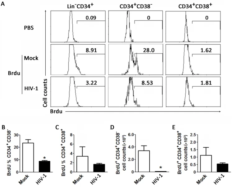
Chronic HIV-1 infection inhibits proliferation of human HPCs in vivo
We next analyzed the effect of chronic HIV-1 infection on the homeostatic proliferation of human HPCs in humanized mice. The results indicate that proliferation of human CD34+ cells in the BM was inhibited by approximately 3-fold in chronic HIV-1 infection compared to the mock animals (Fig 3A). Consistent with the preferential reduction of CD34+CD38-HPCs, BrdU-positive CD34+CD38-early HPCs were significantly decreased by chronic HIV-1 in-fection; meanwhile, the proliferation of CD34+CD38+intermediate HPCs was only mildly reduced by chronic HIV-1 infection (Fig 3B and 3C). In terms of cell numbers, the early HPC counts were significantly decreased by chronic HIV-1 infection compared with mock treat-ment in mice (Fig 3D), while intermediate HPC counts were only slightly reduced (Fig 3E). Thus, while both cell types were less proliferative in the presence of HIV-1, the more marked difference was observed in CD34+CD38-cells. Therefore, chronic HIV-1 infection appeared to suppress human hematopoiesis by inhibiting homeostatic proliferation of human early HPCs in the BM.
Human CD34
+HPCs isolated from BM of mice chronically infected with
HIV-1 display impaired differentiation
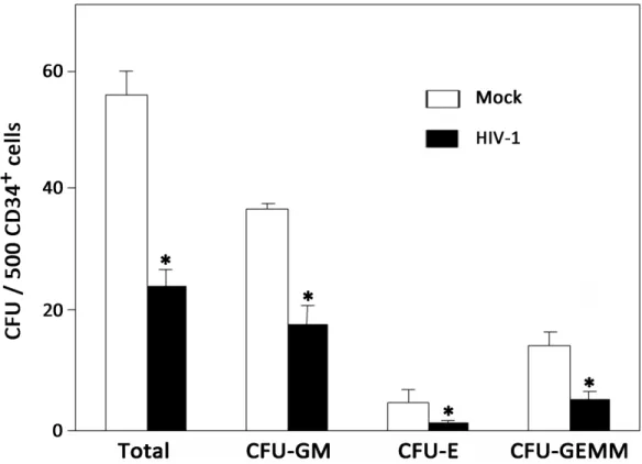
In order to assess the quality ofin vivohuman HPCs during chronic HIV-1 infection, we mea-sured CFU activity of purified human Lin-CD34+HPCs from mock- or HIV-1-infected humanized mice, including GM, E and GEMM (S3 Fig). As shown inFig 4, HPCs isolated from uninfected mice consistently produced over 50 colonies per 500 CD34+cells on average, whereas HPCs derived from HIV-1-infected mice produced less than 30 colonies per 500 CD34+HPCs. Moreover, the ability of HPCs to generate all lineages, including GM, E and GEMM, was also suppressed to some extent in chronically infected mice. Therefore, the results indicated that chronic HIV-1 infection leads to the impaired differentiation of HPCsin vivo.
Decreased quantity and quality of CD34
+CD38
-early HPCs is
dependent on pDCs during chronic HIV-1 infection
Increasing reports have demonstrated that chronic inflammation could lead to the functional exhaustion of BM HPCs [19,22,28]. Our recent study indicated that depletion of pDCs effi-ciently rescued human CD45 cell reconstitution in humanized mice with chronic HIV-1 infec-tion [29]. Thus, we hypothesized that pDCs may be responsible for the depletion of
that both the percentages and cell counts of CD34+CD38-early HPCs were restored by deple-tion of pDCs in HIV-1-infected mice (Fig 5B and 5C). In contrast, pDC depletion did not influence the percentages of CD34+CD38-early HPCs (CD38 expression) and their prolifera-tionin vivoindicated by BrdU expression in the BM in the absence of HIV-1 infection (S4B Fig). In addition, the cell counts of CD34+CD38+intermediate HPCs showed only minor recovery by the depletion of pDCs during chronic HIV-1 infection, which is consistent with the slight decrease in proportion of CD34+CD38+intermediate HPCs (Fig 5B and 5C).
Notably, only half of the animals showed a recovery in the proportion of CD34+CD38-early HPCs after pDC depletion inFig 5B, which prompted us to investigate whether the extent of pDC depletion affected the effectiveness of rescue of CD34+CD38-early HPCs in humanized mice with chronic HIV-1 infection. We therefore divided the 13 animals into two groups;
Fig 3. Chronic HIV-1 infection impairs proliferation of early CD34+CD38-HPCs in vivo of humanized mice. (A) Representative histograms showing BrdU labeling of CD34+HPC subsets from BM of humanized mice. Numbers indicate percentages of BrdU+within the total CD34+ HPCs, early CD34+CD38-HPCs and intermediate CD34+CD38+HPCs. (B-E) Pooled data indicating percentages of BrdU+cells within early CD34+CD38-HPCs (B) and intermediate CD34+CD38+HPCs (C); and absolute cell numbers of BrdU+early CD34+CD38-HPCs (D) and intermediate CD34+CD38+HPCs (E) in BM of mock (n = 4) and HIV-1-infected (n = 6) humanized mice. Data are shown as the mean±s.e.m.
*P<0.05 (two-tailed unpaired Student’s t-test).
https://doi.org/10.1371/journal.ppat.1006505.g003
animals with less than the median percentage of CD34+CD38-HPCs were placed in the "non-rescued" group (n = 6), while the others were included in the "rescued" group (n = 7). The rescued group was found to have significantly more CD34+CD38-early HPCs and fewer CD34+CD38+intermediate HPCs than the non-rescued group (S4C and S4D Fig). Impor-tantly, the rescued mice showed a marked lack of pDCs in the BM and lower levels of IFN-αin plasma compared with non-rescued mice (S4E and S4F Fig). Accordingly, the rescued mice were also characterized by a higher level of HIV-1 replication than that of non-rescued mice (S4G Fig). Thus, we observed that the CD34+CD38-early HPCs was negatively correlated with pDC percentages within CD45+cells in BM of these HIV-1 infected humanized mice with pDC depletion (S4H Fig). These data indicated that the extent of rescue of CD34+CD38-early HPCs was closely linked toin vivopDC depletion rather than other potential causes such as HIV viral load in humanized mice with chronic HIV-1 infection.
To further qualify HPCs after pDC depletion, Lin-CD34+cells were purified for colony-forming assaysex vivo. Cell colonies including GM, E and GEMM were found in culture (S3 Fig). The results demonstrated that pDC depletion could dramatically enhance CFU activity of the Lin-CD34+cell population as well as increase the quantity of each colony type individually as compared with HIV-1-infected mice (Fig 5D).
Depletion of pDCs restores gene expression profile of human CD34
+HPCs
To understand how pDCs contribute to the impairment of HPCs during chronic HIV-1 infec-tion, we analyzed gene expression of human HPCs in BM from HIV-1 chronically infected
Fig 4. Chronic HIV-1 infection impairs colony-forming activity of CD34+HPCs in BM of humanized mice. Pooled data indicate the number of colonies that developed from human CD34+HPCs of mock (n = 4) and HIV-1-infected (n = 5) mice. Data are shown as the mean and s.e.m.*P<0.05 (two-tailed unpaired Student’s t-test).
humanized mice. Human Lin-CD34+cells from BM of humanized mice were isolated and sub-mitted for gene expression analysis by cDNA array (Fig 6A), as described in a previous study [29]. A total of 3114 genes were significantly up-regulated, and 2994 genes were down-regu-lated spontaneously in CD34+HPCs from HIV-1-infected humanized mice as compared to mock-treated mice (fold change>2,Fig 6BandS5 Fig). Astonishingly, pDCs depletion during chronic HIV-1 infection in mice restored most of the interferon-stimulating genes (ISGs) to levels found in non-infected animals (S6A and S6B Fig). Along with the recovery of ISG gene expression, only 924 genes were significantly up-regulated, and 364 genes were down-regu-lated in BM CD34+HPCs with pDCs depletion as compared to mock-treated mice (fold change>2,Fig 6BandS5 Fig). Thus, pDCs depletion resulted in a restoration of a total of 5664 genes among 6108 genes (>92.7%) that changed during HIV-1 infection in humanized mice, drawing back the whole gene expression profile to a pattern quite similar to that in mock-infected mice (Fig 6BandS5 Fig).
We then searched the GeneGo database to identify potentially relevant pathways in the genes influenced by chronic HIV-1 infection. The hematopoietic cell lineage was the most affected pathway induced by chronic HIV-1 infection among the top 15 pathways, whereas
Fig 5. Depletion of pDCs during chronic HIV-1 infection rescues early CD34+CD38-HPCs in humanized mice. (A) Representative dot plots showing the recovery of early CD34+CD38-HPCs in BM of chronic HIV-1-infected humanized mice with pDC depletion. Numbers indicate percentages of early CD34+CD38-HPCs and intermediate CD34+CD38+HPCs in various groups of mice. (B and C) Summary data of percentages (B) and absolute cell numbers (C) of early CD34+CD38-HPCs and intermediate CD34+CD38+HPCs from BM in mock (n = 11), HIV-1-infected (n = 12) and HIV-1-infected humanized mice with pDC depletion (n = 13). Each dot represents one mouse.*P<0.05 and***P<0.001 (two-tailed unpaired Student’s t-test). (D) Summary data of CFU that developed from CD34+HPCs of mock-infected mice (n = 4), HIV-1-infected mice (n = 5) and HIV-1-infected mice with pDC depletion (n = 4).*P<0.05 (two-tailed unpaired Student’s t-test). Error bars, s.e.m
https://doi.org/10.1371/journal.ppat.1006505.g005
Fig 6. Depletion of pDCs reverses the dysregulated gene expression profile of human CD34+HPCs affected by HIV-1 infection. Humanized mice were infected with HIV-1 JR-CSF and treated with 15B or control IgG at 11 weeks post-infection and terminated at 21 weeks post-infection.
those dysregulated genes were also significantly restored to mock levels in pDC-depleted BM CD34+HPCs of humanized mice chronically infected with HIV-1 (Fig 6C). We comprehen-sively analyzed the 88 genes in the hematopoietic cell lineage pathway (S7A Fig) and found that HIV-1 infection induced a significant up-regulation of 27 genes and down-regulation of 28 genes in humanized mice relative to mock controls (S7B Fig). However, depletion of pDCs during HIV-1 infection only induced a significant change in expression of one gene in relation to mock controls (S7B Fig). Summarized data further indicated that although HIV-1 infection led to a significant change of most of the genes with more than a 2-fold change, pDC depletion could attenuate the up-regulation of genes and restored the down-regulated genes to normal levels during chronic HIV-1 infection (Fig 6DandS7C Fig). In particular, some genes related to the HPC quiescent state (DTX3LandCXCR4) [30], colony forming capacity (CD34,CD38, FLT3andTGFBR1-3) [31], self-renewal and expansion capacity (Notch1-2,Jagged1-2and Hes-1) [32,33], as well as several important cell death genes were significantly altered by chronic HIV-1 infection, while pDCs depletion in HIV-1-infected humanized mice largely restored the abnormal expression of these genes to a similar pattern as seen in mock-infected mice (Fig 6E and 6F,S1 Table).
Simultaneously, we also performed mRNA expression analysis using spleen-derived CD45+ cells (S8A Fig). A total of 4757 genes were significantly up-regulated or down-regulated in splenic CD45+cells from HIV-1-infected humanized mice as compared to mock-infected mice (fold change>2,S8B Fig). The depletion of pDCs resulted in a restoration of about 44.0% of genes (2092/4757) of splenic cells changed by HIV-1 infection in humanized mice to a pattern quite similar to that in mock-infected mice (S8B Fig). The pathway analysis indicated that systemic lupus erythematosus (SLE) was the most affected pathway induced by chronic HIV-1 infection (S8C Fig), whereas the dysregulated genes expressed by splenic CD45+cells were not significantly restored to mock levels in pDC-depleted humanized mice chronically infected with HIV-1 (S8D Fig). We also analyzed 127 genes in the SLE pathway changed by HIV-1 infection (S8E Fig) and found that HIV-1 infection induced significant changes of 48 genes relative to mock controls. However, depletion of pDCs during HIV-1 infection also induced significant changes in expression of 115 genes in relation to mock controls (S8E Fig), indicating that pDCs depletion failed to restore the changes in gene expression by spleen CD45+cells to normal levels during chronic HIV-1 infection (S8F Fig).
These data strongly suggest that pDCs has relatively unique effects on the numerical reduc-tion and funcreduc-tional impairment of BM CD34+HPCs in HIV-1 chronically infected humanized mice, contributing to the suppression of hematopoiesis characterized by dysregulation of gene expression profiles.
We finally tested whether IFN-I directly up-regulates CD38 expression on HPCsin vitro. As shown inS9A and S9B Fig, neither IFN-αnor IFN-βshowed any significant effect on CD38 expression on CD34+cellsin vitro. In addition, IFN-I culture did not affect CD34+HPC expansion (S9C Fig). These data indicate that IFN-I did not directly affect CD38 expression and expansion of CD34+HPC cellsin vitro.
color bars in huCD45+Lin-CD34+cells from HIV-1-infected over mock-infected samples (green, suppressed genes; red, induced genes. Fold change2). (B) Table showing numbers of genes significantly up-regulated and down-regulated in humanized mice with HIV-1 infection treated with control IgG or 15B antibody for pDC depletion. (C) Pathway maps representing a set of signaling and metabolic maps. Sorting was performed for statistically significant maps. Experimental data are represented as green (JR-CSF + IgG) and red (JR-CSF + 15B) histograms. The height of the histogram corresponds to the relative expression of particular pathways. (D) Fold changes of 88 genes from hematopoietic cell lineage in HIV-1-infected mice treated with control IgG or 15B antibody for pDC depletion in relation to mock controls. (E) Expression levels of genes associated with HPC proliferation, differentiation and cell death in huCD45+Lin-CD34+cells are indicated by the heat-map color bars (green, suppressed genes; red, induced genes. Fold change2). (F) Summary data of the expression of HPC-associated genes, including CD38, HES1, Notch-1 and AML-1 in huCD45+Lin-CD34+cells from various groups of mice (n = 4 per group). Data are shown as the mean±s.e.m.*P<0.05 (two-tailed unpaired Student’s t-test).
https://doi.org/10.1371/journal.ppat.1006505.g006
Discussion
The present study demonstrates, for the first time, that human CD34+CD38-early HSCs are sub-ject to preferential depletion and functional impairmentin vivoin the BM of humanized mice with chronic HIV-1 infection, in a pDC-dependent fashion. This study thus reveals a new target for the development of novel drugs targeting pDC activity to treat hematopoietic disorders dur-ing chronic HIV-1 infection and also demonstrates the utility of humanized mice to investigate important questions on HIV-1-mediated hematological abnormalities in the BMin vivo.
Previous studies have attempted to delineate the mechanism by which HIV-1 infection induces the impairment of HPCs, but it remains an intractable question due to the lack of a suitable experimental animal model that closely mimics human hematopoiesis during an ongoing HIV-1 infectionin vivo. Here, we provide evidence that both early and intermediate HPCs are functionally developed and maintained for long-term self-renewal in the BM of humanized micein vivo. This animal model has been demonstrated to be persistently infected by various strains of HIV-1 and develop major immune pathogenesis induced by acute or chronic HIV-1 infection as observed in HIV-1 patients [29,34–36]. Importantly, Nixonet al. made initial advances toward adopting a humanized mouse model to investigate an HIV-1 infection-induced hematopoiesis disorder [13]. Our present study underscores the rationale for utilizing a humanized mouse model to study the impact of HIV-1 infection on hematopoi-esisin vivo. Taken together, these studies support the humanized mouse model as an experi-mentally amenablein vivosystem for investigating HIV-1-associated pathogenesis.
Accumulating evidence has demonstrated that patients with long-term HIV-1 infection exhibit a deficiency in hematopoiesis [1,4,5], although the stage at which HPCs are impaired by HIV-1 infection is unclear. Nixonet al. showed that HPCs were susceptible to HIV-1 infection in vitroandin vivoin humanized mice and concluded that direct infection of intermediate CD34+CD38+HPCs by HIV-1 adversely affected their hematopoietic potential and correlated with the observed pancytopenia in HIV-1 infected patients [13]. In this study, we found that CD34+CD38-early HPCs were preferentially depleted during chronic HIV-1 infection, which correlated with the depression of hematopoiesis development and dysregulated gene expression in bulk Lin-CD34+HPCs. However, our study failed to detect significant productive infection of Lin-CD34+HPCs in the BMin vivoeven with pDC depletion, as the depletion of pDCs led to dramatically increased viral replication. One possible explanation for this finding is that different HIV-1 strains may exhibit discrepancies during infection of HPCsin vivo. Future studies should examine the effect on HPC subsets by infection with other HIV-1 strains with different tropisms.
IFN-induced HPC depletion and will be investigated in future study. Of course, we could not exclude the possibility that HIV-1 products are involved in the dysregulation of hematopoietic development. For example, HIV-1 Nef has been found to be responsible for hematopoietic defects of the BM in HIV-1 infection, dependent on the presence and activation of the PPARγ signaling pathway [17]. Thus, pDCs may contribute to abnormal hematopoiesis during chro-nic HIV-1 infection directly through viral infection or indirectly via diverse cytokines. Taking these studies into consideration, we also propose that HIV-1 infection may affect HPC func-tion through multiple mechanisms (S10 Fig). Future studies should investigate in detail the individual factors responsible for compromising hematopoietic activity.
In summary, pDCs play a pivotal role in the immune-pathogenesis and hematopoiesis depression induced by chronic HIV-1 infection. This study, therefore, provides new insight into HIV-1-induced dysregulation of hematopoiesis and provides a novel strategy for treating abnormal hematopoiesis during chronic HIV-1 infection.
Materials and methods
Ethics statement
Approval for animal work was obtained from the University of North Carolina Institutional Animal Care and Use Committee (IACUC ID: 14–100). The study protocol on human samples was approved by the Institutional Review Board and the Ethics Committee of Beijing 302 Hos-pital in China. The written informed consent was obtained from each subject. Human BM samples were obtained from adult donors with liver transplantation as healthy controls and from HIV-1-infected adult patients for pathological diagnosis. Human fetal livers and thy-muses (gestational age 16 to 20 weeks) were obtained from medically indicated or elective ter-mination of pregnancies through a non-profit intermediary working with outpatient clinics (Advanced Bioscience Resources, Alameda, CA). Written informed consent from the maternal donor was obtained in all cases under regulations governing the clinic. All animal studies were conducted following NIH guidelines for housing and care of laboratory animals. The project was reviewed by the University’s Office of Human Research Ethics, which determined that this submission does not constitute human subjects research as defined under federal regulations [45 CFR 46.102 (d or f) and 21 CFR 56.102(c)(e)(l)].
Construction of humanized mice
We constructed humanized Balb/crag2-γc(DKO) mice andNod-rag1-γc(NRG) mice (The Jackson Laboratory) in a similar manner as previously reported [36]. Briefly, human CD34+ cells were isolated from 16- to 20-week-old fetal liver tissues (Advanced Bioscience Resources, Alameda, CA). Tissues were digested with liver digest medium (Invitrogen, Frederick, MD). The suspension was filtered through a 70-μm cell strainer (BD Falcon, Lincoln Park, NJ) and centrifuged at 150×gfor 5 minutes to isolate mononuclear cells by Ficoll. After selection with the CD34+magnetic-activated cell sorting (MACS) kit, CD34+HPCs (0.5×10⁶) were injected into the liver of each 2- to 6-day-old DKO or NRG mice, which had been previously irradiated at 300 rad. More than 95% of the humanized mice were stably reconstituted with human leu-kocytes in the blood (60–90% at 12–14 weeks). Each cohort had similar levels of engraftment. All mice were housed at the University of North Carolina at Chapel Hill.
HIV-1 virus stocks and infection of humanized mice
An R5-tropic strain of HIV-1, JR-CSF, was used for chronic HIV-1 infection. All viruses were generated by transfection of 293 T cells (SIGMA-ALORICH, Cat#12022001-1VL) with
pYK-JRCSF (NIH AIDS reagents program, Cat# 2708). Humanized mice with stable human leukocyte reconstitution were infected with JR-CSF at a dose of 10 ng p24/mouse, through intravenous (i.v.) injection. Humanized mice infected with 293 T mock supernatant were used in control groups (S2 Fig). Viral genomic RNA in plasma was extracted using the QIAamp Viral RNA Mini Kit (QIAGEN, Cat# 52904) according to the manufacturer’s instruction. HIV-1 replication (genome copies/ml plasma) was measured by real-time PCR (ABI Applied Biosystem) or by p24-FACS detection of productively infected human T cells.
Depletion of human pDCs in humanized mice
A monoclonal antibody specific to blood dendritic cell antigen-2 (BDCA2), 15B, was used to treat humanized mice through intra-peritoneal (i.p.) injection (4 mg/kg) as previously reported [29]. Briefly, 15B was applied to mice at 7 weeks post-infection by injecting twice every week for another 4 weeks.
Animal termination and tissue processing
For chronic JR-CSF infection, mice were terminated at 12 week post-infection. On termina-tion, total leukocytes were isolated from mouse lymphoid organs as previously described [29,34–36]. Lymphoid tissues, including peripheral blood (PB), peripheral lymph nodes (pLN), mesenteric lymph nodes (mLN), spleen and BM were harvested for analysis. Red blood cells were lysed with ACK buffer, and the remaining cells were stained and fixed with 1% (wt/ vol) formaldehyde before FACS analysis. The total cell number was quantified by using Guava Easycytes with Guava Express software (Guava). Human BM cells were isolated by ficoll-hypa-que density gradient centrifugation and collected for further analysis.
Antibodies and flow cytometry
Surface and intracellular fluorochrome-conjugated antibodies or reagents from Biolegend, BD Bioscience, eBioscience and R&D Systems were used in this study. For humanized mice, live human leukocytes (Y7-mCD45-hCD45+) were analyzed for HPC subsets or phenotypic expres-sion by using the CyAn FACS instrument (Dako). Live/dead fixable violet dead cell dye (LD7) was purchased from Molecular Probes (Eugene, OR). For intracellular p24 staining, freshly isolated cells were collected for surface staining, followed by cell permeabilization using a Cytofix/Cytoperm kit (BD Bioscience) and intracellular staining and washing. The data were analyzed using Summit Software.
BrdU labeling in vivo
Colony-forming assays
The EasySep human CD34+selection kit (Cat#:18056, StemCell Tech, Canada) was used to iso-late CD34+cells from frozen BM cells. The purity of CD34+cells was greater than 90%. The CD34+cells were then counted and seeded in complete methylcellulose (Methocult H04034; Stem Cell Technologies) at a concentration of 500 cells/mL and plated in 35-mm grid plates, 1 mL/plate, in triplicate per mouse according to the manufacturer’s instructions. Colonies were counted 2 weeks later in a blinded fashion using a QImaging Micropublisher 3.3 CCD digital camera and QCapture software version 3.0 (QImaging, Surrey, BC).
Cell purification by FACS sorting
BM cells were pooled by mouse groups for human CD45+cells sorting. Cells were stained with
human CD45, mouse CD45 and 7-Aminoactinomycin D (7-AAD). For human CD34+HSC
sorting, lineage (CD3, CD14, CD16, CD19, CD20 and anti-CD56) and anti-CD34 antibodies were added to the antibody mix. Cell sorting was performed by the UNC Flow Cytometry Core.
In vitro HPC maturation and expansion experiments
CD34+cells were isolated from human fetal liver tissues. Then the cells were cultured in Stem-Span SFEM medium (Stem Cell Technologies) with heparin (10μg/ml, Sigma), recombinant human SCF (20 ng/ml, R&D), thrombopoietin (40 ng/ml, Cell Sciences) and CHIR99021 (GSK3 inhibitor, 250 nM, STEMGENT) for 48 hours in the presence of IFN-αor IFN-βat the dose of 20 IU/ml and 200 IU/ml, respectively. The cells were counted and collected for the detection of CD38 expression on HPCs.
Agilent microarray assay
RNA purification was carried out using the RNeasy Plus Mini Kit (Cat# 74134, QIAGEN, Venlo, Limburg, Netherlands) according to the manufacturer’s instructions. DNase (QIA-GEN) treatment was added to the column to eliminate any potential DNA contamination dur-ing RNA preparations. Total RNA was checked for quantity, purity and integrity by capillary electrophoresis. RNA was amplified with Cy3- and Cy5-labeled CTP in separate reactions to produce differentially labeled samples and reference cDNAs. Total RNA (200 ng to 400 ng) was used as the starting material to prepare cDNA. Both samples were hybridized to the same microarray (UNC Genomic and Bioinformatics Core) using SurePrint G3 Human Gene Expression 8^60K Microarray Kit (Agilent). Agilent Feature Extraction v18 software was used to analyze all images. Gene expression values were quantified by the log2 ratio of the red channel intensity (mean) vs. green channel intensity (mean), followed by LOWESS normaliza-tion to remove the intensity-dependent dye bias.
Statistical analysis
Data were analyzed using GraphPad Prism software version 5.0 (GraphPad software, San Diego, CA). Data from different cohorts of mice were compared using a 2-tailed unpaired T test. All results were considered significant forPvalues<0.05.
Supporting information
S1 Table. The expression of HSC-associated genes in HIV-infected mice with or without pDCs depletion.
(DOCX)
S1 Fig. Gating strategy for CD34+HPCs from BM in humanized mice. After gating on
lym-phocytes (FSC-SSC), singlets and live human CD45+cells, the lineage-CD34+cells that remained were identified as total HPCs. Based on CD38 expression, HPCs were further divided into CD38-early and CD38+intermediate HPC subpopulations. The lineage markers included CD3, CD14, CD16, CD19, CD20 and CD56.
(TIF)
S2 Fig. Establishment of persistent HIV-1 infection in humanized mice (n = 9).
(TIF)
S3 Fig. Representative pictures showing morphology of colonies differentiated from CD34+HPCs in BM of humanized mice. CFU-GM, colony-forming unit-granulocyte,
mac-rophage. CFU-E, colony-forming unit-erythroid. CFU-GEMM, colony-forming unit-granulo-cyte, erythroid, macrophage, megakaryocyte.
(TIF)
S4 Fig. Dependence of recovery of CD34+CD38-early HPCs on pDC depletion status. (A)
Plasma HIV-1 load at the termination of humanized mice with HIV-1 infection with or with-out pDC depletion. (B) Depletion of pDCs did not influence the CD38 expression and BrdU expression on HPCs in the BM from humanized mice (each group, n = 3) in the absence of HIV-1 infection. (C-G) Differences in CD34+CD38-early HPC proportion (C), CD34+CD38+ intermediate HPC proportion (D), BM pDC percentage (E), plasma IFN-αlevel (F) and plasma HIV-1 load (G) between rescued (n = 7) and non-rescued (n = 6) groups in humanized mice with HIV-1 infection after pDC depletion. Mice with less than the median percentage of CD34+CD38-HPCs were defined as the non-rescued group (n = 6), while others were defined as the rescued group (n = 7). (H) Correlation analysis between the percentage of CD34+CD38 -HPCs and the percentage of pDCs among CD45+cells in BM of HIV-1 infected humanized mice with pDC depletion (Spearman correlation test). r, correlation coefficient;Pvalues are shown.
(TIF)
S5 Fig. Distribution of significantly up-regulated or down-regulated gene numbers in BM CD34+HPC cells from HIV-1-infected mice with IgG and 15B relative to mock mice.
(TIF)
S6 Fig. Relative expression of ISGs in BM CD34+HPC cells from HIV-1-infected mice treated with IgG and 15B over mock mice. (A) Heat map showing the relative expression of
ISGs indicated by the color bars in huCD45+Lin-CD34+cells from HIV-1-infected mice treated with IgG and 15B relative to mock samples (green, suppressed genes; red, induced genes. Fold change2). (B) Table showing relative expression levels of ISGs in HIV-1-infected mice treated with IgG and 15B relative to mock mice.
(TIF)
S7 Fig. Expression of HPC-associated genes in BM CD34+HPC cells from HIV-infected mice with or without pDC depletion. (A) Heat map showing relative expression of 88 genes
pDC depletion in relation to mock mice. (TIF)
S8 Fig. Depletion of pDCs slightly changes the dysregulated gene expression profile of splenic CD45+cells affected by HIV-1 infection. (A) Heat map showing the relative gene
expression indicated by the colored bars in huCD45+cells from the spleen of HIV-1-infected humanized mice over mock samples (green, suppressed genes; red, induced genes). (B) Table showing the number of genes significantly up-regulated and down-regulated in splenic CD45+cells from HIV-1-infected humanized mice with or without pDC depletion. (C) Table showing the KEGG pathway analysis of the top 8 most altered pathways in splenic CD45+cells from HIV-1-infected humanized mice relative to mock samples. (D) Heat map showing the relative gene expression of the SLE pathway indicated by the colored bars in splenic huCD45+cells of HIV-1-infected humanized mice over mock samples (green, sup-pressed genes; red, induced genes). (E) Table showing numbers of significantly up-regulated or down-regulated genes in SLE pathway in HIV-1-infected mice with IgG and 15B relative to mock mice.Δchange, D-value between HIV-1+IgG minus HIV-1+15B. (F) Fold changes of 88 genes from SLE pathway in HIV-1-infected mice with or without pDC depletion in relation to mock mice.
(TIF)
S9 Fig. IFN-I slightly up-regulated CD38 expression on HPCsin vitro. (A and B)
Represen-tative dot plots (A) and pool data (n = 3, B) indicated CD38 expression on purified CD34+ fetal liver-derived HPCs in vitro of 24- and 48-hours in the presence of IFN-αand IFN-βat 20 IU/ml and 200 IU/ml doses. The numbers in (A) indicated that the CD38 percentages on HPCs.p<0.05. (C) Pool data indicated the cell counts of CD34+ HPCs in vitro after 24- and
48-hour culture in the presence of IFN-I (n = 3). (TIF)
S10 Fig. A model of HIV-1-induced hematopoietic suppression via pDC-dependent mech-anisms. HIV-1 infection activates pDCs, possibly through type I IFNs, IL-6 and TNF-α, suppresses self-renewal proliferation of early CD34+CD38-HPCs and dysregulates gene expression profile in HPCs leading to their depletion and functional impairment. This effect on early HSCs subsequently contributes to HIV-1-induced pathogenesis such as loss of all human leukocyte cells (pancytopenia). Suppression of HIV-1 replication by①cART,②
depletion of pDC or③blocking reagents against pDC-derived cytokines will restore HSC number and function.
(TIF)
Acknowledgments
We thank all the patients, study-site staff, and all participating consultants for their contribu-tions, which made this study possible.
Author Contributions
Conceptualization: Liguo Zhang, Lishan Su, Zheng Zhang.
Data curation: Guangming Li, Juanjuan Zhao, Qi Jiang, Liguo Zhang, Zheng Zhang.
Formal analysis: Guangming Li, Sheng Kan, Zheng Zhang.
Funding acquisition: Juanjuan Zhao, Lishan Su, Zheng Zhang.
Investigation: Guangming Li, Juanjuan Zhao, Qi Jiang, Liguo Zhang, Zheng Zhang.
Methodology: Guangming Li, Juanjuan Zhao, Liang Cheng, Zheng Zhang.
Project administration: Lishan Su, Zheng Zhang.
Resources: Enqiang Qin, Bo Tu, Xin Zhang.
Software: Guangming Li, Sheng Kan.
Supervision: Zheng Zhang.
Validation: Guangming Li, Juanjuan Zhao, Zheng Zhang.
Visualization: Guangming Li, Juanjuan Zhao, Qi Jiang, Liguo Zhang, Zheng Zhang.
Writing – original draft: Guangming Li, Juanjuan Zhao, Zheng Zhang.
Writing – review & editing: Lishan Su.
References
1. Moses A, Nelson J, Bagby GC Jr. (1998) The influence of human immunodeficiency virus-1 on hemato-poiesis. Blood 91: 1479–1495. PMID:9473211
2. Zon LI, Arkin C, Groopman JE (1987) Haematologic manifestations of the human immune deficiency virus (HIV). Br J Haematol 66: 251–256. PMID:3606961
3. Burstein Y, Rashbaum WK, Hatch WC, Calvelli T, Golodner M, et al. (1992) Alterations in human fetal hematopoiesis are associated with maternal HIV infection. Pediatr Res 32: 155–159.https://doi.org/10. 1203/00006450-199208000-00006PMID:1508604
4. Donahue RE, Johnson MM, Zon LI, Clark SC, Groopman JE (1987) Suppression of in vitro haematopoi-esis following human immunodeficiency virus infection. Nature 326: 200–203.https://doi.org/10.1038/ 326200a0PMID:2434864
5. Treacy M, Lai L, Costello C, Clark A (1987) Peripheral blood and bone marrow abnormalities in patients with HIV related disease. Br J Haematol 65: 289–294. PMID:3567082
6. Sloand E (2005) Hematologic complications of HIV infection. AIDS Rev 7: 187–196. PMID:16425959
7. Marandin A, Katz A, Oksenhendler E, Tulliez M, Picard F, et al. (1996) Loss of primitive hematopoietic progenitors in patients with human immunodeficiency virus infection. Blood 88: 4568–4578. PMID:
8977248
8. Sloand EM, Young NS, Sato T, Kumar P, Kim S, et al. (1997) Secondary colony formation after long-term bone marrow culture using peripheral blood and bone marrow of HIV-infected patients. AIDS 11: 1547–1553. PMID:9365758
9. Huang SS, Barbour JD, Deeks SG, Huang JS, Grant RM, et al. (2000) Reversal of human immunodefi-ciency virus type 1-associated hematosuppression by effective antiretroviral therapy. Clin Infect Dis 30: 504–510.https://doi.org/10.1086/313714PMID:10722435
10. Thiebot H, Vaslin B, Derdouch S, Bertho JM, Mouthon F, et al. (2005) Impact of bone marrow hemato-poiesis failure on T-cell generation during pathogenic simian immunodeficiency virus infection in macaques. Blood 105: 2403–2409.https://doi.org/10.1182/blood-2004-01-0025PMID:15388577
11. Mercer EM, Lin YC, Murre C (2011) Factors and networks that underpin early hematopoiesis. Semin Immunol 23: 317–325.https://doi.org/10.1016/j.smim.2011.08.004PMID:21930392
12. Novershtern N, Subramanian A, Lawton LN, Mak RH, Haining WN, et al. (2011) Densely interconnected transcriptional circuits control cell states in human hematopoiesis. Cell 144: 296–309.https://doi.org/ 10.1016/j.cell.2011.01.004PMID:21241896
13. Nixon CC, Vatakis DN, Reichelderfer SN, Dixit D, Kim SG, et al. (2013) HIV-1 infection of hematopoietic progenitor cells in vivo in humanized mice. Blood 122: 2195–2204. https://doi.org/10.1182/blood-2013-04-496950PMID:23886835
14. Durand CM, Ghiaur G, Siliciano JD, Rabi SA, Eisele EE, et al. (2012) HIV-1 DNA is detected in bone marrow populations containing CD4+ T cells but is not found in purified CD34+ hematopoietic progenitor cells in most patients on antiretroviral therapy. J Infect Dis 205: 1014–1018.https://doi.org/10.1093/ infdis/jir884PMID:22275402
16. Pace M, O’Doherty U (2013) Hematopoietic stem cells and HIV infection. J Infect Dis 207: 1790–1792.
https://doi.org/10.1093/infdis/jit120PMID:23554379
17. Prost S, Le Dantec M, Auge S, Le Grand R, Derdouch S, et al. (2008) Human and simian immunodefi-ciency viruses deregulate early hematopoiesis through a Nef/PPARgamma/STAT5 signaling pathway in macaques. J Clin Invest 118: 1765–1775.https://doi.org/10.1172/JCI33037PMID:18431514
18. Akkina R (2013) New insights into HIV impact on hematopoiesis. Blood 122: 2144–2146.https://doi. org/10.1182/blood-2013-08-518274PMID:24072846
19. Essers MA, Offner S, Blanco-Bose WE, Waibler Z, Kalinke U, et al. (2009) IFNalpha activates dormant haematopoietic stem cells in vivo. Nature 458: 904–908.https://doi.org/10.1038/nature07815PMID:
19212321
20. Baldridge MT, King KY, Boles NC, Weksberg DC, Goodell MA (2010) Quiescent haematopoietic stem cells are activated by IFN-gamma in response to chronic infection. Nature 465: 793–797.https://doi. org/10.1038/nature09135PMID:20535209
21. MacNamara KC, Jones M, Martin O, Winslow GM (2011) Transient activation of hematopoietic stem and progenitor cells by IFNgamma during acute bacterial infection. PLoS One 6: e28669.https://doi. org/10.1371/journal.pone.0028669PMID:22194881
22. Sato T, Onai N, Yoshihara H, Arai F, Suda T, et al. (2009) Interferon regulatory factor-2 protects quies-cent hematopoietic stem cells from type I interferon-dependent exhaustion. Nat Med 15: 696–700.
https://doi.org/10.1038/nm.1973PMID:19483695
23. Yang L, Dybedal I, Bryder D, Nilsson L, Sitnicka E, et al. (2005) IFN-gamma negatively modulates self-renewal of repopulating human hemopoietic stem cells. J Immunol 174: 752–757. PMID:15634895
24. Dybedal I, Bryder D, Fossum A, Rusten LS, Jacobsen SE (2001) Tumor necrosis factor (TNF)-mediated activation of the p55 TNF receptor negatively regulates maintenance of cycling reconstituting human hematopoietic stem cells. Blood 98: 1782–1791. PMID:11535512
25. Pronk CJ, Veiby OP, Bryder D, Jacobsen SE (2011) Tumor necrosis factor restricts hematopoietic stem cell activity in mice: involvement of two distinct receptors. J Exp Med 208: 1563–1570.https://doi.org/ 10.1084/jem.20110752PMID:21768269
26. Zhang Y, Harada A, Bluethmann H, Wang JB, Nakao S, et al. (1995) Tumor necrosis factor (TNF) is a physiologic regulator of hematopoietic progenitor cells: increase of early hematopoietic progenitor cells in TNF receptor p55-deficient mice in vivo and potent inhibition of progenitor cell proliferation by TNF alpha in vitro. Blood 86: 2930–2937. PMID:7579385
27. Esplin BL, Shimazu T, Welner RS, Garrett KP, Nie L, et al. (2011) Chronic exposure to a TLR ligand injures hematopoietic stem cells. J Immunol 186: 5367–5375.https://doi.org/10.4049/jimmunol. 1003438PMID:21441445
28. Zhao Y, Ling F, Wang HC, Sun XH (2013) Chronic TLR signaling impairs the long-term repopulating potential of hematopoietic stem cells of wild type but not Id1 deficient mice. PLoS One 8: e55552.
https://doi.org/10.1371/journal.pone.0055552PMID:23383338
29. Li G, Cheng M, Nunoya J, Cheng L, Guo H, et al. (2014) Plasmacytoid dendritic cells suppress HIV-1 replication but contribute to HIV-1 induced immunopathogenesis in humanized mice. PLoS Pathog 10: e1004291.https://doi.org/10.1371/journal.ppat.1004291PMID:25077616
30. Tzeng YS, Li H, Kang YL, Chen WC, Cheng WC, et al. (2011) Loss of Cxcl12/Sdf-1 in adult mice decreases the quiescent state of hematopoietic stem/progenitor cells and alters the pattern of hemato-poietic regeneration after myelosuppression. Blood 117: 429–439. https://doi.org/10.1182/blood-2010-01-266833PMID:20833981
31. Cardoso AA, Li ML, Batard P, Hatzfeld A, Brown EL, et al. (1993) Release from quiescence of CD34+ CD38- human umbilical cord blood cells reveals their potentiality to engraft adults. Proc Natl Acad Sci U S A 90: 8707–8711. PMID:7690969
32. Butler JM, Nolan DJ, Vertes EL, Varnum-Finney B, Kobayashi H, et al. (2010) Endothelial cells are essential for the self-renewal and repopulation of Notch-dependent hematopoietic stem cells. Cell Stem Cell 6: 251–264.https://doi.org/10.1016/j.stem.2010.02.001PMID:20207228
33. Kunisato A, Chiba S, Nakagami-Yamaguchi E, Kumano K, Saito T, et al. (2003) HES-1 preserves puri-fied hematopoietic stem cells ex vivo and accumulates side population cells in vivo. Blood 101: 1777– 1783.https://doi.org/10.1182/blood-2002-07-2051PMID:12406868
34. Jiang Q, Zhang L, Wang R, Jeffrey J, Washburn ML, et al. (2008) FoxP3+CD4+ regulatory T cells play an important role in acute HIV-1 infection in humanized Rag2-/-gammaC-/- mice in vivo. Blood 112: 2858–2868.https://doi.org/10.1182/blood-2008-03-145946PMID:18544681
35. Zhang L, Jiang Q, Li G, Jeffrey J, Kovalev GI, et al. (2011) Efficient infection, activation, and impairment of pDCs in the BM and peripheral lymphoid organs during early HIV-1 infection in humanized rag2(-)/(-)
gamma C(-)/(-) mice in vivo. Blood 117: 6184–6192.https://doi.org/10.1182/blood-2011-01-331173
PMID:21505190
36. Zhang L, Kovalev GI, Su L (2007) HIV-1 infection and pathogenesis in a novel humanized mouse model. Blood 109: 2978–2981.https://doi.org/10.1182/blood-2006-07-033159PMID:17132723
37. Zhang Z, Cheng L, Zhao J, Li G, Zhang L, et al. (2015) Plasmacytoid dendritic cells promote HIV-1-induced group 3 innate lymphoid cell depletion. J Clin Invest 125: 3692–3703.https://doi.org/10.1172/ JCI82124PMID:26301812
38. Cheng L, Ma J, Li J, Li D, Li G, et al. (2017) Blocking type I interferon signaling enhances T cell recovery and reduces HIV-1 reservoirs. J Clin Invest 127: 269–279.https://doi.org/10.1172/JCI90745PMID:
27941247
39. Zhen A, Rezek V, Youn C, Lam B, Chang N, et al. (2017) Targeting type I interferon-mediated activation restores immune function in chronic HIV infection. J Clin Invest 127: 260–268.https://doi.org/10.1172/ JCI89488PMID:27941243
40. O’Brien M, Manches O, Wilen C, Gopal R, Huq R, et al. (2016) CD4 Receptor is a Key Determinant of Divergent HIV-1 Sensing by Plasmacytoid Dendritic Cells. PLoS Pathog 12: e1005553.https://doi.org/ 10.1371/journal.ppat.1005553PMID:27082754
41. Beignon AS, McKenna K, Skoberne M, Manches O, DaSilva I, et al. (2005) Endocytosis of HIV-1 acti-vates plasmacytoid dendritic cells via Toll-like receptor-viral RNA interactions. J Clin Invest 115: 3265– 3275.https://doi.org/10.1172/JCI26032PMID:16224540
42. Papatriantafyllou M (2013) Infection: the interferon paradox. Nat Rev Immunol 13: 392.
43. Sandler NG, Bosinger SE, Estes JD, Zhu RT, Tharp GK, et al. (2014) Type I interferon responses in rhe-sus macaques prevent SIV infection and slow disease progression. Nature 511: 601–605.https://doi. org/10.1038/nature13554PMID:25043006
44. Teijaro JR, Ng C, Lee AM, Sullivan BM, Sheehan KC, et al. (2013) Persistent LCMV infection is con-trolled by blockade of type I interferon signaling. Science 340: 207–211.https://doi.org/10.1126/ science.1235214PMID:23580529
45. Wilson EB, Yamada DH, Elsaesser H, Herskovitz J, Deng J, et al. (2013) Blockade of chronic type I interferon signaling to control persistent LCMV infection. Science 340: 202–207.https://doi.org/10. 1126/science.1235208PMID:23580528
46. Kim PG, Canver MC, Rhee C, Ross SJ, Harriss JV, et al. (2016) Interferon-αsignaling promotes embry-onic HSC maturation. Blood 128: 204–216.https://doi.org/10.1182/blood-2016-01-689281PMID: