The Role of Protein Degradation in Maintaining Genome Stability Through Regulation of Origin Licensing
Jonathan Russell Hall
A dissertation submitted to the faculty of the University of North Carolina at Chapel Hill in partial fulfillment of the requirements for the degree of Doctor of Philosophy in the Department of Biochemistry and Biophysics.
Chapel Hill 2008
Approved by:
Dr. Jean Cook
Dr. Brian Kuhlman
Dr. William Marzluff
ABSTRACT
JONATHAN R. HALL: The Role of Protein Degradation in Maintaining Genome Stability Through Regulation of Origin Licensing.
(Under the directions of Dr. Jean Cook)
In eukaryotes cell proliferation requires the formation of a pre-replication complex (preRC) at chromosomal replication origins. PreRCs are constructed from the origin recognition complex (ORC) which recruits Cdc6 and Cdt1, and these three components work together to load the minichromosome maintenance (MCM) DNA helicase at each origin. Once preRC formation is completed the origin is “licensed” and replication of the genome can be initiated. It is important that origin licensing occurs once per cell cycle during G1, and is inhibited outside of G1 and in response to cellular damage. Mechanisms to restrict origin licensing rely on degradation of Cdc6 and Cdt1 and the Cdt1 inhibitor geminin. Failure to properly regulate origin licensing leads to genome instability, contributing to the development of cancer.
Depletion of the Cdt1 inhibitor geminin causes rereplication, a form of
endogenous DNA damage. I find that both Cdt1 and Cdc6 are degraded in geminin-depleted cells. Furthermore, I show that Cdt1 degradation in cells that have
rereplicated requires the PCNA binding site of Cdt1 and the Cul4DDB1 ubiquitin ligase, and Cdc6 degradation requires the Huwe1 ubiquitin ligase. Moreover,
perturbations that disrupt Cdt1 and Cdc6 degradation exacerbate rereplication when combined with geminin depletion. I propose that the degradation of Cdt1 and Cdc6 in rereplicated cells represents an evolutionarily conserved mechanism that
minimizes the extent of rereplication.
ACKNOWLEDGEMENTS
First, I would like to thank Jean for the excellent mentoring she has provided to me over the past four years. She has taught me how to be a scientist, and I have enjoyed all of our discussions and interactions. I cannot thank her enough for all of her teachings.
I would like to acknowledge all of the current and past members of the Cook Lab. They created a great work environment and I enjoyed all of the scientific and non-scientific discussions. I would especially like to thank Kathleen Nevis and Elizabeth Dorn, for their help with experiments, the proofreading of manuscripts, and always being there to brainstorm ideas. In addition, I would like thank my committee members: Dr. Aziz Sancar, Dr. Brian Kuhlman, Dr. Bill Marzluff, and Dr. Yue Xiong for their time, expertise, and enthusiasm. I would also like to acknowledge my collaborators Dr. Bob Duronio, Kate Lee, Dr. Qing Zhong and Chiajung Lu for their many scientific contributions.
graduate school, and I want to thank them for their support and genuine interest in my scientific studies.
Finally, I would like to recognize my wife, Sara. She is the love of my life and has been there for all of the highs and lows of graduate school. She has helped me through the stressful times and the disappointments, and has been there to
TABLE OF CONTENTS
LIST OF FIGURES……….………….x
LIST OF ABBREVIATIONS………...xiii
Chapter 1. Introduction BIOLOGICAL SIGNIFICANCE………2
THE EUKARYOTIC CELL CYCLE……….3
ORIGIN LICENSING……….3
The origin recognition complex (ORC)………..……4
Cdc6………5
Cdt1……….7
The minichromosome maintenance complex (MCM)…………..8
INITIATION OF DNA REPLICATION………...…11
ORIGINS NEED TO BE LICENSED ONCE DURING THE CELL CYCLE………..12
REGULATION OF PreRC ASSEMBLY……….…..13
Regulation of preRC components by the E2F transcription factor………...14
Cell cycle regulation of Cdt1………...19
REGULATION OF ORIGIN LICENSING IN RESPONSE TO DNA DAMAGE………23
DNA damage inactivates Cdks ...………..……23
Cdt1 regulation following DNA damage………24
Cdc6 regulation following DNA damage………...25
REGULATION OF ORIGIN LICESNSING DOES NOT RELY ON A SINGLE MECHANISM………..………27
2. Cdc6 stability is regulated by the Huwe1 ubiquitin ligase after DNA damage………...38
INTRODUCTION………...…...…39
MATERIALS AND METHODS………....41
Growth and manipulation of cells………...……41
Small interfering RNA..……..………..42
Antibodies and immunoblots...……….. 42
Two-hybrid screen………43
Plasmids and viruses………...…43
In vitro protein-binding assay……….44
Co-immunoprecipitation………..44
In vitro ubiquitination assay………45
Chromatin fractionations……….45
RESULTS………..……46
Huwe1-dependent regulation of Cdc6 stability in cells…….….53
Conservation of Huwe1-dependent regulation of Cdc6 ………57
DISCUSSION………58
3. Cdt1 and Cdc6 are destabilized by rereplication-induced DNA damage……….73
INTRODUCTION………...74
MATERIALS AND METHODS………75
Growth of cells………..……75
Small interfering RNA………..75
Plasmids and viruses……….………..76
Cell cycle analysis………76
Antibodies and immunoblots………..76
RESULTS………..77
Rereplication induces the degradation of Cdt1………...77
The rereplication-induced degradation of Cdt1 requires the PCNA binding site and Cul4DDB1……….80
Degradation of Cdt1 limits the extent of rereplication………….82
Rereplication-induced degradation of Cdc6 requires the Huwe1 ubiquitin ligase……….82
Rereplication initiates prior to the degradation of Cdt1 and Cdc6……….84
Background………97 Does the preRC protect Cdc6 from DNA damage-induced
degradation?...97 Is soluble Cdc6 a better substrate for ubiquitination
following DNA damage?...98 Does DNA damage result in a modification of
Cdc6 promoting its release from chromatin?...100 Does DNA damage disrupt the Cdc6-ORC interaction?...103 Potential pitfalls in targeting the ATR/ATM DNA
damage checkpoint pathway………...105 Determine the physiological significance of Cdc6 regulation following DNA damage………..106 FURTHER INVESTIGATE THE SIGNALS REQUIRED
FOR CDT1 DEGRADATION FOLLOWING DNA DAMAGE……...…107 Degradation of Cdt1 requires nucleotide excision repair…….107
UNANSWERED REREPLICAITON QUESTIONS………109
The long term effects of expressing non-degradable
forms of Cdt1 and Cdc6…….………...109 Is rereplication sequence specific?...112
The role of the DNA damage checkpoint in
restricting rereplication………..……112 Loss of the checkpoint protein p53
enhances rereplication………..113
Potential pitfalls of the long-term rereplication studies……….115
LIST OF FIGURES
Chapter 1. Introduction
1.1. The Eukaryotic Cell Cycle………28
1.2. Assembly of the prereplication complex (preRC)……….29
1.3. Origin re-licensing………..30
1.4. Regulation of preRC assembly………31
1.5. Protein ubiquitination by Ring Finger E3 ubiquitin ligases………..32
1.6. Cell cycle regulation of Cdc6………...33
1.7. Cell cycle regulation of Cdt1….………...34
1.8. The DNA damage checkpoint pathway………..35
1.9. DNA damage removes a major mechanism to restrict preRC assembly………...36
1.10. The regulation of Cdt1 and Cdc6 in response to DNA damage………37
Chapter 2. Cdc6 stability is regulated by the Huwe1 ubiquitin ligase after DNA damage 2.1. Cdc6 degradation after DNA damage depends on its abundance but not cell cycle phase………64
2.2. The DNA damage-induced degradation of Cdc6 is independent of cell cycle phase………..65
2.3. Cdc6 degradation after DNA damage is independent of Cdk phosphorylation, APCCdh1, and p53 status………...66
2.7. Huwe1 is required for Cdc6 degradation after DNA damage…….70 2.8. Huwe1 regulates Cdc6 stability following DNA damage………...71 2.9. Conservation of Huwe1 regulation of Cdc6………..……….72
Chapter 3. Cdt1 and Cdc6 are destabilized by rereplication-induced DNA damage
3.1. Cdt1, but not N-terminally tagged Cdt1, is degraded
in geminin depleted cells………..88 3.2. HA-Cdt1 is resistant to UV-induced degradation, but not
cell cycle-dependent degradation……….………..…89 3.3. Rereplication-induced Cdt1 degradation requires
DDB1 and the PIP motif of Cdt1……….90 3.4. Cdt1∆C does not induce rereplication………91
3.5. Cdt1 degradation limits the extent of rereplication………...92 3.6. Geminin depletion induces Huwe1-dependent
Cdc6 degradation………..93 3.7. Geminin depletion induces rereplication in normal and
cancer cells……….94 3.8. Rereplication is limited by the degradation of Cdt1 and Cdc6…...95
Chapter 4. Future directions based on unpublished data from Chapters 2 and 3.
ABBREVIATIONS
APC – anaphase promoting complex ATM – ataxia telangiectasia mutated
ATR – ataxia telangiectasia mutated and Rad3 related BrdU – bromodeoxyuridine
Cdc6 - Cell division Cycle 6 Cdk – cyclin dependent kinase Cdt1 – Cdc10 dependent transcript 1 Chk1 – checkpoint kinase 1
Chk2 – checkpoint kinase 2 Cul4 – cullin 4
DDB1 – DNA damage binding protein 1 FISH- fluorescent in situ hybridization HECT – homologous to E6-AP C-terminus Huwe1 – Hect, UBA, WWE, containing 1 IR – ionizing irradiation
Lasu1 – Larger sequence of UreB1
MCM – minichromosome maintenance complex MMS – methyl methane sulfonate
PIP motif – PCNA interacting protein motif preRC – pre-replication complex
RFC- replication factor C
RING – really interesting new gene siRNA – small interfering RNA UV – ultraviolet radiation
Chapter 1
BIOLOGICAL SIGNIFICANCE
The human cell has the daunting task of completely replicating over a billion base pairs of DNA in a limited amount of time. The process of copying the genome (DNA replication) must not only be fast and efficient, but duplication of the genome must also be extremely accurate and occur only when the cells is prepared for DNA replication. Accurate completion of DNA replication requires that the entire genome is completely copied only once, and is duplicated free of errors and deletions. Furthermore, the copied genome must be precisely segregated into two new cells. Completion of this task is a scientific marvel, and is required for the successful transfer of genetic material and maintenance of genome stability.
Normally, cells can completely replicate the genome without any difficulty; however cancer cells display several characteristics of defects in DNA. First, many cancers arise from gene mutations that affect expression of proteins required to regulate cell proliferation. Mutations in the genome can result in a specific protein being expressed in an inactive form, expressed at the wrong time, or not expressed at all. Improper expression of genes can affect how cell cycle regulatory pathways are activated and inhibited, and contribute to uncontrolled cell proliferation, a second characteristic of cancer. In addition, failure to copy the genome completely and only once can result in the deletion of genetic material, thereby promoting genome
to the development of cancer. This dissertation will explore the mechanisms by which DNA replication is regulated to maintain genome stability
THE EUKARYOTIC CELL CYCLE
When cell proliferation is discussed reference is often made to the four
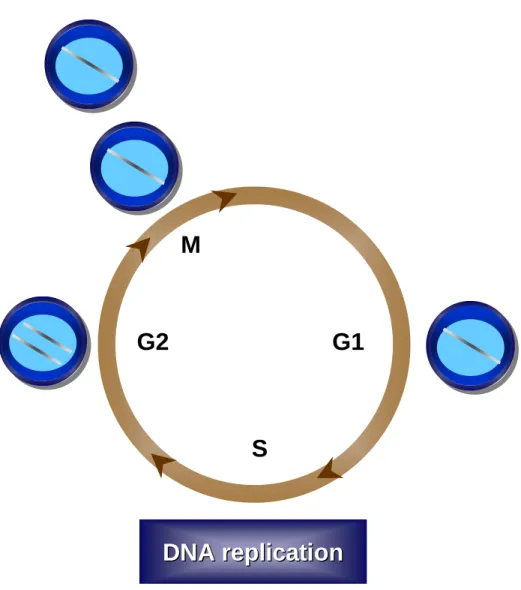
phases of the cell growth cycle (Figure 1.1). The G1 (Gap 1) phase of the cell cycle is considered the first stage of the cell cycle and it is during this phase in which the cell prepares for DNA replication including expression of many components required to initiate DNA replication. S phase is when the cell completely replicates the entire genome. The completion of S phase gives rise to the second gap phase, G2, in which a cell prepares for mitosis (M phase). During mitosis, the replicated DNA is segregated into separate nuclei, followed by cellular division resulting into two new daughter cells (reviewed in[1]).
ORIGIN LICENSING
To complete the extraordinary task of duplicating the genome, cells initiate replication at thousands of sites throughout the genome. Initiation of DNA
replication is the first committed step towards cell proliferation and requires the formation of a multi-protein complex, termed the prereplication complex (preRC) at sites of replication called origins. Construction of the preRC requires four
Formation of the preRC at the site of replication origins, termed “origin licensing”, occurs late in mitosis and early in G1. The binding of ORC (Orc1-6) to DNA is the first step towards preRC formation (Figure 1.2). Once ORC is bound to DNA its associates with the replication proteins Cdc6 and Cdt1. Cdc6 and Cdt1 bind to ORC separately; however the binding of both Cdc6 and Cdt1 to origins is required for the loading of the MCM complex onto DNA (reviewed in [2]and [3], Figure 1.2).
The origin recognition complex (ORC)
The origin recognition complex was identified in yeast for proteins binding to autonomously replicating sequence (ARS) consensus sequence (ACS). ARS
sequences have been shown to act as true origins of replication in the budding yeast chromosome. Within ARS elements is the conserved ACS sequence, consisting of 11 DNA base pairs that are required for ORC binding [4]. ORC is comprised of six subunits (Orc1-6), which are evolutionarily conserved. However, the sequence dependent DNA binding of ORC is lost in most eukaryotes, likely due to the lack of a consensus origin sequence in organisms outside of budding yeast. Additionally, ORC from fission yeast, frog, and human prefer A-T rich DNA consequences [5]. The lack of sequence specificity in ORC DNA binding is confirmed by the fact that human ORC can replace frog ORC in replication assays [5, 6].
bound state can bind to DNA, but hydrolysis of ATP by ORC is not required for ORC binding to DNA [4, 7, 8]. The ATP binding by ORC is required for the ORC-Cdc6 interaction on DNA, with ORC mutants defective in ATP binding failing to recruit Cdc6 [9-11]. In addition, ATP hydrolysis by ORC is required for the loading of the MCM complex onto DNA [12]. Further discussion on the role of ORC in the loading of the MCM complex onto DNA will be discussed later.
Interestingly, ORC ATPase activity is inhibited by single-stranded (ss) and double-stranded (ds) DNA [4]. In addition, Cdc6 can bind to ORC in vitro and influence ORC’s sensitivity to proteases, suggesting Cdc6 binding induces a
conformational change of DNA bound ORC, and activates ORC ATPase activity [11, 13, 14].
Cdc6
of ORC to replication origins, Cdc6 is recruited to ORC. The interaction between Cdc6 and ORC was originally confirmed in budding yeast through 2D gel analysis and other biochemical assays [21]. Cdc6 works in conjunction with Cdt1 to load the MCM complex onto DNA, and Cdc6 interacts with Cdt1 through its N-terminal non-catalytic domain [22]. Immunodepletion of Cdc6 frog extracts inhibits the initiation of DNA replication by interfering with MCM loading [23, 24]
Cdc6 is member of the family of AAA+ ATPases, and has strong amino acid sequence similarity to Orc1 [25, 26]. In addition, recombinant human Cdc6 has been shown to bind and hydrolyze ATP in vitro [27]. The role of Cdc6 in preRC assembly is dependent on its ability to bind and hydrolyze ATP. However, ATP binding and ATP hydrolysis have distinct roles in the functions of Cdc6, and both are required for Cdc6 function. One proposed model is that Cdc6 binds ATP after Cdc6 is bound to ORC [28]. Cdc6 mutants that cannot bind ATP are defective in S phase entry [29, 30], suggesting that ATP bound Cdc6 is required for preRC assembly through recruitment of Cdt1 and the MCM complex. Cdc6 can associate with Cdt1 and the MCM complex independent of ATP hydrolysis; however the ATPase activity of Cdc6 is required for the fixed loading of the MCM complex at replication origins. Cdc6 mutants impaired in their ability to hydrolyze ATP display defects in entry into S phase and fail to initiate replication [22, 28, 31-33].
consists of a bundle of α helices, including a winged-helix (WH) fold that suggests a DNA binding motif, and mutations to the WH domain in Cdc6 from fission yeast results in loss of DNA replication [30]. However, there has been no direct evidence that Cdc6 binds DNA directly. The dual lobe formed by domains I and II are similar to other DNA sliding clamp loading proteins [30]. Replication factor C (RFC), a AAA+ ATPase that acts a clamp loader in the loading of the DNA polymerase processitivity factor PCNA, shares structurally confirmed similarity to Cdc6 [31]. In addition, Cdc6 contains significant sequence similarity to subunits of eukaryotic and prokaryotic clamp loader proteins, such as the dnaC helicase loader, and suggests that Cdc6 may also function as a nucleotide-dependent DNA sliding clamp loader [30-32].
Cdt1
Cdt1 was originally isolated from fission yeast as a gene that was regulated by the Cdc10 transcription factor (Cdc10 dependent transcript 1) [34]. In this screen, cells carrying a null allele of Cdt1 were defective in DNA replication, suggesting an essential role for Cdt1 in DNA replication [34]. The divergence of Cdt1 sequence delayed the discovery of Cdt1 homologs in other eukaryotes, but Cdt1 is
evolutionarily conserved [35].
[36]. This observation was confirmed; when budding yeast Cdt1 mutants fail to assemble the MCM complex and initiate S phase [35]. However, in both of these studies the depletion of Cdt1 abolished the binding of the MCM complex to DNA, but the depletion of Cdt1 had no effect on ORC or Cdc6 association with origins [35, 36]. Cdt1 is required for the loading of the MCM complex, however Cdt1 has no
described enzymatic activity, and it has been proposed that Cdt1 may act as an escort by binding to the MCM complex and bringing the helicase to the origin [28]. The quantification of Cdt1 and MCM association with ORC revealed that these two proteins associated with ORC at similar levels and at an equal molar ratio [28]. This result suggests that in budding yeast that Cdt1 and the MCM complex are loaded together onto the Cdc6-ORC-origin complex [28].
The minichromosome maintenance (MCM) complex
The MCM complex is comprised of six subunits (Mcm2-7) and is conserved in all eukaryotes. The MCM complex was originally discovered in a budding yeast genetic screen for mutants that were defective for the maintenance of
minichromosomes [37, 38]. The rationale was that mutations that reduce the activity of proteins necessary for DNA replication would have more dramatic effects on minichromosomes, which have only one origin, are non-essential, and can be lost from cells.
bacterium, Methanobacteriumthermoautotrophicum, has shown that the MCM
complex exists as a dodecamer formed by two opposed ring-liked hexamers [40]. The crystal structure of the N-terminal domain of the Methanobacterium
thermoautotrophicum MCM complex confirmed the dodecamer structure was the
shape of a dumbbell and a channel large enough to include single-stranded (ss) and double-stranded (ds) DNA [41]. The crystal structure suggests that the MCM
complex wraps around the DNA, with the DNA threaded through the center. Yeast degron studies of the MCM complex revealed that all MCM mutants displayed elongation defects and stopped replication immediately, following subunit degradation at the restrictive temperature [42]. These effects are very similar to the studies done with the E.coli dnaB helicase which display the similar defectives in
elongation [43]. Like ORC and Cdc6, the MCM complex is an ATPase. The MCM complex is required for replication initiation and elongation in all eukaryotes,
suggesting the MCM complex is the replication helicase required for unwinding the DNA during DNA replication. Mcm4/6/7 sub-complexes purified from human cells display ATPase activity, the ability to bind both ssDNA and dsDNA, and weak helicase activity [44, 45]. In addition, these studies have been confirmed in yeast, frog and mouse [46-49].
raises the question of the role of these excess MCM complexes. Further insight into the “MCM paradox” has come from studies in Xenopus and human cells. These
studies have shown that the excess MCM complexes license “dormant” origins that are not required for completion of an unperturbed S phase, and dormant origins typically do not fire during S phase. However, dormant origins are activated within active replicating regions if replication fork progression is inhibited, by chemical treatments that inhibit a DNA polymerase (aphidicolin) or reduce pools of dNTPs (hydroxyurea, HU). Interestingly, replication at dormant origins is initiated despite the activation of S-phase checkpoints [58, 59]. Cells with lower MCM levels still replicate at normal rates, but when challenged with replication inhibitors HU and aphidicolin these cells had reduced rates of DNA synthesis and cell viability. Partial knockdown of MCM expression induces hypersensitivity to otherwise nontoxic levels of HU [58, 59]. These studies suggest a mechanism that cells utilize to continue DNA replication under conditions of replication stress.
mechanism of how the MCM complex is loaded onto DNA has not been completely defined and is an ongoing question in the field.
INITIATION OF DNA REPLICATION
Following the loading of the MCM complex onto DNA, preRC formation is complete and the origin is licensed for replication. In addition, once the MCM complex has been loaded onto DNA, ORC, Cdc6, and Cdt1 are no longer required for replication and to maintain the MCMs on DNA [24, 36, 53, 62]. Studies in
Xenopus and budding yeast have shown that after the loading of the MCM complex
onto DNA, replication is still initiated and the MCMs remain DNA bound even if ORC or Cdc6 have been depleted from extracts or removed from DNA with high salt [24, 36, 51, 53, 62]. Furthermore, fission yeast can still complete S phase, when Cdt1 expression is lost in early S phase arrested cells (arrest with hydroxyurea) [36]. Therefore, once the origin has been licensing the activities of ORC, Cdc6 and Cdt1 are not required to initiate and complete DNA replication.
initiation of replication, such as Cdc45, the GINS complex, and DNA polymerase α
(reviewed in [2, 3].
It has been shown in many organisms, including humans, that Cdc7 directly phosphorylates Mcm2, Mcm4, and Mcm6 [63-69]. The Cdc7-dependent
phosphorylation of the MCM complex induces a conformational change and promotes loading of the replication machinery [70, 71]. Cdc45 binds to the
phosphorylated MCM complex, travels with the replication fork, and is required for DNA elongation [72, 73]. Furthermore, Cdc45 is required to load the replication machinery, and activate the MCM helicase [70, 74]. Studies in Xenopus showed
that isolated MCM complexes displayed helicase activity, only if bound by Cdc45 [49]
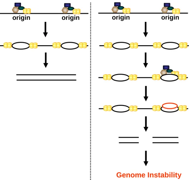
ORIGINS NEED TO BE LICENSINED ONCE DURING THE CELL CYCLE It is critical that origins are only licensed once per cell cycle. Failure to correctly control origin licensing can lead to improper preRC assembly at previously fired origins. The re-initiation of replication at origins that have already fired results in rereplication of regions of the genome (Figure 1.3). The presence of even a small amount of rereplicated DNA can have catastrophic consequences leading to
Furthermore, the expression of Cdc6 and Cdt1 are often elevated in tumors and in tumor-derived cell lines, suggesting elevated expression of Cdc6 and Cdt1 may contribute to tumorigenesis [81-84]. Additional studies have shown that mouse embryonic fibroblasts overexpressing Cdt1 formed tumors when injected into
immune-compromised mice [82]. Mice overexpressing Cdt1 in T-cells develop lymphomas when the p53 tumor suppressor is deleted (these cells develop
lymphomas at a higher frequency than in the p53 null mice) [85]. Taken together, these studies reinforce the need to restrict origin licensing to once per cell cycle and that proper regulation of preRC assembly is required to maintain genome stability.
REGULATION OF PreRC ASSEMBLY
One way in which human cells prevent origin re-licensing is through
regulation of the preRC components. During the cell cycle the activity, expression, degradation, and localization of preRC components are regulated to ensure preRC formation does not occur outside of G1, although the mechanisms for many of these events have yet to be characterized completely [77, 86-90]. Many of the
deregulated in several cancers (such as the E2F/Rb pathway [91]; and cyclins and cyclin-dependent kinases [92]).
Regulation of preRC components by the E2F transcription factor
One method by which preRC assembly is inhibited is through the regulation of the expression of preRC components. The expression of the preRC components is regulated by the E2F family of DNA-binding transcription factors (Figure 1.4A) [93-96]. The E2F transcription factor is regulated by the retinoblastoma tumor
suppressor protein (Rb). The inhibition by Rb of E2F-mediate gene transcription can occur through at least three distinct mechanisms when Rb is recruited to the E2F-regulated gene promoters. First, the Rb protein binds to the activation domain of E2F; repressing transcription of E2F controlled genes [97, 98]. Second, the recruitment of Rb to gene promoters blocks the assembly of transcription initiation complexes, possibly inhibiting the activity of adjacent transcription factors [99]. Third, Rb interacts with complexes that modify chromatin structure, serving as a bridge allowing chromatin modifying enzymes to interact with E2F-regulated promoters, thereby repressing gene transcription [100-103].
1.4A), and this mechanism restricts the expression of preRC components to times suitable for origin licensing.
Cyclin-dependent kinase (Cdk) inhibition of preRC assembly
Cdks plays a complex role in regulation of DNA replication. First, Cdk activity activates origins for replication as cells transition into S phase. Second, the same Cdk activity is required to prevent origin licensing until the subsequent G1 phase [86, 87, 89, 90]. One mechanism by which Cdks prevent rereplication is through
inhibition of preRC assembly (Figure 1.4B).
All eukaryotes rely heavily on Cdk activity to prevent rereplication. The first studies to show Cdks prevent rereplication in any eukaryote were performed in budding yeast. These studies showed that depletion of the mitotic cyclin or mitotic Cdk results in rereplication [107, 108]. Furthermore, inactivation of Cdk activity in G2/M results in yeast results in full rereplication of the genome [109, 110]. The link between inhibition of preRC assembly and Cdk activity is further supported by
results in budding yeast which indicate that elevated expression of Cdk activity in G1 prevents new preRC formation [19, 109]. Xenopus cell free in vitro replication
CDC2 (Cdk1) gene is regulated by an inducible promoter [112]. In the absence of Cdc2 expression cells underwent extensive DNA rereplication, indicating Cdc2 is required for prevention of rereplication [112]. Fluctuations of cyclin E levels are required to drive endoreduplication cycles in Drosophila [113]. Although the components of the preRC are conserved, the effects of Cdk activity on the preRC components vary among organisms [86, 89, 90, 114]. In addition, the roles by which Cdks inhibit origin licensing in metazoans in unclear.
The Cdk2 phosphorylation of the MCM complex may inhibit the ability of the MCM complex to bind ORC, Cdc6, or Cdt1 [86, 87, 89, 90, 114], while the role of Cdks in the regulation of ORC is controversial. It has been reported that the Orc1 subunit is targeted for degradation through an SCF-dependent mechanism that may depend on Cdk activity [115-117], however in some cell lines Orc1 is phosphorylated in S phase but remains stable throughout the cell cycle [118, 119]. In addition, Cdk activates Cdt1 and Cdc6 (only in yeast) for degradation, inhibits Cdc6 degradation in humans, and Cdk activity is required for the regulation of the ubiquitin ligases that regulate the activity of Cdc6 and Cdt1 either directly or indirectly [120-126]. The regulation of Cdt1 and Cdc6 by Cdks will be highlighted later in this chapter when discussing the cell cycle regulation of Cdt1 and Cdc6.
Ubiquitin-mediated protein degradation
E2, and E3) which results in the transfer of the small protein ubiquitin to a target protein. The construction of chains of ubiquitin on a target protein (called poly-ubiquitination) marks that protein for degradation through proteolysis by the 26S proteasome (reviewed in [127, 128]). The E3 ubiquitin ligase is the enzyme that catalyzes the poly-ubiquitination of a target protein, and the activity of E3 ubiquitin ligases play a critical role in regulating origin licensing. The anaphase promoting complex (APC), SCFSkp2 and Cul4DDB1 ubiquitin ligases will be discussed throughout this dissertation and are members of the RING (really interesting new gene) family ubiquitin ligases. RING family E3s commonly exist as large multi-subunit
complexes, and act as a scaffold bringing together the E2 and the target protein (Figure 1.5). SCF (Skp1 – Cullin – F-box protein) and SCF-like complexes are the largest class of ubiquitin ligases. Cullins are a closely related family of 6 proteins that bind a ring finger protein, which bind and activate E2 conjugating enzymes. The Skp protein serves as an adaptor (Skp1 or DDB1), bridging the cullin (Cul1 or Cul4) with the F-box substrate binding protein (Skp2 or Cdt2) (Figure 1.5). Unlike other classes of ubiquitin ligases, RING E3s catalyze the transfer of ubiquitin from the E2 directly to the target protein with no E3-ubiquitin intermediate (Figure 1.5) [127, 128]. The other class of ubiquitin ligases, the HECT family of ubiquitin ligases will be
discussed in Chapter 2 regarding the regulation of Cdc6.
Cell cycle regulation of Cdc6
Cdc6 is a signal for protein degradation [121-123], metazoan Cdc6 is not inhibited by Cdk activity, and Cdc6 remains bound to chromatin after origin licensing until mitosis [119, 129]. Furthermore, overexpression of cyclin A in human cells does not affect Cdc6 nuclear localization, and Cdc6 phosphorylated on serine-54 maintains a high affinity for chromatin during S phase [130]. Evidence suggests that excess Cdc6 that is not contained within preRCs is exported out of the nucleus during S phase through a mechanism dependent on Cdk activity, although there is little evidence to suggest this mechanism inhibits the re-licensing of origins [131, 132]. Therefore, in human cells it appears that unlike other components of the preRC the activity of Cdks does not inhibit Cdc6 function, and suggests that Cdc6 is regulated through a mechanism independent of Cdk activity.
(residues 56-64). The D-box is a sequence that is commonly found in APC substrates and is required for substrate ubiquitination [136]. Deletion of D-box residues 58-61 is sufficient to stabilize Cdc6 during the cell cycle [135]. In addition, Cdc6 also contains a KEN motif (consisting of the amino acid residues K-E-N) that has been described as a Cdh1-targeting motif distinct from the D-box [137]. Mutations to the KEN motif partial stabilized Cdc6, suggesting that both motifs are required for the APC-mediated degradation [135].
Interestingly, the degradation of Cdc6 is inhibited when Cdc6 is phosphorylated by cyclin E/Cdk2 [126, 138]. Mutation of all three Cdk phosphorylation sites on Cdc6 from serine to aspartic acid (mimics the
phosphorylated state, Cdc6DDD) stabilizes Cdc6 in the presence of exogenous cyclin E [126]. However, when the Cdk sites are altered from serine to alanine (mimics the unphosphorylated state, Cdc6AAA), Cdc6 is efficiently degraded in the presences of exogenous cyclin E. The N-terminal Cdk phosphorylation sites of Cdc6 reside within the D-box and KEN motifs, and APCCdh1 recognition of Cdc6 is diminished by the phosphorylation of Cdc6. Both the wild-type Cdc6 and the Cdc6 unphosphorylated mutant (Cdc6AAA) could interact with Cdh1, as measure by co-immunoprecipitation [126]. However, the association between Cdh1 and Cdc6 was lost in the phosphorylated mimic mutant (Cdc6DDD). The Cdk-mediated
Cell cycle regulation of Cdt1
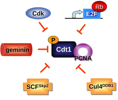
Following origin licensing Cdt1 activity is inhibited through interaction with the geminin protein (Figure 1.6) [22, 139-142]. Geminin was discovered in Xenopus
extract during a screen for substrates of the anaphase promoting complex (APC), and is an inhibitor of DNA replication [120]. In addition, a second geminin cDNA was found that has an alternative role in determination of neural cell fate during
embryonic development [143]. Furthermore, the mechanism of DNA replication inhibition by geminin is only present in metazoans [120]. Geminin is targeted for degradation as cells transition into anaphase and G1 by APC, and therefore is only present in the cell during S, G2, and early mitosis [120].
Geminin inhibits DNA replication by binding to Cdt1 on chromatin, and this interaction blocks preRC formation at previously licensed origins [120, 141, 144, 145]. Geminin inhibits Cdt1 function by blocking Cdt1 binding to both Cdc6 and Mcm2 [22]. However, the geminin protein is only present during S phase and G2 due to ubiquitination by the APC E3 ubiquitin ligase ([120] and APC regulation described above). The high expression of Cdt1 and low expression of geminin during G1, results in a small window of time during the cell cycle in which Cdt1 can participate in origin licensing. Loss of geminin in human and drosophila cells induces rereplication, due to the dysregulation of Cdt1.
component of the SCFSkp2 ubiquitin ligase (Figure 1.6). The ubiquitination of Cdt1 by the SCFSkp2 is dependent on Cdt1 phosphorylation on residue T29, and requires the cyclin binding motif in the N-terminus of Cdt1 [124, 125]. The ubiquitin mediated regulation of Cdt1 by SCFSkp2 is limited to S and G2 phases of the cell cycle due to the activity of Cdk2, and the degradation of Skp2 by APC outside of S and G2[151] .
However, inhibition of Cdk2 activity does not completely stabilize Cdt1 during S phase. Deletion of the cyclin binding (Cy) motif or mutation of T29 to alanine (to mimic the unphosphorylated state) does not stabilize Cdt1 during S phase [147]. In addition, it was also observed that Cdt1 does not accumulate in Skp2-/- mouse embryonic fibroblasts (MEFs; [152]). These observations suggest that Cdt1 stability during S phase may be controlled by more than one mechanism. A study in
Caenorhabditis elegans showed that worms lacking Cul4 fail to degrade Cdt1 during
S phase [153]. In fact, Cdt1 is degraded in Xenopus through a
of Cdt1 relies on two overlapping mechanisms, requiring the SCFSkp2 and the Cul4DDB1 ubiquitin ligases [148].
Evidence in Xenopus demonstrates only chromatin bound Cdt1 is
ubiquitinated by Cul4DDB1, and suggests the interaction between PCNA and Cdt1 may recruit Cdt1 to chromatin for ubiquitination by the Cul4DDB1 ubiquitin ligase [150, 154]. However, the requirement of Cdt1 chromatin association for ubiquitination has not been confirmed in a human model system.
In addition, studies in Caenorhabditis elegans and human cells have shown
that cells lacking Cul4DDB1, re-initiate DNA replication resulting in accumulation of double strand DNA breaks and other markers of genome instability [76, 153]. In
Xenopus, loss of the Cdt1-PCNA interaction induces rereplication [154].
Furthermore, overexpression of Cdt1 is sufficient to induce rereplication in human cells [75]. These studies suggest that the activity of Cul4DDB1 is critical to maintain the integrity of the genome.
geminin, phosphorylated and degraded, thereby preventing origin licensing until next cell cycle.
REGULATION OF ORIGING LICENISNG IN RESPONSE TO DNA DAMAGE
DNA damage inactivates Cdks
It has already been described that eukaryotes rely heavily upon Cdk activity to restrict origin licensing outside of G1, and inhibition of Cdk activity can result in the reloading of the MCM complex and origin re-licensing. However, in response to DNA damage Cdks become inactivated as part of a DNA damage response to stop cell proliferation, and to allow time to repair damage DNA [158, 159].
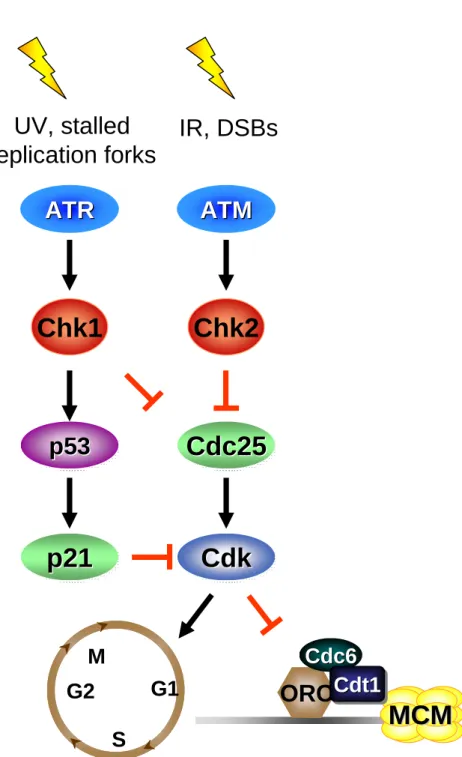
The detection of DNA damage activates a canonical DNA damage pathway requiring the ATM and ATR DNA damage kinases (Figure 1.8).
Ataxia-telangiectasia mutated (ATM) and ATM and Rad3 related (ATR) proteins activate a signaling pathway that ultimately inhibits cell cycle progression [159]. In response to DNA damage ATR and ATM are activated, resulting in the activating
Cdc25 phosphatases (Cdc25A, Cdc25B, and Cdc25C); a family of phosphatases that are required to remove an inhibitory phosphate from Cdks and activate Cdks (Figure 1.8) [161]. The ATM-Chk2 pathway is typically activated in response to formation of DNA double strand breaks. The ATR-Chk1 pathway is activated by other forms of DNA damage such as ultraviolet radiation and DNA modifying agents which result in DNA lesions and adducts which stall replication forks, a form of replication stress [158, 160, 161].
The inhibition of Cdk activity in response to DNA damage outside of G1/S removes one of the key defenses to restrict origin licensing to G1, and creates an environment favorable to re-license fired origins because of low Cdk activity (Figure 1.9). Inhibition of Cdk activity in G2 induces inappropriate MCM chromatin loading, and if Cdk activity is restored these reloaded MCM complexes are sufficient to promote rereplication [107, 162, 163]. If genome stability is to be maintained, then Cdc6 and Cdt1 must be inhibited to prevent preRC formation (MCM chromatin loading) during periods of low Cdk activity. Therefore, preRC components must be regulated throughout the cell cycle both during normal growth conditions and in response to DNA damage, through Cdk-independent pathways, to ensure genome stability is maintained. This dissertation will discuss how Cdt1 and Cdc6 are regulated in response to both exogenous and endogenous forms of DNA damage.
both ultraviolet (UV) and ionizing (IR) irradiation Cdt1 is rapidly degraded through a Cul4DDB1-dependent process (Figure 1.10A) [155, 164-167]. Like the cell cycle regulated ubiquitination of Cdt1 by Cul4DDB1, the DNA damage-induced degradation of Cdt1 requires an interaction between Cdt1 and PCNA [155, 165, 166]. PCNA is loaded onto DNA following DNA damage as part of the DNA repair process [158]. Furthermore, Cdt1 mutants defective in PCNA binding display increased stability following DNA damage [155, 165]. Studies in Xenopus have shown that Cdt1 binds
to PCNA on chromatin and the interaction between Cdt1 and PCNA is required for the ubiquitination of Cdt1 by Cul4DDB1 [150, 154]. However, no interaction between Cdt1 and chromatin has been observed in human cells following DNA damage.
Cdc6 regulation following DNA damage
The anaphase-promoting complex (APCCdh1) also targets Cdc6 for destruction in response to ionizing irradiation (IR) as part of a p53-dependent pathway. In
radiation, and therefore is unlikely the only method for Cdc6 degradation after DNA damage (Figure 1.10B) [138].
In response to treatment with the DNA alkylating agent adozelesin, Cdc6 is destroyed by a p53-independent and proteasome-dependent pathway [168]. In numerous cancers p53 is commonly inactivated; therefore a mechanism by which replication can be inhibited in cancers with deregulated p53 could be an important target for cancer therapeutics. Interestingly, the APC-independent pathway of adozelesin-induced ubiquitination and degradation of Cdc6 has not been characterized, namely the ubiquitin ligase responsible has not been identified. Moreover, the D-Box, the APCCdh1 interacting motif, is not required for the DNA damage degradation in response to treatment with adozelesin, suggesting a mechanism independent of APC [168]. Furthermore, this APC-independent mechanism for Cdc6 degradation following DNA damage is conserved in budding yeast and the plant model, Arabidopsis thaliana, suggesting a conserved
mechanism that uncouples DNA replication from the cell cycle following DNA damage [168]. In Chapter 2 I will further investigate the DNA damage-induced degradation of Cdc6 that is independent of APCCdh1. In addition to
REGULATION OF ORIGIN LICENSING DOES NOT RELY ON A SINGLE MECHANISM
The pathways that prevent re-licensing of fired origins in naive and damaged cells are not completely understood. What is clear is that cells go to great lengths to ensure replication occurs only once in a cell cycle and that improper licensing of origins after DNA damage does not occur. There are many overlapping pathways to control preRC assembly in other organisms and therefore it is likely that there is more than one mechanism to regulate a given preRC component. The existence of multiple mechanisms to inhibit rereplication suggests that any one mechanism by itself is inefficient in maintaining genome stability. Furthermore, it has been shown that perturbations to multiple regulatory mechanisms are required to induce
M
G2
S
G1
DNA replication
DNA replication
Figure 1.1. The Eukaryotic Cell Cycle.The eukaryotic cell cycle is divide into 4
origin
ORC
ORC
Cdc6
Cdc6
Cdt1
Cdt1
Cdt1
Figure 1.2. Assembly of the prereplication complex (preRC). The preRC
assembles during G1 at replication origins. PreRC formation occurs in a step-wise manner and begins with the binding of the origin recognition complex (ORC) at origins. Next, the replication factors Cdt1 and Cd6 are recruited independently to ORC, however both Cdt1 and Cdc6 must be present for the loading of the minichromosome maintenance (MCM) complex, the replication helicase. Once these four components have assembled the origin is termed “licensed” for replication.
MCM
MCM
ORC
Cdc6
Cdc6
origin
origin
origin
origin
Genome Instability
Figure 1.3. Origin re-licensing. (Left Panel) PreRCs license origins during G1, and
DNA replication is initiated as cells transition into S phase. (Right panel) Failure to restrict preRC assembly to G1 can result in the re-licensing of previously fired
ubiquitin
ligase
Cdc6
Cdc6
A. E2F-mediated transcriptional repression
ORC
P
MCM
MCM
P
Cdt1
Cdt1
Cdt1
P
Cdk
Cdk
Cdk
B. Phosphorylation by Cyclin-dependent kinase (Cdk)
C. Ubiquitin-mediated protein degradation
E2F
E2F
Rb
Rb
Cdt1
Cdt1
Cdt1
Cdt1Cdt1
Cdt1
MCM
MCM
ORC Cdc6
Cdc6
Figure 1.4. Regulation of preRC assembly. To restrict preRC formation to G1
E2
E2
E2
Skp2
Skp2
Roc
Roc
Cullin 1
Cullin 1
Ub Ub UbTarget
protein
Target
Target
protein
protein
Ub Ub UbSkp1
Skp1
A. SCFSkp2 E3 ubiquitin ligase
B. Cul4DDB1 E3 ubiquitin ligase
E2
E2
E2
Cdt2
Cdt2
Roc
Roc
Cullin 4
Cullin 4
Ub Ub UbTarget
Protein
Target
Target
Protein
Protein
Ub Ub UbDDB1
DDB1
Figure 1.5. Protein ubiquitination by RING Finger E3 ubiquitin ligases.
APC
Cdh1APC
APC
Cdh1Cdh1Mitosis/G1
E2F
E2F
Rb
Rb
Cdc6
Cdc6
Figure 1.6. Cell cycle regulation of Cdc6. The replication factor Cdc6 is
Cdt1
Cdt1
Cdt1
P
Cdk
Cdk
Cdk
geminin
geminin
E2F
E2F
Rb
Rb
PCNA
PCNA
PCNA
Cul4
DDB1Cul4
Cul4
DDB1DDB1SCF
Skp2SCF
SCF
Skp2Skp2Figure 1.7. Cell cycle regulation of Cdt1. The replication factor Cdt1 is
ATR
ATR
ATM
ATM
Chk1
Chk2
Cdk
Cdk
Cdk
Cdc25
Cdc25
Cdc25
p53
p53
p53
p21
p21
p21
UV, stalled
replication forks
IR, DSBs
Figure 1.8. The DNA damage checkpoint pathway. In response to various
forms of DNA damage the ATR/ATM checkpoint pathway is activated to halt cell cycle progression enabling the cell to repair any damage and prevent the
accumulation of more damage to the genome.
G1
S
G2
M
MCM
MCM
ORC
Cdc6
Cdc6
Cdt1
Cdt1
Cyclin
Cyclin
Cyclin
Cdk
Cdk
Cdk
DNA Damage
Cell cycle progression
Origin re-licensing
Figure 1.9. DNA damage removes a major mechanism to restrict preRC
assembly. Activation of the DNA damage checkpoint results in the inhibition of
Cdt1
Cdt1
Cdt1
Cul4
DDB1Cul4
Cul4
DDB1DDB1PCNA
PCNA
PCNA
APC
Cdh1APC
APC
Cdh1Cdh1?
?
?
other forms of
DNA damage
Cdc6
Cdc6
Ionizing
radiation
B. A.
Figure 1.10. The regulation of Cdt1 and Cdc6 in response to DNA
damage. (A). Cdt1 is targeted for degradation in response to DNA damage
through a mechanism dependent on the Cul4DDB1ubiquitin ligase and an
interaction between Cdt1 and PCNA. (B).In response to ionizing radiation, Cdc6 is targeted for degradation through a mechanism that requires the anaphase promoting complex (APCCdh1). However, Cdc6 is ubiquitinated
and degraded in response to other forms of DNA damage through a
mechanism that is independent of APCCdh1. In addition Cdc6 is targeted for
destruction by caspase-mediated cleavage.
caspase
Chapter 2
Cdc6 stability is regulated by the Huwe1 ubiquitin ligase after DNA damage
Modified from J.R. Hall, Kow, E., Nevis, K.R., C.K. Lu, K.S. Luce, Zhong, Q., and J.G. Cook. Molecular Biology of the Cell. September 2007, Volume 18, pages 3340-3350.
INTRODUCTION
Duplication of large mammalian genomes requires that DNA replication initiate at thousands of chromosomal origins. In order for an origin to be competent for replication, it must first be bound by a multi-protein complex, the prereplication complex (preRC). PreRCs are constructed in a stepwise process through the
chromatin binding of the origin recognition complex (ORC), which then recruits both the Cdc6 ATPase and Cdt1, two proteins that are required for the stable loading of the minichromosome maintenance complex (MCM). The Cdc6 and Cdt1-dependent loading of MCM complexes at origins licenses them for replication during the G1 phase of the cell cycle. Sufficient preRCs must be assembled during G1 to promote complete replication, but new preRCs must not assemble after S phase begins because re-licensing of previously fired origins leads to rereplication and genome instability [75, 80, 172, 173]. For these reasons, preRC assembly is one of the most highly regulated events in the control of DNA replication. Cells restrict preRC
assembly to the G1 period through a combination of overlapping mechanisms that regulate individual preRC components (reviewed in [3, 96, 174-176]).
addition, the high levels of Cdc6 protein observed in multiple cancers may contribute to cell cycle regulation defects and genome instability that consequently promote tumor progression [81, 178-181].
Cdc6 is subject to multiple forms of regulation, including both transcriptional and posttranscriptional mechanisms. The human CDC6 gene is regulated by the Rb-E2F transcriptional program that results in peak CDC6 mRNA levels in late G1 [93, 182, 183]. The Cdc6 protein is degraded each cell cycle in early G1 as a
consequence of ubiquitination by the Cdh1-activated form of the anaphase promoting complex (APC), a cell cycle–regulated ubiquitin E3 ligase [135]. Ubiquitination of Cdc6 by APCCdh1 is regulated not only by cell cycle–dependent fluctuations in APC activity, but also by phosphorylation of Cdc6 by cyclin-dependent kinases, most notably cyclin E/Cdk2. Cdk2-mediated phosphorylation blocks the association of Cdc6 with the Cdh1 protein, thus stabilizing Cdc6 when Cdk2 is active [126, 138] .
Cdc6 is also ubiquitinated and degraded in response to DNA damage [138, 168]. Cells may eliminate Cdc6 to reduce the possibility of rereplicating DNA or to promote checkpoint functions that block mitosis with damaged DNA. One
adozelesin or methyl methane sulfonate (MMS; [168]). Importantly, APC activity itself is inhibited during S phase and G2, times when it may be particularly important to regulate Cdc6 in order to prevent rereplication or to promote checkpoint
activation. These observations implicate an as yet unidentified APC-independent pathway for degradation of Cdc6. We sought to determine this p53- and APC-independent mechanism of Cdc6 degradation, and we report here our finding that Cdc6 stability after DNA damage is controlled by a novel ubiquitin ligase, Huwe1.
MATERIAL AND METHODS
Growth and manipulation of cells
Small interfering RNA
Small interfering (siRNA) targeting Huwe1 (5'-GAGUUUGGAGUUUGUGAAGTT-3'),human Cdh1
(5'-UGUGAAGUCUCCCAGUCAGTT-3'), and the negative controlgreen fluorescent protein (GFP; 5'-GGCUACGUCCAGGAGCGCACCTT-3')were synthesized by Invitrogen and transfected at a final concentrationof 100 nM using Dharmafect Reagent 1 (Dharmacon). DDB1, Cul4A, and Cul4B siRNA were described in Hu et al [164].;geminin siRNA was described in Ballabeni et al. [186].
Antibodies and immunoblots
Anti-Cdc6 (sc-9964), Anti-Cdc6 (D-1), anti-cyclin A (C-19),anti-c-Myc (sc-40), anti-hemagglutinin (Y-11), anti-p53 (D01),anti-ScMcm2 (yN-19), anti-geminin (FL-209) were purchased fromSanta Cruz Biotechnology, anti-cyclin B1 (V152)from Lab Vision, anti-Cdh1 (DH01) from Biomeda, Orc2 from BD PharMingen, and anti-tubulin (DM1A) from Sigma. Phosphospecific antibodiesto p53 phosphorylated on Ser15 and Chk2 phosphorylated on T69were purchased from Cell Signaling Technologies. Anti-Huwe1 (anti-Lasu1, BL671) was purchased from Bethyl Laboratories(for immunoprecipitations) or was the gift ofS. Wing (McGill
National Institutes of Health, Bethesda, MD; http://rsb.info.nih.gov/ij/,1997–2006). The ratio of Cdc6 signal to tubulin signal(after background subtraction) is reported as the mean of twoor more experiments.
Two-hybrid screen2
The Gal4 DNA-binding domain was inserted at the carboxy terminusof human Cdc6 through a PCR strategy and expressed from theGPD promoter in plasmid p2U (gift of D. Picard, Universitéde Genève). Cotransformants of strain PJ69a with one of two cDNA fusion libraries (placentalcDNA or thymus cDNA, Clontech) were selectedon medium containing 3 mM 3-amino-1,2,4 triazole (Sigma). Morethan 8 million co-transformants of each library were screened;Huwe1 clones were identified from both libraries.
Plasmids and viruses
expressesa constitutively cytoplasmic protein that is stable in quiescentcells (Cook, unpublished observations). Cdc6WT bears the sameNLS fusion.
In vitro protein-binding assay
GST-Huwe1C was produced in BL21(DE3) purified on glutathione-Sepharose (GE Healthcare, Waukesha, WI) and incubated with cell lysatesprepared in buffer 1 (50 mM HEPES, pH 7.2, 33 mM potassium acetate,0.5 mM EDTA, 0.5 mM EGTA, 0.1% Nonidet P-40, 10% glycerol, andprotease and phosphatase inhibitors)
essentially as describedin Cook et al. [22]. GST-hCdc6 was produced by infection of SF21 insect cells with GST-hCdc6 baculovirus and purificationof GST-hCdc6 was performed as described in Herbig et al. [27].
Co-immunoprecipitation
Chromatin fractionations
Nuclei were prepared from whole cell lysates by resuspenation of HeLa cells in CSK buffer (10 mM PIPES pH 7.0, 100 mM NaCl, 200 mM sucrose, and 3 mM MgCl2) supplemented with 0.5% Triton-X100, 10 mM AEBSF (CalBiochem), 1 µg/ml pepstatin A (Sigma), 1 µg/ml leupeptin (Sigma), 1 µg/ml aprotinin (Sigma), 0.5 mM sodium orthovanadate (Sigma), 1 mM glycerol 2-phosphate (Sigma), 10 µg/ml phosvitin (Sigma) and 1 mm ATP (Sigma) followed by low-speed centrifugation. Micrococcal nuclease (Roche) digestion was performed at 37°C for 5 minutes to release chromatin bound proteins.
In vitro ubiquitination assay3
RESULTS
An APC-independent mechanism for Cdc6 degradation
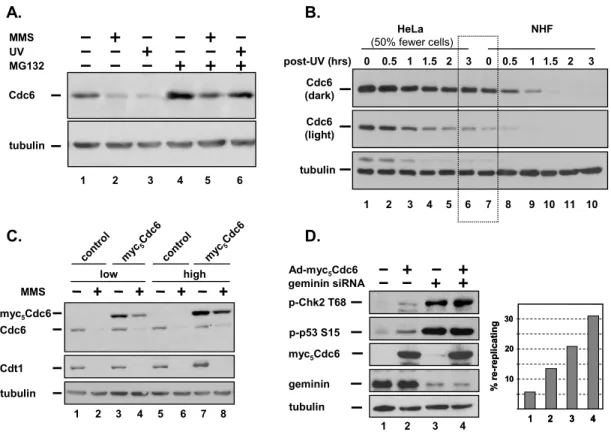
To explore the regulation of Cdc6 after DNA damage, we treatedan
asynchronous population of HeLa cells with MMS or with UVirradiation. Similar to previous results (Blanchard et al.,2002), both kinds of DNA damage resulted in
significant lossof endogenous Cdc6 (Figure 2.1 A, compare lanes 2 and 3 with lane1), and the degradation was sensitive to proteasome inhibitionby MG132 (Figure 2.1A, lanes 4–6). Under these conditions,we did not observe the caspase cleavage product of Cdc6 afterDNA damage (data not shown); this cleavage normally occurs 12h after induction of apoptosis [170].
We next compared the kinetics of Cdc6 degradation in HeLa cellsand in normal human fibroblasts (NHF; Figure 2.1B4) to determineif differences in Cdc6 regulation might contribute—alongwith deregulated transcription—to the high levels of Cdc6
replication; thus cancer cells with highlevels of Cdc6 have a relatively long period of abundant Cdc6after DNA damage.
We specifically tested if Cdc6 overproduction could suppressDNA damage– induced Cdc6 degradation by overwhelming thecapacity of the cells to target the excess protein. We infectedU-2OS cells with either control adenovirus or virus-producingCdc6 at either moderate doses or high doses and then treatedcells with MMS to induce Cdc6 degradation. Both the ectopicand endogenous Cdc6 proteins were detected by immunoblot analysis.At the lower viral dose, ectopic Cdc6
accumulated to three timesthat of endogenous Cdc6 as estimated by densitometry (data notshown), and though it was reduced by DNA damage, some ectopicCdc6 protein still remained (Figure 2.1C, lanes 3 and 4). Increasingthe level of ectopic Cdc6 further to approximately six timesthat of endogenous Cdc6 significantly impaired the ability ofthese cells to degrade Cdc6 in the presence of MMS, but had no effect on the DNA damage–induced degradation of Cdt1(Figure 2.1C, lanes 7 and 8).
To determine if high levels of Cdc6 can induce rereplication,we overproduced Cdc6 in cells arrested in G2. Extensive rereplicationis associated with an activated cell cycle checkpoint characterizedby phosphorylation of both the p53 tumor
suppressor and theChk2 protein kinase. These markers are strongly induced by depletionof geminin, a negative regulator of preRC assembly that inhibitsCdt1 (Zhu
endogenousCdc6 (data not shown). High levels of Cdc6 induced phosphorylationof both Chk2 and p53 (Figure 2.1D, compare lanes 1 and 2). Moreover,Cdc6
overproduction was sufficient to cause rereplication detectableas an increase in the number of cells with greater than 4C DNAcontent (Figure 2.1D). Depletion of
geminin caused robust rereplication as had been reported byothers [80, 189, 190], and the combination of highlevels of Cdc6 with geminin depletion increased the number ofrereplicating cells even further (Figure 2.1D, numbers 3 and 4). We thus conclude that Cdc6 de-regulationcan promote rereplication, and that the
degradation of bothCdc6 and Cdt1 after DNA damage may be important for maintaininggenome integrity.
Because persistent Cdc6 might contribute to genome instabilitywe sought to understand the mechanism of DNA damage-inducedCdc6 degradation. To
determine if Cdc6 degradation is affectedby cell cycle stage, we synchronized U-2OS cells at G2/M bysequential treatment with thymidine and nocodazole followed by release from the arrest; cell cycle position was confirmedby flow cytometry (2.2A). In undamaged cellsendogenous Cdc6 protein was low in G1 and
accumulated throughoutS phase and G2, as expected. Strikingly, DNA damage– inducedCdc6 degradation at every stage of the cell cycle despite thepresumed inhibition of APC after the G1/S transition (Figure 2.2B).
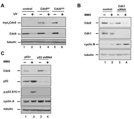
Cdc6 and sensitizing Cdc6 to APC, thenthese aspartic acid substitutions would prevent Cdc6 degradation.To test this possibility, we constructed adenoviral vectors to produce myc-epitope–tagged versions of both normalCdc6 (WT) or Cdc6 in which the Cdk target sites, serines 54,74, and 104, were altered to aspartic acid, Cdc6S3D 5
. Low dosesof these viruses were used to infect HeLa cells such that thelevel of ectopic Cdc6 was less than endogenous Cdc6. UV irradiationcaused the
degradation of not only the endogenous Cdc6 (Figure 2.3A,lanes 1 and 2), but also the ectopically expressed Cdc6 (Figure 2.3A,lanes 3 and 4). Moreover, the S3D mutant showed equal susceptibilityto UV-induced degradation (Figure 2.3A, lanes 5 and 6), indicatingthat phosphorylation at these serine residues does not controlDNA damage-induced Cdc6 degradation.
Cdc6 ubiquitination by APC requires the targeting subunit, Cdh1[135, 138]. It was possiblethat during a DNA damage response, Cdc6 became more APC-sensitive by some mechanism other than dephosphorylation at the knownCdk sites. To
definitively demonstrate that Cdc6 degradationcan occur independently of APCCdh1, we eliminated Cdh1 in cellsby siRNA transfection. Effective knockdown of Cdh1 was confirmedby immunoblot analysis and the resulting accumulation of cyclinB (Figure 2.3B). Despite the depletion of Cdh1, Cdc6 degradationafter MMS treatment was unaffected (Figure 2.3B, compare lanes2 and 4). APC-mediated ubiquitination of Cdc6 after ionizingradiation requires p53 [138], but we observedno difference between
factthat naive p53-deficient cells have higher endogenous Cdc6 levels,presumably because of the combined effects of the p53-dependentderegulation of Cdks on both E2F-dependent CDC6 transcription[93, 191]and APC-dependent Cdc6 protein stability during G1 [126, 138]. We thus concludethat DNA damage induces Cdc6 degradation regardless of cellcycle position, APC activity, or p53 status.
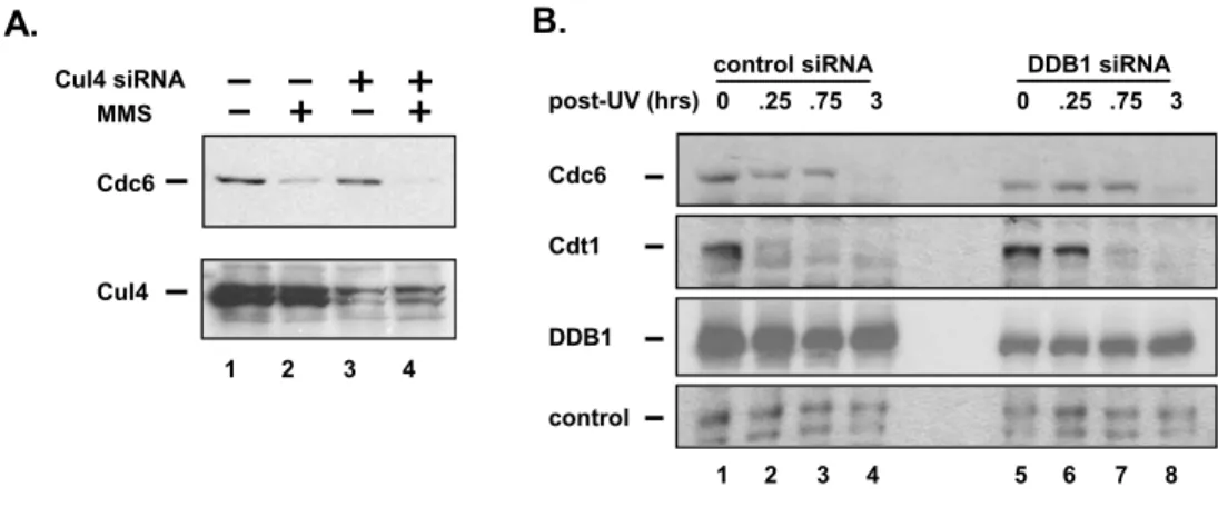
Like Cdc6, Cdt1 is ubiquitinated and degraded after DNA damage.
Ubiquitination of Cdt1 under these conditions is dependent onthe Cul4DDB1 ubiquitin ligase [164, 167]; thus it was possible that Cul4 also ubiquitinated Cdc6after DNA damage. However, cells treated with siRNAs targetingboth Cul4A and Cul4B
showed no difference in the degradationof Cdc6 after MMS treatment (Figure 2.4A, compare lanes 1 and2 with lanes 3 and 4). Reduction of DDB1 by a similar siRNA strategy significantly delayed the normally rapid destructionof Cdt1 (Figure 2.4B, compare lanes 2 and 6), though the remainingDDB1 was capable of eliminating Cdt1 by later time points. (Thishypomorphic phenotype is typical of DDB1-depleted cells; J.Hu and Y. Xiong, personal communication.) Importantly, UV-irradiatedcells in which DDB1 is reduced by siRNA treatment degraded Cdc6with the same kinetics as control cells (Figure 2.4B, comparelanes 1–4 with lanes 5–8). We thus
hypothesizedthat an entirely different ubiquitin ligase plays an importantrole in Cdc6 ubiquitination and degradation in response to DNAdamage.
member of the HECT familyand can ubiquitinate the Mcl-1 anti-apoptotic protein, corehistones, the c-myc transcription factor, and the p53 checkpointmediator. These reports named the enzyme Mule [188, 192], Lasu1 [187], HectH9 [193], and ARF-BP1 [194], respectively.We use the official gene name, HUWE1 for "HECT, UBA and WWEdomain containing 1" (gene ID 10075) and refer to the proteinas Huwe1. All of our two-hybrid isolates contain portions ofthe carboxy-terminal
catalytic domain of Huwe1 (Figure 2.5C).Interestingly, this sequence is distinct from both the reportedmyc-interaction region of Huwe1 just amino-terminal to the HECT domain [193] and the BH3 motif at amino acids1972–1999, which binds Mcl-1 [188, 192]. We constructed derivatives of the Cdc6 two-hybridfusion in which increasing amounts of the amino terminal (non-catalytic)domain were deleted and tested them for interaction with theHuwe1 fusions. Removal of as many as the first 154 amino acids,but not the first 192, from Cdc6 had no effect on the two-hybridinteraction of Cdc6 with Huwe1, demonstrating that this regionis dispensable for the Cdc6-Huwe1 interaction (Figure 2.5B). Wenote that the catalytic domain of Cdc6 is the most highly conserveddomain and is included in the constructs that retain bindingto the most highly conserved domain of Huwe1.
We confirmed that Cdc6 and Huwe1 interact biochemically. Forthis purpose, we produced a fusion of GST to the c-terminaldomain of Huwe1 (amino acids 3987–4374 "GST-Huwe1C") inEscherichia coli. Glutathione beads coated with GST or
insect cellsco-precipitates endogenous full-length Huwe1 from HeLa cell extracts (Figure 2.6B, compare lanes 2 and 3). To determine if these proteinscan bind in the absence of other cellular proteins (human oryeast), we mixed purified recombinant Cdc6 with glutathionebeads coated with purified recombinant GST-Huwe1C. Cdc6 was specifically retained on these beads, indicating that thesetwo proteins can interact directly (Figure 2.6C).
Finally, we tested if Huwe1 and Cdc6 associate when they areexpressed at their endogenous levels. To determine if Huwe1and Cdc6 interact during a DNA damage response, we irradiatedcells with UV (or left them untreated) and then added MG132to block the degradation of Cdc6. We prepared nuclear lysatesfrom HeLa cells and released Cdc6 from chromatin with nuclease.These extracts were subjected to immunoprecipitation with anti-Huwe1antibodies and then probed for endogenous Cdc6. Cdc6 was foundin Huwe1 immunoprecipitates both in the absence and in the presenceof DNA damage (Figure 2.6D, lanes 5 and 6).
If Huwe1 is the ubiquitin ligase that controls Cdc6 degradation,then Huwe1 should be able to ubiquitinate Cdc6. We thereforetested the ability of full-length (492 kDa) recombinant Huwe1purified from insect cells [188] to polyubiquitinate
Huwe1 (Figure 2.6E, comparelanes 2–4). These results suggest that Huwe1 binds Cdc6for the purpose of catalyzing Cdc6 ubiquitination and that Huwe1may play a role in Cdc6 stability in cells.
Huwe1-dependent regulation of Cdc6 stability in cells
chromatin bound Cdc6 was almost undetectable (Figure 2.7A, compare lanes 8 and 14). Importantly, Huwe1 itself is not appreciably associated with chromatin either before or after DNA damage (Figure 2.7A, lanes 8–14). These data suggest that Cdc6 is released from chromatin after DNA damage into the soluble fraction where it can associate with the soluble Huwe1. This mechanism of induced interaction was not detectable in the co-immunoprecipitation experiments in Figure 2.6D because Cdc6 required solubilization by nuclease digestion of the lysates before
immunoprecipitation.
To test if Huwe1 is required for the degradation of Cdc6 incells, we designed siRNA molecules to target the Huwe1 mRNA.Cells transfected with Huwe1 siRNA showed significant knockdownof endogenous Huwe1 protein after 48 h (Figure 2.7B, middle panel,compare lanes 1–3 with lanes 4–6). Asynchronouscells
transfected with control siRNA rapidly degraded Cdc6 afterDNA damage induced by UV, but cells with reduced Huwe1 did soconsiderably less efficiently (Figure 2.7B, top panel, comparelanes 1–3 with lanes 4–6). This result is similarto the effects of DDB1 knockdown on Cdt1 stability after DNAdamage (Figure 2.4B) in that Huwe1-depleted cells are hypomorphicfor Cdc6 degradation.
If reduction of Huwe1 induced a delay or arrest in G2, thenCdc6 levels might simply have been higher as a consequence ofan indirect cell cycle effect. However, duplicate cultures treatedwith Huwe1 siRNA and analyzed by flow cytometry
apparent changesin cell cycle distribution compared with control cultures evenat 5 days after transfection (data not shown). The growth arrestis not associated with robust changes in Cdc6 (data not shown),but may be explained by deregulation of other Huwe1 substratessuch as c-myc and Mcl-1 [188, 192-194]. We thus conclude that the increased Cdc6 in UV-treated cells as a consequenceof the loss of Huwe1 is not a reflection of a cell cycle arrest,but rather a more direct effect on steady-state Cdc6 abundance.
Two-thirds of the asynchronous Huwe1-depleted cells in Figure 2.7B were in G1 (Figure 2.7C). Presumably these cells contained activeAPC, which may have contributed to Cdc6 ubiquitination in theabsence of Huwe1. To focus specifically on the APC-independentdegradation of Cdc6, we arrested cells in S phase by
treatmentwith thymidine. In thymidine-arrested cells in the absence ofDNA damage, Cdc6 is stable because APC in inactive. This assertionis supported by the
observation that treatment of S phase cellswith the proteasome inhibitor MG132 had no effect on Cdc6 abundance. In response to DNA damage,however, S phase cells degraded Cdc6 after UV or MMS treatment(Figure 2.7D, compares lanes 2 and 3 to lane 1). In marked contrast,we observed much less degradation of Cdc6 after DNA damage causedby either UV or MMS in Huwe1-depleted cells (Figure 2.7D,
comparelanes 5 and 6 to lane 4). Reduction of Huwe1 with a differentsiRNA molecule had the same effect (data not shown).