ASSOCIATIONS BETWEEN THE BIOLOGICAL, STRUCTURAL, AND MECHANICAL COMPONENTS OF CARTILAGE HEALTH FOLLOWING ACUTE LOADING IN
HEALTHY PARTICIPANTS
Matthew S. Harkey
A dissertation submitted to the faculty of The University of North Carolina at Chapel Hill in partial fulfillment of the requirements for the degree of Doctor of Philosophy in the
Curriculum of Human Movement Science in the School of Medicine.
Chapel Hill 2017
ABSTRACT
Matthew S. Harkey: Associations Between the Biological, Structural, and Mechanical Components of Cartilage Health following Acute Loading in Healthy Participants
(Under the direction of Brian Pietrosimone)
Context: A systems-based approach to assess cartilage health uses an acute bout of joint loading to signal acute changes in cartilage structure and metabolism. Evaluating how multiple systems, specifically cartilage structure and metabolic outcome measures, are influenced by loading may be a more sensitive measurement approach to
COMP. Results: US provides a reliable and precise modality for detecting the in vivo cartilage deformation and recovery response following walking and drop-landing, but the majority of US measures are not associated with lower extremity loading. COMP
increases following walking and drop-landing in healthy individuals, but these changes are not associated with lower extremity loading measures. While the majority of
ACKNOWLEDGEMENTS
To my advisor Brian Pietrosimone, it has been a long time coming. We are finishing up our 7th year together, and I could not have dreamed it up any better. I don’t know how you did it, but you took an unmotivated kid straight out of undergrad and inspired and pushed me for seven years to get me to the point where I am excited to come in to work and learn something new every day. I will never be able to let you know how much it meant for you to put your faith in Brittney and I when you brought us with you from Toledo. Your passion for what you do and your ability to make people excited about your research is remarkable, and I would not be anywhere if it was not for your guidance over this last 7 years.
To my committee member Randy Schmitz, thank you for you your help and experience with the ultrasound technique. We would not have been able to design and complete this project.
To my committee member Mike Lewek, thank you for your help with the
biomechanical instrumentation and the instruction you provided in your LabView class. To my committee member Anthony Hackney, thank you for the countless hours you spent teaching us how to how to draw blood, not to mention you sacrificing your arms during the phlebotomy training. Also, thank you for the long days in the biochem lab helping run our assays.
knowledge for the biomechanics, ultrasound, LabVIEW, and basically anything else regarding how anything in the lab worked.
To my research assistants, you devoted hours upon hours of your time helping me collect and process the data for this project. This project definitively could not have happened without each and every one of yours help. (Daniel Farrell, Gabrielle Smith, Nicole Thomas, Arianna Douglas, Ryan Fockler, Leslie Sierra-Arevalo, Kyle Wolfe, Samantha Hammock).
To my funding sources from my last year, thank you for the funding you provided over the last year to allow me to successfully complete my dissertation. To the UNC HMSC program, thank you for selecting me as the recipient of the Duquette
Scholarship. To the Royster Society of Fellows, thank you for selecting me as a
recipient of a Dissertation Completion Fellowship. To the NATA Research & Education Foundation, thank you for selecting me as a recipient of a Doctoral Scholarship.
To all the other doctoral students, thank you for helping me successfully navigate through the program. Whether it was struggling in class, figuring out stuff in the lab, or just having a little fun. This process was a lot easier and a lot more fun having all of you around to lean on.
Last but not least, to Dr. Brittney Harkey, I would not have been able to make it through this without you there every step of the way. One of the most common
TABLE OF CONTENTS
LIST OF TABLES ... XI
LIST OF FIGURES... XII
CHAPTER I ... 1
1.1INTRODUCTION ... 1
1.2 STATEMENT OF PURPOSE... 6
1.3 SPECIFIC AIMS ... 7
Specific Aim 1 - Cartilage Ultrasound and Biomechanics ... 7
Specific Aim 2 - Cartilage Metabolism Biomarkers and Biomechanics ... 8
Specific Aim 3 - Cartilage Ultrasound and Cartilage Metabolism Biomarkers ... 8
1.4 OPERATIONAL DEFINITIONS ... 9
CHAPTER II ... 10
2.1 PATHOGENESIS OF OSTEOARTHRITIS DEVELOPMENT ... 10
Epidemiology of Knee Osteoarthritis ... 10
Post-traumatic Osteoarthritis Pathogenesis ... 10
Anatomy of Articular Cartilage ... 12
2.2 STRUCTURAL COMPONENT OF CARTILAGE HEALTH ... 14
Problems with Current OA Management ... 14
Evidence of MRI Cartilage Alterations Following ACL Injury and
Reconstruction – A Model for Structural Post-traumatic Osteoarthritis
Development... 18
Musculoskeletal Ultrasound Imaging and Cartilage Health ... 21
Limitations of the Structural Component of Cartilage Health ... 24
2.3 BIOLOGICAL COMPONENT OF CARTILAGE HEALTH ... 24
Biomarkers of Osteoarthritis... 25
Cartilage Oligomeric Matrix Protein as a Biomarker of Osteoarthritis ... 28
Osteoarthritis-Related Biomarkers Following ACL Injury and Reconstruction - A Model for Biological Post-traumatic Osteoarthritis Development ... 29
Cartilage Compositional Imaging as a Measure of Cartilage Biology ... 30
Declines in Cartilage Composition Following ACL Injury ... 31
Use of Ultrasound as a Cartilage Compositional Imaging Modality ... 32
2.4 MECHANICAL COMPONENT OF CARTILAGE HEALTH ... 33
2.5 INTERACTION BETWEEN MECHANICS AND CARTILAGE STRUCTURE – CARTILAGE DEFORMATION FOLLOWING ACUTE DYNAMIC LOADING ... 37
2.6 INTERACTION BETWEEN MECHANICS AND CARTILAGE BIOLOGY – CHANGE IN COMP CONCENTRATION AND COMPOSITIONAL IMAGING FOLLOWING ACUTE DYNAMIC LOADING ... 42
2.7 INTERACTION BETWEEN MECHANICS, BIOLOGY, AND STRUCTURE – PROVIDING THE LINK BETWEEN THE MAIN COMPONENTS OF CARTILAGE HEALTH ... 45
CHAPTER III ... 48
3.1 OVERVIEW: AIMS 1-3 ... 48
3.2 PARTICIPANTS: AIMS 1-3 ... 48
Research Design ... 50
Screening Session ... 51
Data Collection Overview ... 51
Ultrasonographic Assessment of the Femoral Articular Cartilage ... 52
Ultrasonographic Image Acquisition ... 52
Ultrasonographic Imaging Processing ... 53
Quantifying Cartilage Metabolism ... 55
Blood Sample Collection ... 55
Analysis of Serum Cartilage Oligomeric Matrix Protein ... 56
Biomechanical Assessment of Lower Extremity Loading ... 56
Participant Preparation for Biomechanical Assessment ... 56
Walking Biomechanical Assessment ... 57
Drop-Landing Biomechanical Assessment ... 58
Analysis of Lower Extremity Loading ... 58
Joint Center Calculations and Alignment ... 60
Kinematic Calculations ... 60
Kinetic Calculations ... 60
Biomechanics Data Reduction ... 61
Walking, Drop-Landing, and Control Conditions ... 61
Walking Condition ... 61
Drop-Landing Condition ... 62
Control Condition ... 62
Aim 1: Cartilage Ultrasound and Biomechanics ... 62
Aim 2: Cartilage Biomarkers and Biomechanics ... 64
Aim 3: Cartilage Ultrasound and Cartilage Biomarkers ... 66
CHAPTER 4: RESULTS ... 68
SPECIFIC AIM 1 RESULTS - CARTILAGE ULTRASOUND AND BIOMECHANICS ... 68
SPECIFIC AIM 2 RESULTS - CARTILAGE BREAKDOWN BIOMARKERS AND BIOMECHANICS ... 73
SPECIFIC AIM 3 RESULTS - CARTILAGE ULTRASOUND AND CARTILAGE BREAKDOWN BIOMARKERS ... 74
CHAPTER 5 - MANUSCRIPT 1 ... 77
OVERVIEW ... 77
INTRODUCTION ... 79
METHODS ... 82
RESULTS ... 94
DISCUSSION ... 100
CHAPTER 6 - MANUSCRIPT 2 ... 115
OVERVIEW ... 115
INTRODUCTION ... 116
METHODS ... 119
RESULTS ... 129
DISCUSSION ... 131
CHAPTER 7 - MANUSCRIPT 3 ... 143
INTRODUCTION ... 144
METHODS ... 147
RESULTS ... 156
LIST OF TABLES Manuscript 1 Tables
Table 1: Demographics ... 107 Table 2: Reliability, Precision, and Minimal Detectable Change ... 108 Table 3: Ultrasonography Outcome Measure Percent Change Scores
Following Activity Conditions ... 109 Table 4. Ultrasonographic Cartilage Outcome Measure Response and
Lower Extremity Loading during Walking and Drop-Landing Conditions ... 110
Manuscript 2 Tables
Table 5: Demographics ... 136 Table 6: Baseline and COMP Response Following Activity Conditions ... 137 Table 7: Association between COMP Response and Lower Extremity
Loading During Activity Conditions ... 138 Table 8: Differences in Lower Extremity Loading Between Increased
and Decreased COMP Responders ... 139
Manuscript 3 Tables
Table 9: Demographics ... 162 Table 10: Baseline, Post, and Percent Change of COMP and US Measures ... 163 Table 11: Associations Between Baseline COMP and Baseline US Measures ... 165 Table 12: Associations between COMP Percent Change and US Measure
LIST OF FIGURES
Figure 1: Anatomy of Articular Cartilage ... 13
Figure 2: Radiographic Signs of Osteoarthritis ... 15
Figure 3.Morphological MRI Processing ... 16
Figure 4. Subcompartmentalization of Cartilage ... 17
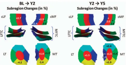
Figure 5. Baseline to Year 2 and Year 2 to Year 5 thickness changes ... 21
Figure 6. Microscopic Stages of OA ... 25
Figure 7. Continuum of OA Stages... 26
Figure 8. Hypothetical Development of OA Biomarkers ... 26
Figure 9: Knee adduction moment and OA Severity ... 36
Figure 10: Cartilage Response to the Knee Adduction Moment During Gait ... 36
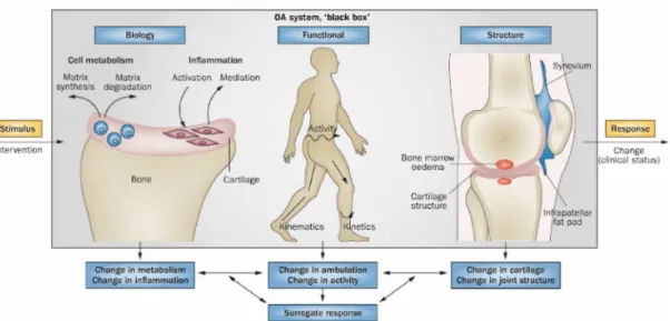
Figure 11: Utilizing a system-based stimulus-response model to determine the interaction of components of cartilage health. ... 37
Figure 12. Pre-clinical stages of OA ... 38
Figure 13. Comparison of Cartilage Deformation following drop landing and running ... 41
Figure 14: Maintenance of Healthy Cartilage ... 45
Figure 15: Maintenance of Cartilage Health displayed as a Slot Machine ... 46
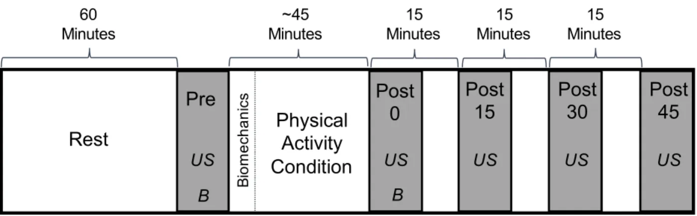
Figure 16: Within study design for the walking, drop-landing, and control Conditions ... 50
Figure 17: Femoral Cartilage Ultrasonography Setup and Participant Positioning ... 55
Figure 18: Femoral Cartilage Ultrasonography Outcome Measures ... 55
Manuscript 1 Figures
Figure 20: Study Design ... 111 Figure 21: Femoral Cartilage Ultrasonography Setup and Participant
Positioning ... 112 Figure 22. Femoral Cartilage Ultrasonography Outcome Measures ... 113 Figure 23. Walking and Drop-Landing Biomechanical Assessment Setup ... 114
Manuscript 2 Figures
Figure 24: Study Design ... 140 Figure 25: Walking and Drop-Landing Biomechanical Assessment Setup ... 141 Figure 26: Associations between Walking COMP Response and
Drop-Landing COMP Response ... 142
Manuscript 3 Figures
Figure 27: Study Design ... 166 Figure 28: Femoral Cartilage Ultrasonography Setup and Participant
CHAPTER I 1.1 Introduction
Osteoarthritis (OA) is one of the most common joint diseases worldwide, affecting an estimated 10% of men and 18% of women.1 OA is characterized by progressive degradation of cartilage, subchondral bone, and synovium that ultimately lead to synovial joint failure.2 Specifically, knee OA leads to impaired mobility and decreased quality of life and is the sixth leading cause of years lived with disability worldwide.3 With OA expenses estimated at approximately $81 billion per year in direct medical costs in the United States,4 being able to effectively prevent OA is needed to limit its increasing financial burden. However, despite efforts to prevent OA
progression,5 the incidence of OA is on the rise. The lack of preventative strategies is in part due to the maintenance of cartilage health being controlled by a complex,
multifactorial process that is dependent on three primary components: mechanical,6 biological,7 and structural.8 Rather than the classical approach that seeks to
independently treat either the mechanical, biological, or structural factors associated with OA,9,10 a novel systems-based view of OA states that the development of disease is due to a continuously shifting interaction of each component that will determine the development of irreversible clinical OA.11,12 Healthy cartilage homeostasis is maintained when each of the primary components of joint health is operating within “normal
understanding the relative state and interaction between each measure of joint health in healthy cartilage will be imperative for determining the “normal ranges” of a healthy joint. Once these normal ranges are established we can monitor individuals at risk for OA development with a goal of better identifying the early stages of cartilage decline.
Structural Component of Cartilage Health
The first component of cartilage health is structural, which relates to cartilage morphology, as measured by different imaging modalities.8 Alterations in cartilage morphology are considered the hallmark sign of the disease,2 and radiographic evidence of joint space narrowing is most commonly used to diagnosis of knee OA.13 However, classic radiographs are not capable of providing insight to very early declines in joint health because they are unable to directly visualize cartilage and lack sensitivity to capture early changes in cartilage structure,14 Therefore, more cartilage specific imaging modalities (i.e. magnetic resonance imaging [MRI] and ultrasound) are needed to closely monitor joint health to determine who may be at risk for OA development.8 While MRIs are capable of visualizing articular cartilage, MRIs are costly, time
consuming, and not easily accessible.15 Within recent years, ultrasound has become an assessable, cost-effective, and easy to conduct alternative to MRI.16 Additionally,
previous studies have demonstrated high agreement with ultrasound measured cartilage thickness when compared to cross-sectional cadaver measurements17 and MRI,18,19 indicating that ultrasound is a valid tool for quantitatively assessing knee cartilage structure. Ultrasound has been used extensively in populations with
is capable in detecting earlier subtle changes in cartilage health that may be indicative of future OA development. Additionally, while end-stage OA is characterized by
decreased cartilage thickness, there is evidence that the earliest stages of OA result occur to the cartilage biology prior to overt changes in cartilage thickness.20 Therefore, utilizing measures of cartilage structure, while also monitoring changes in cartilage biology will be very important in understanding the relationship between structure and biology in the early changes in cartilage health.
Biological Component of Cartilage Health
The biological component of cartilage health is defined by cartilage metabolism and composition factors that are involved in the maintenance of normal tissue.21 The development of OA is often considered a slow-progressing continuum that ultimately leads to decreased cartilage thickness and eventually joint failure.2 However, the
body.21 Depending on the biomarker being tested, tissue metabolism ranging from type II collagen,23 proteoglycan,24 and bone25 can be quantified to provide information of subtle changes in joint metabolism that may eventually lead to cartilage degradation. Cartilage oligomeric matrix protein (COMP) is a specific biomarker of cartilage
degradation and is important in organization of the cartilage-collagen matrix.25 COMP is one of the most studied biomarkers of cartilage metabolism and is one of the most promising biomarker candidates for assessing early OA risk.26,27 Besides assessing biomarker concentrations, novel compositional imaging techniques are capable of providing insight into the biology of cartilage by allowing for characterization and
quantification of the composition of cartilage.20 While ultrasound has not been utilized as a cartilage compositional imaging technique, the echo-intensity of ultrasound may be able to assess cartilage water content.28 Since early proteoglycan depletion and collagen disorganization results in an influx of water into the cartilage,29 echo-intensity of the cartilage may be a surrogate measure of cartilage composition. While assessing biomarkers of cartilage health and compositional imaging techniques provide evidence of early biological declines in cartilage health, there is evidence that coupling these tools with a mechanical stimulus (i.e. loading) may enhance the diagnostic specificity to
discover even earlier alterations in cartilage health.30,31
Mechanical Component of Cartilage Health
developed and progressed due to abnormal cyclic loading that occurs during walking gait.6 Cartilage health is not only susceptible to an abnormal increase in the magnitude of joint loading, but is also highly vulnerable to an increased rate of loading during gait. Due to the viscoelastic properties of cartilage, rapidly applied loads (i.e. impulsive
loading) decrease the ability of cartilage to dampen loads during gait.32 These increases in magnitude and rate of loading (i.e. vertical ground reaction force [vGRF]) during gait have been observed in patients with clinically diagnosed OA,33 as well as a population at risk for OA development (i.e. anterior cruciate reconstructed patients).34 While the vGRF provides a measure of gross impact force during gait, OA often times affects mainly the medial tibiofemoral compartment. Quantification of the knee adduction moment (KAM)35 and KAM loading rate36 during gait are used as a more specific gait variable to estimate the magnitude and rate of medial compartment loading,
respectively. Alterations in both the KAM and KAM loading rate are observed in patients with OA, and have been linked to disease progression.35,36 Understanding how these specific knee gait biomechanics affect both the structure and biology of the cartilage will be important in determining who may be at risk and which prevention interventions may best slow the progression of the disease.
Interaction Between the Structural, Biological, and Mechanical Components of Cartilage Health
medial-to-lateral cartilage thickness ratio was positively associated with KAM in healthy individuals, while medial-to-lateral cartilage thickness ratio and KAM was negatively associated in individuals with OA.37 This indicates that healthy cartilage may be positively conditioned to the cyclical loading of gait, while OA cartilage responds negatively to load. Additionally, COMP has been described as mechano-sensitive as it plays a role in transducing mechanical forces in the cartilage, and there is evidence that subjecting the cartilage to a mechanical stimulus (i.e. gait) is effective at elevating the sensitivity of COMP to provide an indicator of cartilage health.38 Using a stimulus-response study design, OA subjects completed a 30 min walk (i.e. mechanical component) that stimulated a change in COMP concentration (i.e. biological component), and this change in COMP was significantly correlated with five year longitudinal declines in cartilage thickness (i.e. structural component).31 Similar to the purpose of a cardiac stress test, in which controlled exercise is used to produce a physiological response that is used to reveal underlying pathology that is not observed at rest, determining changes in both cartilage structure and biology following a
mechanical stimulus may reveal early declines in cartilage health.
1.2 Statement of Purpose
to be used in an attempt to prevent OA development in patients following acute injury. Thus, the following specific aims have been developed to establish the relative state and interaction of the mechanical, biological, and structural components of joint health in healthy individuals.
1.3 Specific Aims
Specific Aim 1 - Cartilage Ultrasound and Biomechanics
The purpose of specific aim 1 was to compare the acute response and recovery of US measures of cartilage health (i.e. thickness, area, and echo-intensity) between a walking, drop-landing, and control condition in healthy participants. Additionally, we sought to determine the associations between a change in US measures of cartilage health and lower extremity loading
biomechanics during the walking and drop-landing condition.
We hypothesized that cartilage thickness and area will decrease, while echo-intensity will increase following the walking and drop-landing condition when compared to the control condition. We hypothesized that the deformation created by the drop-landing condition will take longer to recover when compared to the walking cartilage deformation recovery. Additionally, we hypothesized that lower extremity loading
Specific Aim 2 - Cartilage Metabolism Biomarkers and Biomechanics
The purpose of specific aim 2 was to compare the acute serum COMP response between walking, drop-landing, and control in healthy individuals. Secondarily, we sought to determine the association between the COMP
response and lower extremity loading biomechanics during the walking and drop-landing conditions.
We hypothesized that we would see an increase in COMP concentration following the walking and drop-landing conditions when compared to the control
condition, as well as a larger magnitude COMP response in the drop-landing compared to the walking condition. Additionally, we hypothesized that lower extremity loading biomechanics would be associated with a greater COMP response following the walking and drop-landing conditions.
Specific Aim 3 - Cartilage Ultrasound and Cartilage Metabolism Biomarkers The purpose of specific aim 3 was to determine the association between baseline US measures of cartilage health (i.e. thickness, area, and echo-intensity) and baseline serum COMP. The second purpose of this study was to determine the association between the change in US measures of cartilage health and the serum COMP response following walking and drop-landing in healthy individuals.
1.4 Operational Definitions
1. Loading Protocol – a loading protocol will be considered the walking and drop-landing protocols.
2. Cartilage Health – The health of the joint is dependent on the interaction between three main components: structural, biological, and mechanical.
3. Structural Component of Cartilage Health – The structural component of cartilage health relates to cartilage morphology as measured by different imaging modalities. Within this study, the structural component of cartilage health is defined using ultrasound.
4. Biological Component of Cartilage Health – The biological component of cartilage health is defined by measures of cartilage metabolism. Within this study the
biological component of cartilage health is quantified using serum concentrations of COMP.
5. Mechanical Component of Cartilage Health - The mechanical component of cartilage health describes the joints ability to withstand and cope with forces applied to the joint, most notably how the joint functions during walking. The mechanical
component of cartilage health will be quantified by the magnitude and rate of the vertical ground reaction force, internal knee extension moment, internal knee abduction moment, and the internal knee adduction moment during walking and drop-landing.
CHAPTER II 2.1 Pathogenesis of Osteoarthritis Development Epidemiology of Knee Osteoarthritis
Knee osteoarthritis (OA) affects 29 million Americans at an annual cost of $165 billion.39,40 OA is a leading cause of disability by deteriorating quality of life41,42 and leading to comorbidities such as obesity, depression, and cardiovascular disease.43,44 The World Health Organization reports that OA is the 4th leading cause of years of life lost due to disability.45 Currently, osteoarthritis treatment is palliative rather than preventative, with joint replacements being the primary end-stage treatment for OA.46 Joint replacements are utilized to alleviate pain, but no interventions have been established to prevent OA development or progression. One reason for the lack of an effective prevention strategy is because OA is not diagnosed until later stages of joint breakdown has occurred. Since the majority of OA phenotypes are idiopathic (i.e. uncertain cause and timing of the disease origin), this complicates early detection and treatment at the earliest stages of disease development. Therefore, further research is necessary to better identify early changes in joint health, which will hopefully allow us to prevent the progression of knee OA and reduce the associated disability.
Post-traumatic Osteoarthritis Pathogenesis
injury.47 Due to the known “inciting event” that triggers PTOA development, researchers and clinicians are able to monitor patients’ joints following acute injury to better
understand early PTOA pathogenesis.48 Since approximately one-third of patients following anterior cruciate ligament (ACL) injury and reconstruction (ACLR) develop knee OA within the first decade following acute injury, these patients serve as a good model for PTOA development.49 While the median age of individuals with idiopathic OA is 55 years of age,50 ACL injuries occur primarily in patients between the ages of 15 – 2451 Thus, patients following an ACLR are ideal for the prospective study of PTOA development because of 1) their significant patient population (~250,000 occurring annually52), 2) they are already seeking medical attention prior to the development of OA, and 3) PTOA is a rapidly advancing form of OA that may allow for shorter follow times to determine progression. Therefore, utilizing ACLR patients as a model for PTOA development, we are able to monitor changes in cartilage health to determine very early risk factors that may lead to the future development of OA.
Anatomy of Articular Cartilage
Articular cartilage is a dynamic tissue that plays an important role in the
protection of synovial joints. While healthy cartilage may only be ~2mm thick,53 cartilage is imperative for distributing load and minimizing stresses placed on the subchondral bone, as well as providing a low friction environment within synovial joints.29 Cartilage is both avascular and aneural. Due to a lack of blood supply, cartilage receives its
nutrients from mechanical movement of synovial fluid in and out of the structure. The absence of a nerve supply is important because degeneration can occur to the tissue the occurrence of pain. Combined, this makes the tissue susceptible to early damage without many warning signs. Additionally, once damage has occurred to the tissue, cartilage has very limited healing capacity.
The extracellular matrix is comprised mainly of collagen and proteoglycans. Collagen accounts for 25% of the entire structure of articular cartilage and the bulk of the extracellular matrix, with type II collagen being the most prevalent (95% of total cartilage collagen).54 Proteoglycans are negatively charged hydrophilic molecules that consist of aggregates of glycosaminoglycans (GAGs) bonded together by link proteins. The major proteoglycan in articular cartilage is aggrecan, which consists of the following GAGs: chondroitin sulfate and keratin sulfate.29 Collagen restricts the hydration of these hydrophilic molecules to 40-60% and variation from this homeostatic balance can
change the mechanical qualities of the cartilage and lead to damage. As cartilage is compressed during movement the water is propelled out of the cartilage, which forces the like charged GAGs to come in close proximity to each other.29 The closely oriented GAGs repel each other and create the majority of the compressive strength seen in articular cartilage.
Since healthy cartilage is imperative to sustaining the health of the entire joint, monitoring cartilage structure for subtle declines in cartilage health is important at determining patients at risk for future OA development.
2.2 Structural Component of Cartilage Health Problems with Current OA Management
The structural component of cartilage health is quantified by imaging modalities that provide information regarding the structural integrity of cartilage.8 Monitoring structural changes in cartilage is one of the primary factors used to diagnose OA, and the amount of structural cartilage degradation is utilized to grade the severity of the disease. However, radiographs (i.e. X-rays) are currently used to diagnose OA with a semi-quantitative grading scale (i.e. the Kellgren and Lawrence [KL] Grade55) that is less than adequate for assessing early cartilage alterations.56 The first issue with radiographical imaging of OA is that X-rays only indicate changes in bone (i.e.
osteophyte formation, sclerosis, reduced joint space), and indirectly imply declines in cartilage health (Figure 2). Joint space narrowing is determined by a decline in the space between the tibia and femur, and is theorized to indicate cartilage breakdown, however, this measurement can easily be confounded by meniscal cartilage legions and meniscal extrusion.57 Another issue with radiographs is that evidence of cartilage
Magnetic Resonance Imaging and Cartilage Health
Magnetic resonance imaging (MRI) has emerged as the gold standard for non-invasively monitoring early structural cartilage changes.61 Unlike radiography, MRI is capable of direct imaging of many of the joint structures involved in OA pathogenesis such as the cartilage, meniscus, bone marrow, tendon and ligaments.62 There are multiple semi-quantitative MRI assessments that provide a global OA score: the Whole Organ Magnetic Resonance Imaging Score (WORMS) and the Knee Osteoarthritis Scoring System (KOSS).63 Additionally, a more recent ACL specific OA scoring system (ACLOAS64) takes in to account baseline structural damage following acute injury. While these scoring systems are a powerful tool at providing a global view of the different aspects of OA, they lack a specific direct quantification of cartilage structure.
Quantitative MRI is capable of providing objective three-dimensional
measurements of cartilage structure.61 Figure 4 displays the basic procedures involved in quantifying cartilage structure. Step 1 (Figure 3A) is acquiring the MR image. Step 2 (Figure 3B) is termed segmentation, which involves tracing the articular cartilage of interest through serial images of the entire knee joint (i.e. femoral, tibial, patellar). One
measure commonly used to classify cartilage structure is the volume of the cartilage. Another similar measurement used is the mean thickness of the cartilage, which is defined as the ratio of the previously mentioned cartilage volume divided by the underlying subchondral bone area.65
Instead of utilizing entire joint volumetric measurements of cartilage, many researchers separate the knee into subcompartments in order to get a better spatial distribution of the morphological cartilage alterations. Usually the knee is divided into functional compartments: medial tibiofemoral (medial femoral condyle and medial tibial surface), lateral tibiofemoral (lateral femoral condyle and lateral tibial surface), and the patellar compartments. However, these compartments are more commonly subdivided with the femoral condyles being separated into five different subcomponents (FC-1 through FC-5) and the tibias separated into three compartments (T-1 through T-3) (Figure 4).66-68 Division of these subcompartments is determined by the position of the meniscus. FC-3 and T-2 are regions of the femoral and tibial cartilage that are in contact at the middle of the knee. FC-2 and T-1 are the regions above and below the anterior horn of the menisci, while FC-4 and T-3 are above and below the posterior horn of the menisci. Finally FC-1 and FC-5 are the anterior and posterior non-weightbearing sections of the femoral cartilage, respectively. However, researchers have been even
more thorough, by dividing the MR images according to the Outerbridge Scoring sheet and dividing a single knee in to 49 separate compartments (9 patella, 18 tibia, 18 femoral condyle, and 4 trochlea);69 providing even more complexity into the spatial alterations in cartilage structure.
Cartilage degradation (i.e. thinning of cartilage structure) has been extensively studied and validated as a marker of disease progression in patients with diagnosed knee OA. Systematic reviews have established the concurrent and predictive validity of MRI,70 as well as its responsiveness and reliability in monitoring changes in cartilage structure.71 Additionally, members of the Osteoarthritis Research Society International working with the US Food and Drug Administration have recommended that MRI is now recommended for clinical OA trials assessing cartilage structure.72 This evidence
indicates that a decline in cartilage volume/thickness measured with MRI provides insight into the progression of the disease. Yet, all of the patients utilized in these studies already have radiographic evidence of the disease. Therefore, monitoring the changes in MRI cartilage structure following ACLR may provide evidence of early structure changes following may provide a structural link between acute injury and PTOA development.
Evidence of MRI Cartilage Alterations Following ACL Injury and Reconstruction – A Model for Structural Post-traumatic Osteoarthritis Development
There is mounting evidence that cartilage degradation following ACL injury and reconstruction is eminent with approximately one-third of patients presenting with radiographic OA within the first decade following injury.49 However, it remains unclear how early cartilage breakdown begins following injury. This section will outline the current evidence regarding alterations in MRI measured cartilage thickness and volume following injury/surgery.
Within 1 Year of Injury
The earliest quantification of cartilage structure following ACL injury is at a mean of 3.4 months following injury, and provides insight in the early cartilage alterations following injury.67 The total cartilage volume from all subjects was significantly less than the their contralateral knee. Further sub-compartment analyses indicate that these overall decreases were driven by a decrease in both the lateral femur and tibia cartilage volume, while the medial compartments were no different than the contralateral limb. This initial decline in the lateral joint may be contributed to the initial impact of injury; since the valgus/internal rotation mechanism of injury seen in the majority of ACL tears results in large compressive forces being placed on the posterior lateral tibia and femur.73 The contralateral limb was utilized as a control rather than the knee of a healthy control participant because of the large inter-subject variability noted between participants. Van Ginckel et al found that ACLR patients at 6 months following
control group.74 These studies within the first year following ACL injury indicate that there may be within subject declines in cartilage volume, but comparing these declines may be masked when comparing to a healthy control group.
Between 1 and 2 years following injury
The first longitudinal evaluation of cartilage structure compares the volume and thickness at three months post injury to outcomes at one year following injury and highlights the spatially different cartilage responses to ACL injury.75 While a majority of the sub-compartments indicate a decline in both volume and thickness (femoral trochlea demonstrating the greatest decline), other compartments resulted in increased
thickness of cartilage (medial femoral cartilage demonstrating greatest increase). This thickening of cartilage initially appears contradictory to diagnosed MRI studies, as cartilage decline is the end stage of OA. However, there is evidence that the earliest stages of OA actually result in an increase in cartilage thickness.76,77 Apparently, OA is not a one-way road to cartilage loss, as there appears to be spatial differences in cartilage adaptation with some cartilage exhibiting cartilage thinning, while some cartilage thickening.77 This thickening is theorized to be due to two potential mechanisms: cartilage matrix hypertrophy or cartilage swelling. Cartilage matrix hypertrophy is though to be a protective mechanism to the altered stresses being
continuing trend observed with an increase in the thickness of the central medial femur and thinning of the femoral trochlea. However, there was also additional thinning of the posterior medial femur and posterior lateral femur between year one and year two following injury. Interestingly, the magnitude of cartilage morphology changes over this year is comparable to (or greater) than the annual change observed in patients with diagnosed OA, potentially demonstrating signs of rapidly progressing PTOA.
Greater than 5 years Following ACL Injury
Despite all the MRI’s ability to provide a valid and reliable tool for specifically visualizing cartilage, MRIs are extremely expensive, the availability of machines is very low, and extensive specialized training is needed to operate. Thus, limiting the ability of MRIs to provide routine use in the clinic to monitor cartilage health. Therefore, additional imaging modalities are needed to provide the same sensitivity to visualize cartilage, while also allowing for accessible clinical use.
Musculoskeletal Ultrasound Imaging and Cartilage Health
Musculoskeletal ultrasound has recently become very popular within research laboratories and clinics due to the potential for ultrasound to be a robust imaging modality to monitor joint changes following acute injury.81 First, ultrasound is a safe, radiation-free and non-invasive technique that allows for accurate measurement of cartilage thickness, as well as other aspects of the joint indicative of OA.16 Additionally, the equipment is widely available due to its cost-effectiveness compared to the other imaging modalities. Due to the accessibility of ultrasound, this technique has the
potential to be used as a bedside procedure for both researchers and clinicians, allowing for a quick and accurate measurement of joint structure that can easily occur during a more inclusive orthopedic assessment. Also, with the other imaging modalities, the patient is required to sit in an uncomfortable machine/position for an extended period of time to only get an image in one plane; however, the ultrasound allows for a multiregional evaluation in a very short period of time.
Validity and Reliability of Ultrasound Measured Cartilage Thickness
Naredo et al conducted a validity study by comparing ultrasound measured cartilage thickness to anatomical measured thickness.17 Following an ultrasound thickness measurement in a cadaver knee, the knee was dissected and the cartilage thickness was assessed via a cross-section view of the same location.17 There was high agreement between the ultrasound and cross-sectional cadaveric measurements of cartilage thickness for the medial and lateral condyles (ICC= 0.732 – 0.883), indicating that the ultrasound measurement is a valid tool for measuring anatomical cartilage thickness. However, this anatomical measurement of cartilage thickness is highly laboratory based, so there is also a need to compare ultrasound to MRI, which is considered the in vivo “gold-standard” measurement of cartilage thickness. In a study comparing ultrasound and MRI measured cartilage thickness, there was an observed strong association between the two imaging modalities (ρ=0.82).82
between raters (ICC=0.86-0.94); however, this study was completed on human cadavers, and may not represent the ability to reproduce the measure on a patient population.17 Bevers et al demonstrated good reproducibility of cartilage thickness in OA patients (κ=0.62-0.68), as well as good reproducibility for other measures of OA
ultrasound like protrusion of medial meniscus, infrapatellar bursitis, effusion, and Baker’s Cyst.83 Additionally, Abraham et al demonstrated that in a clinic setting, there was good reliability between two different raters separated by two weeks (ICC=0.50-0.67).84
Ultrasound has demonstrated the ability to produce a reliable and valid quantification of the femoral articular cartilage thickness, and multiple systematic
reviews have described the use of ultrasound to quantify declines in cartilage thickness in patients with OA.81,85,86 However, no studies have been conducted examining
participants following ACLR, indicating that more work is needed to determine if ultrasound is sensitive enough to determine early changes in cartilage thickness in people at risk for PTOA development. Due to the importance of cartilage in attenuating excessive energy at the knee during gait, ultrasound may be able to provide us with invaluable information as we are treating patients at high risk for developing OA. As there is a huge push in OA care to move from palliation to prevention, a readily
available bed-side tool that can accurately monitor the progression of the disease will be beneficial in determining the effectiveness of treatments aimed to slow disease
Limitations of the Structural Component of Cartilage Health
While the structural component of cartilage health plays a very important role in the diagnosis and progression of OA, overt structural damage is occurring following a long-term latent period of compositional breakdown of cartilage. Thus, coupling
structural measurements with biological outcome measures that provide insight into the cartilage metabolism and composition will be important into understanding very early changes in cartilage breakdown.
2.3 Biological Component of Cartilage Health
Biomarkers of Osteoarthritis
Biomarkers reflect dynamic alterations in joint metabolism allowing insight into joint remodeling and disease progression.14 Utilizing samples of either blood, urine and synovial fluid, a biomarker can either quantify an operator of joint damage/synthesis or products of joint damage/synthesis.21 OA has been described as a disease on a
continuum that begins with a prolonged asymptomatic period that is characterized by molecular changes to the joint that are unable to be detected with structural imaging modalities (Figure 7).87 By monitoring alterations in biomarkers of cartilage metabolism, we may be able to identify individuals at risk for OA development.88 Knowledge of the individuals at most risk for OA development will allow for more targeted prevention programs, in hopes of normalizing cartilage metabolism prior to the development of radiographic OA.48
The OA Research Society International has established a classification system for OA biomarkers, BIPEDS, which is intended to allow for a central language and structure with which to communicate knowledge and advances related to OA biomarkers.87 The acronym BIPEDS (Burden of Disease, Investigative, Prognostic, Efficacy of Intervention, Diagnostic, and Safety) is used to describe the six potential categories an OA biomarker may belong to, which is theorized to aid in the validation of future OA biomarkers (Figure 8).
Figure 7. Continuum of OA Stages; Kwoh 2012
Burden of Disease14
These biomarkers are used to indicate severity of the disease, which is useful in categorizing the different stages of the disease. They can only be used to represent the extent of the disease at the time of assessment, and are not able to denote whether there is any potential for progression of the disease.
Investigative14
This category is for novel biomarkers that have the potential for utilization with an OA population, but there is not enough evidence for the biomarker to fit into a particular category. These will be used along with validated biomarkers to hopefully in the future be able to provide a greater understanding of the metabolic processes within OA.
Prognostic14
Efficacy of Intervention14
These are investigating for drug therapies and investigating whether or not the drug is reaching the desired target and if it is having the desired effect. Helps
understand the pharmacodynamics and pharmacokinetics of the drug interventions.
Diagnostic14
A diagnostic biomarker is indicative of whether or not the disease is present within an individual, but not necessarily the severity of OA. It also possesses the strength to identify people who may be at risk for OA. This is a more promising means of diagnosing an injury than the current radiographic gold standard because the biomarkers will be more sensitive than the imaging technique at initial discovery of the disease and tracking changes over time. These will also be able to identify true controls to use during future OA studies.
Safety87
Safety biomarkers could be used in preclinical and clinical applications to monitor the health of joint tissues, in an attempt to detect pathological changes and cytotoxicity.
Cartilage Oligomeric Matrix Protein as a Biomarker of Osteoarthritis
matrix, and by monitoring concentrations of COMP in serum, we are able to monitor alterations in cartilage metabolism.89 COMP has been established as one of the best candidates as a marker to monitor the progression of OA.90 Multiple studies have demonstrated that serum COMP was able to distinguish between an OA and control group, with the OA group presenting with significantly greater concentrations of serum COMP than individuals without OA.91-95 Additionally, COMP levels were also able to distinguish between severity of OA, with greater KL Grades presenting with greater concentrations of serum COMP.91 The change between serum concentration at
baseline and one96 and three year25 follow ups have demonstrated to be a longitudinal predictor of joint space narrowing over 5 years. Also, using the Outerbridge Score (i.e. an arthroscopic cartilage grading scale), serum COMP is significantly positively
correlated with greater cartilage damage, indicating that greater COMP concentrations reflects greater cartilage damage.27 Therefore, COMP appears to reflect changes in molecular cartilage metabolism that if left unchanged, will eventually lead to greater concentration and greater breakdown. Thus, monitoring concentrations of serum COMP following acute injury may allow for us to determine who may be at risk for future OA development.
Osteoarthritis-Related Biomarkers Following ACL Injury and Reconstruction - A Model for Biological Post-traumatic Osteoarthritis Development
a better understanding of the very early pathogenesis of OA. In the previous paragraph we eluded that serum COMP is one of the better OA biomarkers for detecting early changes in cartilage metabolism; however, serum COMP has yet to be investigated in patients following ACL injury or reconstruction. Yet, there is evidence that following ACL injury there is a significant increase in synovial fluid concentrations of COMP when compared to healthy controls, indicating that there is potentially early alteration in cartilage metabolism following acute injury.98 While not much research has been done utilizing COMP following ACL injury, there is evidence that there are alterations in cartilage metabolism and inflammation following both ACL injury and reconstruction.99 Currently, synovial fluid concentrations of cartilage extracellular matrix degradation (i.e. type II collagen and proteoglycans) are the most consistently increased OA biomarker following injury and reconstruction. Additionally, following ACLR there is an increased early inflammatory cytokine response in the synovial fluid that may contribute to altered tissue turnover in the joint.99 However, more research needs to be done to link early increases in OA-related biomarkers following ACL injury to future development of OA development.
Cartilage Compositional Imaging as a Measure of Cartilage Biology
molecular changes in the cartilage that are potentially reversible, and may help to identify individuals at risk for OA development prior to overt joint damage (Figure 7).100 T1rho MRI is one of these compositional imaging modalities that probes at the slow motion interactions between motion-restricted water molecules and the extracellular matrix.101 Since the hydration of the proteoglycans of the extracellular matrix is so important to the function of the cartilage, this measure of the motion of the water within the tissue (i.e. T1rho relaxation times) provides us with a quantification of the
proteoglycan density of the structure. Initial in vitro studies provided early evidence that increases in T1rho relaxation times were correlated with depletion of proteoglycan.101 Additionally, in vivo studies have provided evidence that patients with OA have
significantly higher relaxation times when compared to control participants, as well as the ability of T1rho to distinguish between stages of cartilage degradation.102 While T1rho has demonstrated a good ability to detect cartilage declines in patients with established OA, the real utility of T1rho will be to discover early declines in cartilage health in patients following acute injury, prior to severe degradation.
Declines in Cartilage Composition Following ACL Injury
knees, indicating a decrease in proteoglycan density early following surgery.68 At two years following ACLR, the declines in cartilage health persisted as the ACLR individuals presented with greater T1rho relaxation times in the posterolateral tibia and medial femur when compared to a healthy control group.103 One of the theories behind the early declines in cartilage health is that the initial mechanical trauma sustained during the injury may alter the metabolism of the cartilage and ultimately lead to the cartilage breakdown.75 Bone marrow lesions are present in up to 80% ACL injured knees and are theorized to be due to the translational impact during the injury itself. Interestingly, the cartilage overlying bone marrow lesions immediately presents with increased T1rho relaxation times; as well as increased relaxation times at one-year following injury, despite resolution of the bone marrow lesion.104 Therefore, it appears that compositional imaging modalities are capable of detecting very early changes in cartilage health
following acute injuries; however, due to the expense, the use of T1rho MRI may not be as clinically feasible as ultrasound.
Use of Ultrasound as a Cartilage Compositional Imaging Modality
Currently, ultrasound has not been used as a cartilage compositional imaging modality, but has been used as a compositional imaging modality for other
theorized to be due to increased fat and fibrous tissue infiltration into the tissue.106 Due to the different composition of fat and fibrous tissue, the echo-intensity of the muscle will be altered. Using this notion that a change in echo-intensity is due to a change in tissue composition, we believe that we will be able to detect composition changes in cartilage composition utilizing ultrasound echo-intensity. Early changes in cartilage composition will result in an influx of water due to disruption of the extracellular matrix, and this increase in water content will be reflected by a change in ultrasound echo-intensity. Therefore, we believe that utilizing cartilage ultrasound echo-intensity will allow accessible monitoring of cartilage composition following acute injury.
2.4 Mechanical Component of Cartilage Health
target when attempting to slow the progression of the disease. Determining how the mechanics are associated with both cartilage structure and biology will be important to understanding how mechanics are involved in the early pathogenesis of the disease.
One quantifier of mechanical loading during walking gait in the lower extremity is the vertical ground reaction force (vGRF), which is simply the force applied to the body by the ground. While the vGRF may not be specific to the knee and may affect many joints within the lower extremity, it is theorized that greater vGRF is indicative of greater mechanical loading at the knee.34 Greater vGRF in ACL transected dogs has been associated with greater depletion of cartilage proteoglycans, indicating that greater mechanical load of the knee resulted in osteoarthritic changes in the knees.110
Additionally, patients with OA demonstrate greater bilateral vGRF during walking when compared to healthy control participants.111 With respect to PTOA, patients soon after ACLR presented with greater magnitude of vGRF during both walking and running; indicating that if left unchanged, this elevated impact loading during gait may be a gait deviation responsible for the greater risk of early cartilage breakdown.34
In addition to the increase in magnitude of the vGRF, increases in the rate of loading is theorized to be just as detrimental to the cartilage.112 Animal studies suggest that higher loading rates are more important than the magnitude of loading as faster loading affects the viscoelastic properties of cartilage and decreases the ability of cartilage to dampen loads.32 Specifically, when testing rabbits in vivo, higher loading rates led to greater cartilage degradation than in animals with lower loading rates, even though the lower loading rates were subjected to greater magnitudes of load.113
with an elevated vGRF loading rate when compared to healthy individuals. Therefore, monitoring both the magnitude and loading rate of the vGRF will provide us
characteristics of the mechanical loading during gait. In addition to the vGRF being an important indicator of mechanical loading itself, vGRF also influences many other kinetic biomechanical variables that are important to joint loading linked to OA development (i.e. external knee adduction moment).
Current literature on gait biomechanics in individuals with knee OA focus on the external knee adduction moment (KAM), as this is a surrogate measure of medial compartment knee joint compressive loading. Greater KAM has been associated with medial compartment knee OA,114 as well as greater OA severity115 and disease
progression (Figure 9).35 The risk of progression of knee OA increased 6.46 times with every 1% increase in KAM.35 Healthy cartilage appears to be conditioned to KAM, as the medial to lateral femoral compartment cartilage thickness ratio is positively
associated with an increase in KAM, indicating that increased medial compartment loading leads to an increase in cartilage thickness (Figure 10, blue line).116 However, in patients with knee OA this association between KAM and cartilage thickness is
reversed, with an increased KAM being negatively associated with medial to lateral cartilage thickness (Figure 10, red line).In addition to the association with cartilage thickness, an increased baseline KAM predicts declines in cartilage thickness after five years, indicating that this increased load during gait is very important for declining future cartilage health.117 Similarly to the vGRF, determining the loading rate of KAM will
cartilage degeneration, the KAM loading rate was significantly positively correlated to cartilage degeneration in transfemoral amputees.118 Interestingly, in the same study, the peak KAM (when controlling for KAM rate) was not significantly related to cartilage degeneration, providing initial evidence that the rate of medial compartment loading may be more influential for cartilage health than magnitude of loading.
While the individual components of cartilage health (i.e. structural, biological, and mechanical) are important for the development of OA, this complex disease does not
Figure 9: Knee adduction moment and OA Severity. Mundermann 2005
originate from a single factor. Rather, the interactions between the components drives the multifactorial etiology of the disease.11 Therefore, an inter-disciplinary systems-based approach is needed to determine how the continuously shifting balance of these factors ultimately leads to OA development.119 Using a systems-based approach
involves a stimulus-response model by introducing the “system” (i.e. participant) to a mechanical stimulus (i.e. walking/drop-landing) and determining how this affects multiple aspects of cartilage health (i.e. structure and biology) (Figure 11). Thus, the following sections discuss how previous work has used this approach to determine the interaction of mechanics on cartilage structure and biology.
2.5 Interaction Between Mechanics and Cartilage Structure – Cartilage Deformation Following Acute Dynamic Loading
While static measures of morphology provide an approximate estimation of the cartilage anatomy, they may not provide the most robust functional assessment of cartilage.30 The cartilage is responsible for facilitating the transmission of loads to underlying subchondral bone, while providing a low friction surface between articulating
bones.29 Alterations in how the cartilage deforms in response to load will alter these shock absorption capabilities, and potentially result in damage to the cartilage and surrounding structures. Cartilage deformation is directly linked to the biochemical composition of the extracellular matrix and may be a more sensitive marker of early declines in cartilage health than the baseline structural measures like volume and thickness. As discussed previously, the earliest alterations in cartilage are the disorganization of the collagen network and depletion of proteoglycans. These
alterations in the cartilage structure decreases the ability of the cartilage to withstand normal loads, but this occurs without a gross change in the morphological
characteristics of the cartilage. Figure 12 displays the different levels of OA depending on the type of imaging modality used to assess the cartilage.30 While static cartilage thickness measures are more sensitive of detecting early changes in cartilage than radiography, the use of functional imaging procedures (i.e. stimulus-response model119) are more capable of detecting early changes in cartilage composition.120
To understand unhealthy cartilage loading, we first need to gather an
understanding of how normal healthy cartilage responds to various dynamic loads. Eckstein et al produced one of the earliest studies investigating how different dynamic loads affected patellar, tibial, and femoral cartilage deformation.120 Using trained healthy
volunteers, a “dose-dependent” response to loading was observed, with more intense loads leading to the most cartilage deformation. The dynamic activities that created the most patellar deformation were running and deep knee bends, while the isometric squats and walking resulted in very limited deformation.120 Interestingly, there were not much cartilage deformation observed in the tibiofemoral joint during these loading protocols, but this may be due to the short transient loads applied during this study (i.e. 200 meter run and 30 deep knee bends). Further studies investigating more sustained loading will be needed to understand the true physiological effects of loading on femoral cartilage.
Another early study regarding exercise and knee cartilage deformation sought to determine how MRI volume would respond to a 1 hour run; as well as how multiple biomechanical and electromyographic variables during running associated with the change in MRI volume.121 Overall cartilage volume was decreased by 3% following the run, with decreases in the femur and lateral tibia being the greatest changes; however, no changes were observed in the medial tibia. The only biomechanical variable that associated with greater cartilage deformation was time in co-contraction of the knee extensors and flexors. This indicating that greater co-contraction requires greater
contraction of the agonist and resulting in a soft tissue force applied to the knee, a force large enough to greatly deform the cartilage at the joint. However, in this study the participants were highly trained, and higher straining status may be related to the cartilage better able to withstand the loading.
determine how more recreational runners would respond to load. Boocock et al utilized recreational runners during a loading procedure that was loading dependent (i.e. 5000 steps) versus time-dependent (i.e. 30 minute run), as they believed this would better control the load being applied to the knee between participants.122 These recreational runners presented with greater cartilage deformation than previously observed in highly trained runners, even though the recreational runners ran for approximately have the duration of the trained runners. Providing early evidence that the composition of the cartilage in trained runners may be conditioned to respond to load better than observed in recreational runners.
More recently, Niehoff et al123 completed a study to compare the change in cartilage volume following a running and drop-landing protocol; in an attempt to
compare a high frequency/small amplitude and a small frequency/high amplitude task, respectively. These authors provide a biomechanical justification for the magnitude and duration of each of the protocols in an attempt to normalize the total kinetic energy between the two interventions. The running intervention resulted in a mean of 4,262 footfalls during the 30 minute running intervention which resulted in a loading frequency of 1.2 Hz per leg, while the 100 drop landings in 30 minutes equated to a loading
drop landing produced significant decreases in cartilage volume when compared to baseline measurements. However, the results indicate that running produced a greater deformation than drop landing, with the greatest observed in the lateral tibia.
The previous studies120,122-124 have provided some interesting insights into the healthy cartilage response to dynamic loads; however, one study has provided an investigation regarding the acute deformation in cartilage of ACLR patients compares to healthy controls.74 This is important as this may provide insights into how cartilage adapts early following an acute injury, prior to the development of PTOA. The ACLR patients were only 6 months out from surgery and completed the same 30 minutes running protocol as the healthy controls to determine how the cartilage of each group responded to loading. Interestingly, baseline MRI revealed no differences in cartilage volume between the ACL and healthy participants; however, there were differences observed in between groups using a compositional MRI technique (i.e. T2 mapping). This indicates that while the ACL participants presented with similar cartilage structure when compared to control participants, there were declines in the cartilage composition at baseline. Following baseline MRI, both the healthy and ACLR subjects completed a similar 30 minute run in an attempt to stress the articular cartilage immediately followed
by four successive MRIs separated by 15 minutes. This study design allows for the investigation of both the acute deformation following loading, but also the temporal sequence of cartilage reformation following load. When compared to baseline, the ACLR participants presented with significant deformation of the medial femur, lateral femur, and lateral tibia cartilage; however, the deformation observed was no different that the deformation observed in the healthy individuals. Yet, the serial MRIs following the run demonstrated that the ACLR participants presented with slower recovery of cartilage volume following load. This delay in cartilage recovery following load may potentially be due to the differences in cartilage composition (i.e. T2 mapping)
measured at baseline, indicating that the disruption of cartilage composition results in in the tensile strength of the tissue and impaired its ability to reform following load. These results provide evidence for measuring a wide range of outcome measures
encompassing both cartilage biology and structure to gain a better understanding of the interaction of each component of cartilage health.
2.6 Interaction Between Mechanics and Cartilage Biology – Change in COMP Concentration and Compositional Imaging Following Acute Dynamic Loading
walking/running/drop-landing) is effective at elevating the sensitivity of COMP to provide an indicator of cartilage health.38
In a study of healthy individuals, 30 minutes of walking (i.e. average of 3507 steps) on an outdoor track resulted in an immediate increase in serum COMP that returned to baseline levels within 30 minutes.125 Interestingly, there was an additional increase in COMP at 5.5 hours following the walking indicating a potential delayed response of cartilage turnover in healthy individuals. In a similar study design utilizing older participants both with and without OA, the authors reported a similar initial increase in COMP between the aged matched older adults.126 While two different studies, the healthy and older individuals serum was processed in the same laboratory with the same enzyme-linked immunoassay, and interestingly the older individuals did not present with the same 5.5 hour increase in COMP concentration following the 30 minutes walking. The authors suggest that the exercise influence on cartilage turnover may be dependent on age, potentially indicating that the younger population is more metabolically active than the older individuals.
Utilizing compositional imaging techniques, we can determine the affect of loading on cartilage composition. Previously, researchers investigated how axial compression device was used to simulate axial compression of the knee to determine how compression of 50% of body weight would affect T2 values during imaging.127 This study determined that during loading, the knee was affected differentially with T2
decreases observed in the femoral loading occurring primarily in the medial
decrease femoral T2 (i.e. collagen cartilage composition) preferentially near the articular surface of the cartilage,128 while an additional study determined similar reductions in T1rho following a similar jogging procedure.129 Collectively, these studies indicate that compositional imaging techniques are sensitive enough to detect cartilage composition response to mechanical load.
In addition to the cartilage composition responses to acute loading, cartilage composition is associated with knee biomechanics during drop-landing130 and walking131. Peak KAM during a drop-landing in healthy individuals was positively associated with the medial/lateral T1rho relaxation time ratio, indicating that increased medial compartment loading (i.e. greater KAM) was associated with lesser medial compartment proteoglycan density.130 Similarly, when ACLR participants were separated into a group with low KAM and high KAM, the group with higher KAM had elevated T1rho relaxation times than the low KAM group.131 Thus, providing data that individuals that have undergone ACLR and have greater medial compartment loading are at increased risk for knee OA.
Collectively, these studies indicate that mechanics and cartilage biology are associated. Combining the use of COMP concentration with compositional imaging may provide interesting evidence combining complementary measures of cartilage biology and determining the interaction between cartilage metabolism and cartilage
2.7 Interaction Between Mechanics, Biology, and Structure – Providing the Link Between the Main Components of Cartilage Health
As mentioned throughout this document, the biological, structural, and
mechanical components are intricately related to the risk of future OA development. The maintenance of healthy cartilage is dependent on each of the three main components of cartilage health being maintained within a normal homeostatic range (Figure 14).12 To best describe the complex interaction between the components of cartilage health, the maintenance of healthy cartilage has been compared to a slot machine (Figure 15).11 With healthy cartilage, there is a large “homeostatic envelope” for each of the
components to fit within, and as activity occurs the cartilage is not at risk for injury. However, this homeostatic range can expand or contract based on the interaction
between the cartilage components, and whenever either of the components falls outside of this range, activity has the possibility of creating cartilage damage. Thus, studying this complex interactions within healthy cartilage will be imperative for understanding a healthy “homeostatic envelope”, which can then be used to determine which people outside of these healthy ranges that may be at risk for OA development.