Characterization of the SecA2 Protein Export Pathway of Mycobacteria
Nathan William Rigel
A dissertation submitted to the faculty of the University of North Carolina at Chapel Hill in partial fulfillment of the requirements for the degree of Doctor of Philosophy in the
Department of Microbiology and Immunology, School of Medicine
Chapel Hill 2008
©2008
ABSTRACT
NATHAN WILLIAM RIGEL: Characterization of the SecA2 Protein Export Pathway of Mycobacteria
(Under the direction of Miriam Braunstein)
Nearly one-third of the world’s population is infected with Mycobacterium
tuberculosis, the bacterium that causes tuberculosis. To establish and maintain infection, M. tuberculosis uses surface and secreted proteins to modulate the host immune response. There are several dedicated export machines that transport surface and secreted proteins from their site of synthesis in the cytoplasm across the bacterial cytoplasmic membrane. The bulk of protein export across the cytoplasmic membrane is carried out by the Sec pathway. Energy for Sec-dependent protein export is provided by the essential ATPase SecA. Recently, a small subset of Gram positive bacteria and mycobacteria were found to possess two SecA homologs, SecA1 and SecA2. In M. tuberculosis and the non-pathogenic model
mycobacterium M. smegmatis, SecA1 is essential for general protein export and is the presumed “housekeeping” SecA, while SecA2 is an accessory SecA specific for a subset of exported proteins. In this work, we describe our initial attempts to characterize the
mechanism behind SecA2-mediated protein export in mycobacteria. We began by
identified putative structural differences between the two SecAs. Both SecA1 and SecA2 have predicted ATP binding sites. We showed that M. tuberculosis SecA1 and SecA2 bind and hydrolyze ATP in vitro. By constructing a secA2 mutant that encodes a protein defective in ATP binding, we also demonstrated that ATP binding is required for normal SecA2
function in vivo. Upon subsequent analysis, we found that SecA2 mutants unable to bind ATP were dominant negative. We used this dominant negative phenotype as a tool to study SecA2 by performing a suppressor screen. We isolated intragenic suppressors and used them to identify structural subdomains of SecA2 that are important for function. While we have not yet identified the extragenic suppressors, we believe they represent a powerful tool for identifying SecA2-interacting proteins. Identifying SecA2-interacting proteins will be crucial to fully understand the accessory SecA2 protein export pathway of mycobacteria. Finally, we show for the first time in any bacterium with two SecA proteins that the canonical SecA1 protein is also required to export SecA2-dependent substrates. By
TABLE OF CONTENTS
LIST OF TABLES………..………..vi LIST OF FIGURES………...………..vii LIST OF ABBREVIATIONS………x Chapter
I. Introduction………1
II. Comparative Analysis of the Properties of
SecA1 and SecA2 of Mycobacteria……….29 III. Construction and Characterization of
Walker Box Mutant Alleles of secA2………..……75 IV. ATPase Activity of Mycobacterium tuberculosis
SecA1 and SecA2 Proteins and Its Importance for
SecA2 Function in Macrophages………..144 V. Examining the Relationship Between the General
Sec Pathway (SecA1/SecYEG) and the Accessory
SecA2 Pathway……….……….172 VI. Efforts to Identify SecA2-Interacting Proteins:
SecA2-HIS and SecA2 K129R-HIS………..192
LIST OF TABLES Table
1.1 Percent amino acid similarity to B. subtilis SecA………....17 1.2 Homologous proteins encoded by the accessory SecA2
locus of mycobacteria………..…....18 3.1 Intragenic suppressors of M. smegmatis secA2 K129R
LIST OF FIGURES Figure
1.1 The general Sec pathway……….…………19
1.2 Evolutionary relationships of SecA2 proteins……….20 1.3 Organization of the genes encoding the accessory SecA2 system…………..21 2.1 SecA2 from M. tuberculosis and M. smegmatis both
restore the macrophage growth defect and altered colony morphology of a M. tuberculosis
∆secA2 mutant……….………61 2.2 Construction of NR116, a new in-frame and
unmarked M. smegmatis ∆secA2 mutant……….………62 2.3 Comparison of phenotypes of two ∆secA2 mutants:
mc22522 and NR116………63
2.4 Overexpression of SecA1 fails to complement and exacerbates phenotypes associated with ∆secA2
null mutant NR116………...………64 2.5 Identification of new ∆secA2 mutant phenotypes…………...……….65 2.6 SecA1 is found equally distributed between cell
envelope and soluble fractions, while SecA2 is found
predominantly in the soluble fraction……….…….66 2.7 A computer-generated homology model of
M. tuberculosis SecA2 is missing domains found
in M. tuberculosis SecA1……….67 3.1 The M. tuberculosis secA2 (K115R) allele
does not complement the M. tuberculosis ∆secA2
mutant phenotype in macrophages………...…..122 3.2 SecA2 Walker box mutants fail to restore the
rough colony morphology in ∆secA2 M. tuberculosis………...……123 3.3 Construction and analysis of a M. smegmatis
strain expressing secA2 K129R from the
3.4 M. tuberculosis Walker Box mutant
secA2 K115R allele fails to complement and exacerbates the M. smegmatis ∆secA2 mutant
rich agar growth defect and azide hypersensitivity………..…………..125 3.5 M. smegmatis SecA2 K129R is unable to support
the export of Msmeg1704-HA and Msmeg1712-HA………....126 3.6 M. smegmatis Walker Box mutant allele
secA2 K115R fails to complement and exacerbates the M. smegmatis ∆secA2 mutant rich agar growth
defect and azide hypersensitivity……….……..127 3.7 SecA2 K129R is dominant negative……….………….128 3.8 Model depicting SecA2 K129R interfering
with the essential Sec pathway……….…….129 3.9 Mutation of the SecA2 Walker Box changes
localization of SecA2 from the cytoplasm to the cell envelope, while the localization of SecA1
remains unchanged……….130 3.10 Spontaneous suppressors of the SecA2 KR rich agar
growth defect can be isolated………...131 3.11 ClustalW alignment of E. coli SecA and M. smegmatis SecA2………..…..132 3.12 Phenotypes of representative suppressors of SecA2 K129R………….……133 3.13 A model to explain how intragenic suppressors
of SecA2 K129R alleviate the exacerbated rich agar
growth defect caused by SecA2 K129R………..…..134 3.14 Subcellular localization of SecA1 and SecA2
is altered in intragenic suppressors of SecA2 K129R………....135 3.15 Intragenic suppressor mutations of SecA2 K129R
also render wild-type SecA2 nonfunctional………...……136 3.16 The localization of SecA1 and SecA2 is altered in
two different extragenic suppressors compared to
wild-type M. smegmatis……….137 3.17 Msmeg1712-HA is not exported to the cell wall
4.1 Purification of M. tuberculosis
SecA1 and SecA2………..…………163 4.2 M. tuberculosis SecA proteins have decreased
thermostability compared to that of E. coli SecA………..……164 4.3 M. tuberculosis SecA1 and SecA2
bind and hydrolyze ATP………...….165 4.4 Effect of the K115R and K115A substitutions
in the Walker A motif of SecA2 in vitro………..…..166 4.5 A substitution in the Walker A motif of SecA2
affects biological activity in M. tuberculosis……….………167 5.1 A SecA1 depletion strain was constructed
in a ∆secA2 mutant background……….186 5.2 Expression of both SecA1 and SecA2 is
required for export of Msmeg1712-HA……….…187 5.3 Proposed model for SecA2-mediated protein export……….188 6.1 SecA2-HIS is highly expressed and fully
functional in M. smegmatis………206
6.2 SecA2-HIS forms multimers………..………207
6.3 SecA2 K129R-HIS can be purified from
ABBREVIATIONS
2D Two-dimensional
32P phosphorus
A alanine
ABC ATP binding cassette ADP adenosine diphosphate
AIDS Acquired Immune Deficiency Syndrome Asp aspartic acid
ATP adenosine triphosphate BCG bacillus Calmette-Guerin BME beta-mercapto ethanol
bp base pair
C cysteine
CFP-10 culture filtrate protein 10 kilodalton CFU colony forming units
CW cell wall fraction
Cys cysteine
D aspartic acid
dCTP deoxycytidine triphosphate DNA deoxyribonucleic acid
DSP dithiobis(succinimidyl propionate)
E. Escherichia
ESAT-6 early secreted antigen target 6 kilodalton ESX-1 ESAT-6 secretion system
F phenylalanine
G glycine
Glu glutamic acid
H hour
HA hemagglutinin
His histidine
HIS hexahistidine tag
HIV Human Immunodeficiency Virus hyg hygromycin resistance gene
I isoleucine
Ile isoleucine
K lysine
kan kanamycin resistance gene kbp kilobasepair
kDa kilodalton
L leucine
L. Listeria
Leu leucine
M. Mycobacterium
MEM membrane fraction
mg milligram
min minute
ml milliliter mM millimolar
MOI multiplicity of infection
ng nanogram
nm nanometer
OD600 optical density at 600 nanometers
ORF open reading frame
PAGE polyacrylamide gel electrophoresis PCR polymerase chain reaction
PEL pellet fraction Prl protein localization
R arginine
S serine
S. Streptococcus
Suc sucrose
T threonine
Thr threonine
SDS sodium dodecyl sulfate
Sec secretion
SRP Signal Recognition Particle
TB tuberculosis
V valine
WCL whole cell lysate
XDR extensively drug resistant
Y tyrosine
β beta
∆ gene deletion
Chapter 1 INTRODUCTION
Tuberculosis (TB) is caused by the acid-fast bacillus Mycobacterium tuberculosis. By current estimates, up to one-third of the world’s population is infected with M.
tuberculosis (71). In 2006, 1.7 million people died from TB while an additional 9.2 million new cases of TB were reported. TB is the leading cause of death from a bacterial disease (72). TB is also a leading cause of death of HIV positive individuals (71). As with other infectious diseases, the burden of TB is highest in developing countries, especially in Africa and Eastern Asia.
Spread of M. tuberculosis and development of TB disease. While public health
As of February 2008, 45 countries, including the US, reported confirmed cases of XDR-TB (70). 40,000 new cases of XDR-TB are expected to emerge each year,
underscoring the urgent need to devise new anti-TB treatments.
M. tuberculosis is spread person to person via aerosols that are expelled when a person with an active case of TB coughs or sneezes. M. tuberculosis is contained within these aerosols, which are then inhaled into the alveolar space of the lung. The bacteria are then phagocytosed by macrophages. M. tuberculosis is an intracellular pathogen, and as such, it has developed the means to survive inside host cells. The mechanism by which M. tuberculosis survives inside macrophages is complex and is a matter of active investigation (35, 55).
For individuals with a healthy immune system, primary infection with M.
Identification of the molecular mechanisms that M. tuberculosis uses to gain a foothold in the host is an active area of research. Protein export systems of M. tuberculosis are of particular interest given the importance of such systems to virulence in numerous and diverse bacterial pathogens (28, 40). Exported proteins are defined as any protein located beyond the cytoplasmic membrane and includes proteins attached to the bacterial cell surface or secreted into the extracellular environment. The extracytoplasmic location of exported proteins makes them ideally positioned to interact with host cells. Consequently, exported proteins and the requisite systems for exporting them often play important roles in bacterial pathogenesis.
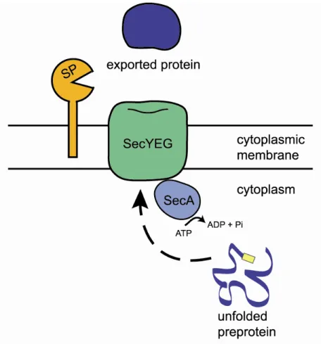
The general Sec system of protein export. In bacteria, the bulk of protein export from the cytoplasm across the cytoplasmic membrane is carried out by the universally conserved general secretion (Sec) pathway (Figure 1.1) (22, 47). The Sec pathway is essential in all organisms tested. Much of the detailed characterization of the Sec pathway was performed using the model organisms E. coli and B. subtilis (22, 50).
The central component of the Sec apparatus is the membrane-bound channel called the Sec translocase. The translocase is composed of the integral membrane proteins SecY, SecE, and SecG (12). Together, these three proteins form a complex that spans the
53). SecA also binds with low affinity to acidic phospholipids in the cytoplasmic membrane and with high affinity to SecYEG (34). SecA is an ATPase and the energy from ATP hydrolysis is required for Sec export (46). With each cycle of ATP binding and hydrolysis, SecA undergoes conformational changes that allow it to feed nascent pre-proteins through the SecYEG translocase, 20 amino acids at a time (58). Given the essential roles SecA plays in protein export, it is no surprise that in all cases tested SecA is essential for viability.
ATP binding and ATPase activity are essential for SecA function (46). SecA
contains ATP-binding Walker Box motifs, a structural feature found in many ATPases (69). Mutations in the Walker Box motifs of E. coli SecA destroy the ability of SecA to bind ATP (46). Furthermore, mutations in the SecA Walker Box are nonfunctional as shown by the inability to complement temperature sensitive secA mutants. SecA Walker Box mutants are also nonfunctional for in vitro pre-protein translocation assays. Additionally, disruption of ATPase activity interferes with the ability of SecA to cycle between the cytoplasmic
membrane and the cytoplasm (25). Normally, SecA is found evenly distributed between the cytoplasmic membrane and cytoplasm (14). However, a mutated Walker Box results in a SecA that is predominantly cell membrane-associated (46). In fact, treatment with carbonate is unable to dissociate Walker Box mutant SecA from the cytoplasmic membrane, indicating that this protein is permanently lodged in the membrane and unable to cycle upon ATP binding and hydrolysis. This fixed localization helps explain why a SecA Walker Box mutant is unable to complement temperature sensitive secA mutants.
that contains a cleavage site (22). During the process of export, a periplasmic signal peptidase, either LepB or LspA, cleaves the signal sequence to release the exported mature protein (66, 67, 77). LepB cleaves standard signal sequences while LspA specifically cleaves signal sequences of lipoproteins by recognizing a specific lipobox motif (L-A/S-G/A-C+1).
The Sec pathway also participates in delivering integral membrane proteins to the cytoplasmic membrane (22, 76). This class of proteins is delivered to the SecYEG
translocase cotranslationally with the aid of the Signal Recognition Particle (SRP). SRP is a ribonucleoprotein composed of a 4.5S RNA and a GTPase Ffh (fifty-four homolog). As a newly synthesized pre-protein emerges from the ribosome, SRP recognizes the signal sequence or transmembrane domains of the nascent polypeptide chain. This complex then interacts with FtsY, which in turn interacts with both anionic phospholipids and the SecYEG translocase. Upon GTP hydrolysis, SRP and FtsY are released from the membrane, leaving the pre-protein associated with the SecYEG translocase.
The Sec pathway also plays a role in delivery of integral membrane protein into the cytoplasmic membrane (22, 76). SecA delivers some, but not all transmembrane proteins to the SecYEG translocase. Once inserted into the SecYEG pore, hydrophobic transmembrane domains are believed to exit the translocase through a lateral gate. The precise mechanism behind lateral exit and integration of transmembrane proteins into the cytoplasmic membrane is not fully understood. Furthermore, insertion of a subset of transmembrane proteins
YidC can function independently of the Sec pathway and directly insert transmembrane domains into the cytoplasmic membrane (76).
Specialized protein secretion systems have roles in bacterial pathogenesis. In addition to the universally conserved Sec pathway, many bacterial pathogens use specialized secretion systems to transport virulence factors (28). There are now six named specialized secretion systems in Gram negative bacteria (30). Some of these pathways export proteins in two steps. Such proteins first cross the cytoplasmic membrane through the Sec system and are then secreted across the Gram negative outer membrane by a separate export apparatus. Other pathways secrete proteins in one-step by a process that is completely independent of the Sec pathway. One of the best-characterized specialized secretion systems is the Type III secretion system (T3SS) found in bacteria including Salmonella and Yersinia (38). The T3SS spans the entire bacterial cell envelope (comprised of the cytoplasmic membrane and the cell wall) and forms a needle complex that extends beyond the bacterial cell surface. The T3SS directly injects proteins synthesized in the bacterial cytoplasm through the needle complex and into host cells. Once inside the host, these bacterial effector proteins can function in a wide variety of ways to mediate bacterial infection.
constitute a specialized secretion system (11). Several Gram positive bacteria encode ESX secretion systems including Staphylococcus aureus, Corynebacterium diphtheriae, and Listeria monocytogenes (1, 62).
Another example of specialized protein secretion in Gram positive bacteria is the cytolysin-mediated translocation pathway. Examples of this pathway are found in Streptococcus pyogenes and Listeria monocytogenes (44, 52). In this system, a soluble cytolysin released from the bacterium forms a large oligomer within the membrane of a eukaryotic host cell. Once this pore is formed, other effector proteins exported from the bacterial cytoplasm can directly enter the host cell cytoplasm. In the S. pyogenes example, streptolysin O (SLO) forms a pore in membrane of human keratinocytes (44). Then, SPN (S. pyogenes NAD-glycohydrolase) translocates into the host cytoplasm. The combined activity of SLO and SPN is required for full cytotoxicity.
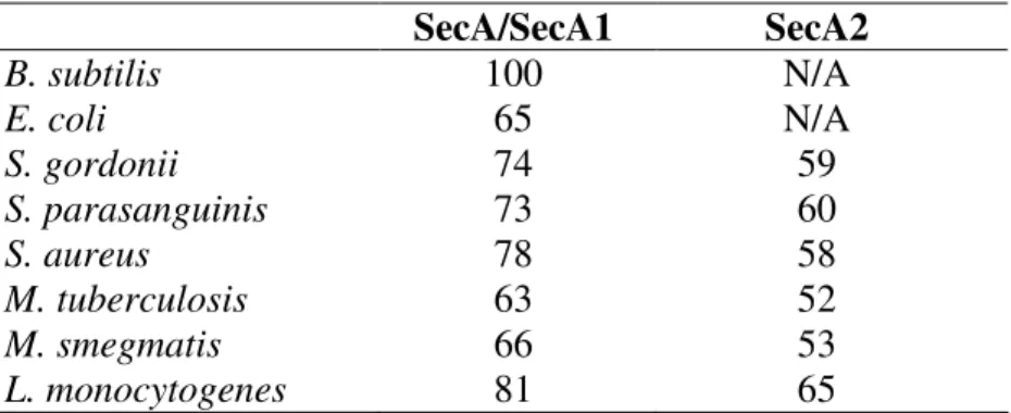
Accessory SecA2 protein export systems. It was long thought that all bacteria possess a single and essential SecA (24). Recently, mycobacteria and a small subset of Gram positive bacteria were found to encode two SecA homologs (5, 9, 10, 15, 18, 42, 54, 59). Included in this list are pathogens (M. tuberculosis, Listeria monocytogenes, Streptococcus gordonii, Streptococcus parasanguinis, Staphylococcus aureus, and Bacillus anthracis) and nonpathogens (M. smegmatis, L. innocua, and Corynebacterium glutamicum). In these bacteria, the SecA with highest homology to the essential SecA of B. subtilis is called SecA or SecA1 (Table 1.1). The other SecA is called SecA2. Bacteria with an accessory SecA2 can be divided into two groups: those that also possess an accessory SecY2 protein
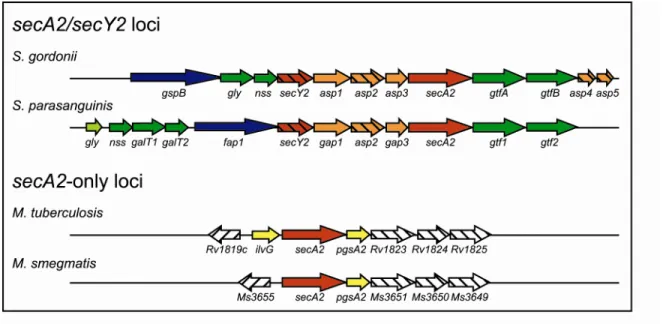
The accessory SecA2/Y2 systems of S. gordonii and S. parasanguinis. The
best-studied SecA2/SecY2 systems are those of S. gordonii and S. parasanguinis, two organisms found in the oral cavity (5, 18). The components and the mechanism of the accessory SecA2/SecY2 systems in these two bacteria are very similar. In both cases, the secA2 and secY2 genes are encoded in a locus that additionally possesses a suite of similarly arranged genes that include a SecA2-exported substrate and proteins involved in glycosylation and export of this substrate (Figure 1.3). In S. gordonii, the SecA2 system exports the serine-rich glycoprotein GspB (5). After crossing the cytoplasmic membrane, GspB becomes anchored to the cell wall where the protein is implicated in platelet binding. Platelet binding by S. gordonii is believed important to development of endocarditis (3). Similarly, the SecA2 system of S. parasanguinis exports a serine-rich glycoprotein called Fap1 to the cell wall (18). Fap1 forms fimbriae that are needed for attachment of S. parasanguinis to the surface of teeth and subsequent accumulation of dental plaque leading to periodontal disease (74). It seems highly likely that the proper export of these serine-rich glycoproteins by SecA2 is important to development of disease.
the absence of Gap3, Fap1 is still exported but the pattern of glycosylation is different (51). The significance of the difference between Gap3 and Asp3 function is unclear. There are other differences between the SecA2/SecY2 systems of S. gordonii and S. parasanguinis. In S. gordonii, asp4 and asp5 encode proteins with homology to B. subtilis SecE (52%
similarity) and SecG (55% similarity), respectively (65). Both proteins are required for GspB export. However, there are no obvious Asp4 or Asp5 homologs in S. parasanguinis. The significance of the slight differences between S. gordonii and S. parasanguinis is not clear.
Both GspB and Fap1 are heavily glycosylated proteins. In both the S. gordonii and S. parasanguinis secA2/Y2 loci there are multiple genes encoding proteins with roles in
glycosylation of the exported serine-rich proteins (13, 63, 73). Downstream of secA2 are gtf genes required for glycosylation of GspB and Fap1. Although the genomic organization is slightly different, both S. gordonii and S. parasanguinis secA2/Y2 loci also encode additional genes with homology to glycosyltransferase (gly) and nucleotide sugar synthetase (nss). Gly and Nss specify the types of carbohydrate linkages that are added to GspB. S. parasanguinis encodes two additional glycosylation factors, GalT1 and GalT2. GalT2 is required for full glycosylation of Fap1 (75).
There is evidence that GspB and Fap1 are glycosylated prior to export. While GspB export is blocked in S. gordonii secA2 and secY2 mutants, the GspB that is retained in the cytoplasm is still glycosylated (2). In S. parasanguinis cytoplasmic fractions, Fap1 is detected with both anti-peptide and anti-glycan antibodies, suggesting glycosylation occurs prior to export (18). This finding is unique because the known glycoproteins in M.
membrane (48, 68). It is possible that the accessory SecA2 system is important for exporting proteins with post-translational modifications.
However, data from both S. gordonii and S. parasanguinis indicates glycosylation is not required for SecA2-mediated export. Fap1 is still exported in a S. parasanguinis gtf1 mutant although it lacks any detectable carbohydrate modifications, and unglycosylated GspB is still exported in S. gordonii gtf mutants (6, 73). Interestingly, glycosylation status partially explains why the GspB and Fap1 are not exported by the canonical Sec pathway. In S. gordonii gtf mutants, GspB is not glycosylated but is still exported, and this export is reduced in a gtf secA2 double mutant. One demonstration of the effect of glycosylation comes from experiments showing more GspB is exported by a gtf secA2 double mutant than a secA2 single mutant (6). This difference is attributed to export of unglycosylated GspB via the canonical Sec pathway. In support of this idea, treatment with azide, a known SecA inhibitor, diminishes the residual GspB export in the double mutant. Thus, it appears that glycosylation can block GspB export by the canonical Sec pathway. Perhaps, fully
glycosylated GspB is too large for export through the SecYEG translocase. These findings also suggest that the accessory SecA2/Y2 systems are adapted to export proteins that are modified post-translationally.
In addition to glycosylation, the signal sequence is also important for directing GspB and Fap1 export (4, 6, 17). Both GspB and Fap1 have atypically long N-terminal signal sequences required for export. In S. gordonii, three key glycine residues (G3) are required for export of fully glycosylated GspB (4). The G3 residues serve a dual function by
signal sequence does not have G3 residues, but there may be other features that specify export via SecA2.
SecA1 is the essential housekeeping SecA protein of mycobacteria. Genomic analysis shows that M. tuberculosis encodes all of the essential proteins of the Sec pathway, including the ATPase SecA1 and the translocase components SecY, SecE, and SecG (19). Notably, SecB is not encoded by any mycobacteria. The SecB chaperone is not required for export of all Sec-dependent proteins, even in E. coli. In mycobacteria, there is ample
evidence that SecA1 functions as the housekeeping secretion factor, analogous to E. coli SecA. SecA1 is essential for growth in M. tuberculosis and M. smegmatis; secA1 cannot be deleted from the M. smegmatis chromosome unless a copy of secA1 is expressed from a plasmid (9, 57). Also in M. smegmatis, depletion of SecA1 leads to a loss of viability and prevents export of the MspA porin, which possesses a predicted Sec signal sequence (32). In contrast, the secA2 gene is not essential as ∆secA2 mutants have been constructed in M. tuberculosis and M. smegmatis (9, 10).
or gap genes found in S. gordonii and S. parasanguinis, respectively. Additionally, there is no known SecA2 substrate encoded in the accessory secA2 locus of mycobacteria.
On the other hand, the accessory SecA2 locus contains similar genes organized in the same way across all species of mycobacteria. Sequence alignments show that the proteins encoded by the genes in the secA2 locus of M. smegmatis are >70% similar to the homologs encoded in the M. tuberculosis secA2 locus. This degree of similarity is comparable for any protein conserved between these two organisms. Three genes downstream of secA2 in both M. smegmatis and M. tuberculosis encode proteins with predicted transmembrane domains (Table 1.2), but the function of these proteins has not yet been determined. One possibility is that these proteins form a membrane-embedded translocase to export SecA2-dependent pre-proteins.
The gene immediately downstream of secA2 encodes PgsA2, a
phosphatidylglycerolphosphate homolog predicted to function in acidic phospholipid synthesis (21, 39). Since E. coli SecA interacts with acidic phospholipids, perhaps PgsA2 synthesizes phospholipids that specifically interact with SecA2. In M. tuberculosis, but not M. smegmatis, the gene immediately upstream of secA2 encodes IlvG, a probable
acetolactate synthase, a key enzyme for biosynthesis of branched chain amino acids.
species, including both pathogens and nonpathogens alike. However, genomic analysis does not support the idea of the mycobacterial SecA2 systems functioning similarly to the
accessory SecA2/SecY2 systems in Gram positive bacteria.
Mycobacterial proteins dependent on SecA2 for export. In M. smegmatis, two proteins that require SecA2 for export to the cell wall were identified by 2D-PAGE.
Msmeg1704 and Msmeg1712 (YtfQ) are encoded by genes in a putative operon, and possess N-terminal signal sequences (31). Unlike the SecA2-dependent proteins of S. gordonii and S. parasanguinis, both Msmeg1704 and Msmeg1712 are predicted lipoproteins and have
homology to periplasmic sugar binding proteins. These proteins appear to be genuine lipoproteins, as both proteins are enriched by Triton X-114 extraction, a method used to isolate lipoproteins. Additionally, processing of these proteins is inhibited by treatment with globomycin, an inhibitor of the lipoprotein signal peptidase (31). It is worth noting that the effect of SecA2 on these lipoproteins is specific; other mycobacterial lipoproteins do not require SecA2 for their export. There are no homologs of Msmeg1704 or Msmeg1712 in M. tuberculosis. However, Msmeg1704 is not correctly processed when expressed in a M. tuberculosis ∆secA2 mutant. This result indicates that the SecA2 systems are functionally conserved among mycobacterial species.
In M. tuberculosis, SecA2 substrates were discovered by comparing proteins secreted into the culture medium from cultures of wild-type and ∆secA2 mutant bacteria (10). Three exported proteins are underrepresented in the ∆secA2 mutant culture filtrate: SodA
further. In the absence of SecA2, the amount of SodA exported into the culture medium is greatly reduced as shown by immunoblot and enzymatic activity assays (10, 36).
Additionally, SodA accumulates in the cytoplasm of a ∆secA2 mutant as expected of a true SecA2-dependent protein. KatG (catalase-peroxidase) is also exported into the culture medium by M. tuberculosis. Immunoblot assays show that KatG export depends on SecA2. Both SodA and KatG are antioxidant enzymes and could be important for protecting
intracellular M. tuberculosis from the oxidative burst of macrophages. Taken together, the data obtained from studies of both M. tuberculosis and M. smegmatis consistently shows that SecA2 plays a role in exporting a select subset of proteins.
It is unclear why the SecA2-dependent proteins identified in M. smegmatis have signal sequences and the proteins identified in M. tuberculosis do not. One possibility is that proteins recognized by SecA2 include examples with and without signal sequences.
Alternatively, SecA2 could recognize features of the mature protein, thereby eliminating the need for a signal sequence. It is also possible that role of SecA2 in exporting unconventional exported proteins like SodA is indirect. In this case, SecA2 exports proteins (not yet
Role of SecA2 in pathogenesis of M. tuberculosis. SecA2 is important for virulence
of M. tuberculosis in both mouse and macrophage infection models. Mice infected with the M. tuberculosis ∆secA2 mutant survive longer than mice infected with wild-type M.
tuberculosis (10, 41). Specifically, the ∆secA2 mutant has a growth defect during the early part of infection, prior to establishment of the cell-mediated immune response. Eventually, growth of the ∆secA2 mutant plateaus and the bacteria persist for the duration of the
infection. The bacterial burden of the ∆secA2 mutant in lungs is lower than that of wild-type M. tuberculosis. During the early phase of infection when the ∆secA2 mutant exhibits a defect in mice, M. tuberculosis is growing in macrophages. In macrophage cell culture, the ∆secA2 mutant is defective for intracellular growth in comparison to wild-type M.
tuberculosis (41). We hypothesize that the role of SecA2 is to promote export of proteins important to growth in macrophages. As mentioned above, the ∆secA2 mutant is defective in exporting antioxidant enzymes. This export defect might partially account for the observed macrophage growth defect. However, the ∆secA2 mutant also has a growth defect in macrophages that do not generate an oxidative burst (41). Therefore, SecA2 must export other proteins important for surviving inside host cells. Since macrophage infected with the ∆secA2 mutant release more proinflammatory cytokines, one role of SecA2 may be to export proteins that modulate the host immune system in response to infection.
Summary. It is clear that the mycobacterial accessory SecA2 system is responsible for exporting a select subset of proteins and it plays a role in the virulence of M. tuberculosis. Nevertheless, little is known about the mechanism of SecA2-dependent protein export in mycobacteria. A better understanding of the SecA2 protein export system has the potential to reveal new drug targets or vaccine strategies to combat infection with M. tuberculosis.
In this work, we undertook experiments to characterize the mechanism of SecA2-mediated protein export in mycobacteria. In the following chapters, we discuss biochemical and genetic analyses of the accessory SecA2 pathway of M. tuberculosis and M. smegmatis. In Chapter 2, we explore similarities and differences between the essential SecA1 protein and the accessory SecA2 protein. We demonstrate that SecA2 is an ATPase, and that ATP binding is required for normal SecA2 function in vivo (Chapter 3 and 4). Using a dominant negative secA2 allele, we perform a suppressor screen in M. smegmatis and identify
Protein sequences were obtained from TIGR and NCBI and aligned using ClustalW.
Table 1.1 Percent amino acid similarity to B. subtilis SecA SecA/SecA1 SecA2
B. subtilis 100 N/A
E. coli 65 N/A
S. gordonii 74 59
S. parasanguinis 73 60
S. aureus 78 58
M. tuberculosis 63 52
M. smegmatis 66 53
Table 1.2 Homologous proteins encoded by the accessory SecA2 locus of mycobacteria
M. tuberculosis M. smegmatis % similarity TM domain
Rv1819c Msmeg3655 77 yes
IlvG N/A N/A no
SecA2 SecA2 90 no
PgsA2 PgsA2 82 no
Rv1823 Msmeg3651 71 yes
Rv1824 Msmeg3650 94 yes
Rv1825 Msmeg3649 78 yes
Protein sequences were obtained from TIGR and NCBI. Alignments were performed using ClustalW.
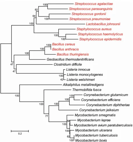
Figure 1.2. Evolutionary relationships of SecA2 proteins. The phylogenetic tree was generated in MEGA4 using the neighbor-joining method. The length of the branches reflects the number of amino acid changes between different SecA2s, as indicated by the bar.
References
1. Abdallah, A. M., N. C. Gey van Pittius, P. A. Champion, J. Cox, J. Luirink, C. M. Vandenbroucke-Grauls, B. J. Appelmelk, and W. Bitter. 2007. Type VII secretion--mycobacteria show the way. Nat Rev Microbiol 5:883-91.
2. Bensing, B. A., B. W. Gibson, and P. M. Sullam. 2004. The Streptococcus gordonii platelet binding protein GspB undergoes glycosylation independently of export. J Bacteriol 186:638-45.
3. Bensing, B. A., J. A. Lopez, and P. M. Sullam. 2004. The Streptococcus gordonii surface proteins GspB and Hsa mediate binding to sialylated carbohydrate epitopes on the platelet membrane glycoprotein Ibalpha. Infect Immun 72:6528-37.
4. Bensing, B. A., I. R. Siboo, and P. M. Sullam. 2007. Glycine residues in the
hydrophobic core of the GspB signal sequence route export toward the accessory Sec pathway. J Bacteriol 189:3846-54.
5. Bensing, B. A., and P. M. Sullam. 2002. An accessory sec locus of Streptococcus gordonii is required for export of the surface protein GspB and for normal levels of binding to human platelets. Mol Microbiol 44:1081-94.
6. Bensing, B. A., D. Takamatsu, and P. M. Sullam. 2005. Determinants of the streptococcal surface glycoprotein GspB that facilitate export by the accessory Sec system. Mol Microbiol 58:1468-81.
7. Binet, R., S. Letoffe, J. M. Ghigo, P. Delepelaire, and C. Wandersman. 1997. Protein secretion by gram-negative bacterial ABC exporters. Folia Microbiol 42:179-83.
8. Bochkareva, E. S., M. E. Solovieva, and A. S. Girshovich. 1998. Targeting of GroEL to SecA on the cytoplasmic membrane of Escherichia coli. Proc Natl Acad Sci U S A 95:478-83.
9. Braunstein, M., A. M. Brown, S. Kurtz, and W. R. Jacobs, Jr. 2001. Two
nonredundant SecA homologues function in mycobacteria. J Bacteriol 183:6979-90. 10. Braunstein, M., B. Espinosa, J. Chan, J. T. Belisle, and W. R. J. Jacobs. 2003.
SecA2 functions in the secretion of superoxide dismutase A and in the virulence of Mycobacterium tuberculosis. Mol Microbiol 48:453-64.
12. Brundage, L., J. P. Hendrick, E. Schiebel, A. J. Driessen, and W. Wickner. 1990. The purified E. coli integral membrane protein SecY/E is sufficient for reconstitution of SecA-dependent precursor protein translocation. Cell 62:649-57.
13. Bu, S., Y. Li, M. Zhou, P. Azadin, M. Zeng, P. Fives-Taylor, and H. Wu. 2008. Interaction between two putative glycosyltransferases is required for glycosylation of a serine-rich streptococcal adhesin. J Bacteriol 190:1256-66.
14. Cabelli, R. J., K. M. Dolan, L. P. Qian, and D. B. Oliver. 1991. Characterization of membrane-associated and soluble states of SecA protein from wild-type and
SecA51(TS) mutant strains of Escherichia coli. J Biol Chem 266:24420-7. 15. Caspers, M., and R. Freudl. 2008. Corynebacterium glutamicum possesses two
secA homologous genes that are essential for viability. Arch Microbiol 189:605-10. 16. Chen, M., K. Xie, F. Jiang, L. Yi, and R. E. Dalbey. 2002. YidC, a newly defined
evolutionarily conserved protein, mediates membrane protein assembly in bacteria. Biol Chem 383:1565-72.
17. Chen, Q., B. Sun, H. Wu, Z. Peng, and P. M. Fives-Taylor. 2007. Differential roles of individual domains in selection of secretion route of a Streptococcus parasanguinis serine-rich adhesin, Fap1. J Bacteriol 189:7610-7.
18. Chen, Q., H. Wu, and P. M. Fives-Taylor. 2004. Investigating the role of secA2 in secretion and glycosylation of a fimbrial adhesin in Streptococcus parasanguis FW213. Mol Microbiol 53:843-56.
19. Cole, S. T., R. Brosch, J. Parkhill, T. Garnier, C. Churcher, D. Harris, S. V. Gordon, K. Eiglmeier, S. Gas, C. E. Barry, 3rd, F. Tekaia, K. Badcock, D. Basham, D. Brown, T. Chillingworth, R. Connor, R. Davies, K. Devlin, T. Feltwell, S. Gentles, N. Hamlin, S. Holroyd, T. Hornsby, K. Jagels, and B. G. Barrell. 1998. Deciphering the biology of Mycobacterium tuberculosis from the complete genome sequence. Nature 393:537-44.
20. Cosma, C. L., D. R. Sherman, and L. Ramakrishnan. 2003. The secret lives of the pathogenic mycobacteria. Annu Rev Microbiol 57:641-76.
21. de Vrije, T., R. L. de Swart, W. Dowhan, J. Tomassen, and B. de Kruijff. 1988. Phosphatidylglycerol is involved in protein translocation across Escherichia coli inner membranes. Nature 334:173-5.
22. Driessen, A. J., and N. Nouwen. 2008. Protein translocation across the bacterial cytoplasmic membrane. Annu Rev Biochem 77:643-67.
24. Economou, A. 1999. Following the leader: bacterial protein export through the Sec pathway. Trends Microbiol 7:315-20.
25. Economou, A., J. A. Pogliano, J. Beckwith, D. B. Oliver, and W. Wickner. 1995. SecA membrane cycling at SecYEG is driven by distinct ATP binding and hydrolysis events and is regulated by SecD and SecF. Cell 83:1171-81.
26. El-Sadr, W. M., and S. J. Tsiouris. 2008. HIV-associated tuberculosis: diagnostic and treatment challenges. Semin Respir Crit Care Med 29:525-31.
27. Fekkes, P., C. van der Does, and A. J. Driessen. 1997. The molecular chaperone SecB is released from the carboxy-terminus of SecA during initiation of precursor protein translocation. Embo J 16:6105-13.
28. Finlay, B. B., and S. Falkow. 1997. Common themes in microbial pathogenicity revisited. Microbiol Mol Biol Rev 61:136-69.
29. Flynn, J. L., and J. Chan. 2001. Tuberculosis: latency and reactivation. Infect Immun 69:4195-201.
30. Gerlach, R. G., and M. Hensel. 2007. Protein secretion systems and adhesins: the molecular armory of Gram-negative pathogens. Int J Med Microbiol 297:401-15. 31. Gibbons, H. S., F. Wolschendorf, M. Abshire, M. Niederweis, and M.
Braunstein. 2007. Identification of two Mycobacterium smegmatis lipoproteins exported by a SecA2-dependent pathway. J Bacteriol 189:5090-100.
32. Guo, X. V., M. Monteleone, M. Klotzsche, A. Kamionka, W. Hillen, M. Braunstein, S. Ehrt, and D. Schnappinger. 2007. Silencing essential protein secretion in Mycobacterium smegmatis using tetracycline repressors. J Bacteriol 189:4614-23.
33. Hartl, F. U., S. Lecker, E. Schiebel, J. P. Hendrick, and W. Wickner. 1990. The binding cascade of SecB to SecA to SecY/E mediates preprotein targeting to the E. coli plasma membrane. Cell 63:269-79.
34. Hendrick, J. P., and W. Wickner. 1991. SecA protein needs both acidic phospholipids and SecY/E protein for functional high-affinity binding to the Escherichia coli plasma membrane. J Biol Chem 266:24596-600.
36. Hinchey, J., S. Lee, B. Y. Jeon, R. J. Basaraba, M. M. Venkataswamy, B. Chen, J. Chan, M. Braunstein, I. M. Orme, S. C. Derrick, S. L. Morris, W. R. Jacobs, Jr., and S. A. Porcelli. 2007. Enhanced priming of adaptive immunity by a
proapoptotic mutant of Mycobacterium tuberculosis. J Clin Invest 117:2279-88. 37. Houben, E. N., L. Nguyen, and J. Pieters. 2006. Interaction of pathogenic
mycobacteria with the host immune system. Curr Opin Microbiol 9:76-85. 38. Hueck, C. J. 1998. Type III protein secretion systems in bacterial pathogens of
animals and plants. Microbiol Mol Biol Rev 62:379-433.
39. Jackson, M., D. C. Crick, and P. J. Brennan. 2000. Phosphatidylinositol is an essential phospholipid of mycobacteria. J Biol Chem 275:30092-9.
40. Kurtz, S., and M. Braunstein. 2005. Protein secretion and export in Mycobacterium tuberculosis, p. 71-138. In T. Parish (ed.), Mycobacterium molecular biology.
Horizon bioscience, Norfolk, UK.
41. Kurtz, S., K. P. McKinnon, M. S. Runge, J. P. Ting, and M. Braunstein. 2006. The SecA2 secretion factor of Mycobacterium tuberculosis promotes growth in macrophages and inhibits the host immune response. Infect Immun 74:6855-64. 42. Lenz, L. L., and D. A. Portnoy. 2002. Identification of a second Listeria secA gene
associated with protein secretion and the rough phenotype. Mol Microbiol 45:1043-56.
43. Lewis, K. N., R. Liao, K. M. Guinn, M. J. Hickey, S. Smith, M. A. Behr, and D. R. Sherman. 2003. Deletion of RD1 from Mycobacterium tuberculosis mimics bacille Calmette-Guerin attenuation. J Infect Dis 187:117-23.
44. Madden, J. C., N. Ruiz, and M. Caparon. 2001. Cytolysin-mediated translocation (CMT): a functional equivalent of type III secretion in gram-positive bacteria. Cell 104:143-52.
45. Meya, D. B., and K. P. McAdam. 2007. The TB pandemic: an old problem seeking new solutions. J Intern Med 261:309-29.
46. Mitchell, C., and D. Oliver. 1993. Two distinct ATP-binding domains are needed to promote protein export by Escherichia coli SecA ATPase. Mol Microbiol 10:483-97. 47. Murphy, C. K., and J. Beckwith. 1996. Export of proteins to the cell envelope in
48. Nita-Lazar, M., M. Wacker, B. Schegg, S. Amber, and M. Aebi. 2005. The N-X-S/T consensus sequence is required but not sufficient for bacterial N-linked protein glycosylation. Glycobiology 15:361-7.
49. Papanikou, E., S. Karamanou, C. Baud, M. Frank, G. Sianidis, D. Keramisanou, C. G. Kalodimos, A. Kuhn, and A. Economou. 2005. Identification of the
preprotein binding domain of SecA. J Biol Chem 280:43209-17.
50. Papanikou, E., S. Karamanou, and A. Economou. 2007. Bacterial protein secretion through the translocase nanomachine. Nat Rev Microbiol 5:839-51.
51. Peng, Z., H. Wu, T. Ruiz, Q. Chen, M. Zhou, B. Sun, and P. Fives-Taylor. 2008. Role of gap3 in Fap1 glycosylation, stability, in vitro adhesion, and fimbrial and biofilm formation of Streptococcus parasanguinis. Oral Microbiol Immunol 23:70-8. 52. Pizarro-Cerda, J., and P. Cossart. 2006. Subversion of cellular functions by
Listeria monocytogenes. J Pathol 208:215-23.
53. Randall, L. L., S. J. Hardy, T. B. Topping, V. F. Smith, J. E. Bruce, and R. D. Smith. 1998. The interaction between the chaperone SecB and its ligands: evidence for multiple subsites for binding. Protein Sci 7:2384-90.
54. Rigel, N. W., and M. Braunstein. 2008. A new twist on an old pathway--accessory Sec systems. Mol Microbiol 69:291-302.
55. Rohde, K., R. M. Yates, G. E. Purdy, and D. G. Russell. 2007. Mycobacterium tuberculosis and the environment within the phagosome. Immunol Rev 219:37-54. 56. Russell, D. G. 2007. Who puts the tubercle in tuberculosis? Nat Rev Microbiol
5:39-47.
57. Sassetti, C. M., D. H. Boyd, and E. J. Rubin. 2003. Genes required for
mycobacterial growth defined by high density mutagenesis. Mol Microbiol 48:77-84. 58. Schiebel, E., A. J. Driessen, F. U. Hartl, and W. Wickner. 1991. Delta mu H+ and
ATP function at different steps of the catalytic cycle of preprotein translocase. Cell 64:927-39.
59. Siboo, I. R., D. O. Chaffin, C. E. Rubens, and P. M. Sullam. 2008.
Characterization of the accessory Sec system of Staphylococcus aureus. J Bacteriol 190:6188-96.
61. Sonnhammer, E. L., G. von Heijne, and A. Krogh. 1998. A hidden Markov model for predicting transmembrane helices in protein sequences. Proc Int Conf Intell Syst Mol Biol 6:175-82.
62. Sundaramoorthy, R., P. K. Fyfe, and W. N. Hunter. 2008. Structure of Staphylococcus aureus EsxA suggests a contribution to virulence by action as a transport chaperone and/or adaptor protein. J Mol Biol 383:603-14.
63. Takamatsu, D., B. A. Bensing, and P. M. Sullam. 2004. Four proteins encoded in the gspB-secY2A2 operon of Streptococcus gordonii mediate the intracellular glycosylation of the platelet-binding protein GspB. J Bacteriol 186:7100-11.
64. Takamatsu, D., B. A. Bensing, and P. M. Sullam. 2004. Genes in the accessory sec locus of Streptococcus gordonii have three functionally distinct effects on the
expression of the platelet-binding protein GspB. Mol Microbiol 52:189-203. 65. Takamatsu, D., B. A. Bensing, and P. M. Sullam. 2005. Two additional
components of the accessory sec system mediating export of the Streptococcus gordonii platelet-binding protein GspB. J Bacteriol 187:3878-83.
66. Tokunaga, M., J. M. Loranger, and H. C. Wu. 1984. A distinct signal peptidase for prolipoprotein in Escherichia coli. J Cell Biochem 24:113-20.
67. van Roosmalen, M. L., N. Geukens, J. D. Jongbloed, H. Tjalsma, J. Y. Dubois, S. Bron, J. M. van Dijl, and J. Anne. 2004. Type I signal peptidases of Gram-positive bacteria. Biochim Biophys Acta 1694:279-97.
68. VanderVen, B. C., J. D. Harder, D. C. Crick, and J. T. Belisle. 2005. Export-mediated assembly of mycobacterial glycoproteins parallels eukaryotic pathways. Science 309:941-3.
69. Walker, J. E., A. Eberle, N. J. Gay, M. J. Runswick, and M. Saraste. 1982. Conservation of structure in proton-translocating ATPases of Escherichia coli and mitochondria. Biochem Soc Trans 10:203-6.
70. WorldHealthOrganization. 2008. Global Tuberculosis Control 2008: Surveillance, Planning, Financing. World Health Organization, Geneva.
71. WorldHealthOrganization 2007, posting date. WHO Information tuberculosis fact sheet. [Online.]
73. Wu, H., S. Bu, P. Newell, Q. Chen, and P. Fives-Taylor. 2007. Two gene determinants are differentially involved in the biogenesis of Fap1 precursors in Streptococcus parasanguis. J Bacteriol 189:1390-8.
74. Wu, H., K. P. Mintz, M. Ladha, and P. M. Fives-Taylor. 1998. Isolation and characterization of Fap1, a fimbriae-associated adhesin of Streptococcus parasanguis FW213. Mol Microbiol 28:487-500.
75. Wu, H., M. Zeng, and P. Fives-Taylor. 2007. The glycan moieties and the N-terminal polypeptide backbone of a fimbria-associated adhesin, Fap1, play distinct roles in the biofilm development of Streptococcus parasanguinis. Infect Immun 75:2181-8.
76. Xie, K., and R. E. Dalbey. 2008. Inserting proteins into the bacterial cytoplasmic membrane using the Sec and YidC translocases. Nat Rev Microbiol 6:234-44. 77. Zwizinski, C., and W. Wickner. 1980. Purification and characterization of leader
Chapter 2
Comparative Analysis of the Properties of SecA1 and SecA2 of Mycobacteria
Nathan W. Rigel, Jessica R. McCann, Henry S. Gibbons†, and Miriam Braunstein
Department of Microbiology and Immunology, University of North Carolina School of Medicine, Chapel Hill, North Carolina
† Edgewood Chemical Biological Center,
Aberdeen Proving Ground, MD
The Sec-dependent translocation pathway is used to export most proteins across the cytoplasmic membrane. Recently, several bacteria, including the pathogen Mycobacterium tuberculosis and the non-pathogen M. smegmatis, were shown to possess two SecA
homologs, SecA1 and SecA2. SecA1 is essential for general protein export. SecA2 is
fractions, while SecA2 was predominantly in the cytoplasmic fraction. Here, we used M. smegmatis as a model to characterize the accessory Sec system of mycobacteria. We redesigned a ∆secA2 mutant of M. smegmatis for use in further genetic and biochemical analyses. This mutant had a larger in-frame, unmarked deletion and had the same
phenotypes as the previously published ∆secA2 mutant. These results are the first step in understanding the unique functions of the two SecA proteins of mycobacteria.
Introduction
In all bacteria, the general Sec pathway is used to export the bulk of proteins from the cytoplasm across the cytoplasmic membrane (15, 42). Given the substantial role the Sec pathway plays in exporting proteins to their proper location, it is not surprising that the Sec pathway is essential in all bacteria in which it has been tested.
Proteins destined for export via the Sec pathway are synthesized as pre-proteins with a characteristic N-terminal sorting signal, called a signal sequence. Signal sequences have a tripartite structure: a positively charged N domain, a hydrophobic H domain, and a polar C domain (15). The signal sequence is cleaved off by one of two periplasmic signal peptidases (LepB or LspA) during or immediately after transport across the cytoplasmic membrane (62, 64, 72). Only unfolded pre-proteins are compatible with the Sec pathway.
binding and hydrolysis, SecA ratchets pre-proteins through the SecYEG pore (19, 58). Subcellular localization experiments show that SecA is evenly distributed between the cell envelope (comprised of membrane and cell wall) and the cytoplasm (7, 18). This reflects the dynamic nature of SecA function in protein export. SecA interacts with lipids and the Sec translocase at the membrane (20, 28). It also interacts with chaperones, such as SecB, and newly synthesized pre-proteins in the cytoplasm (21, 22, 48). Thus, the presence of SecA in both membrane and cytosolic containing fractions is not surprising. In E. coli, mutations that destroy ATPase activity in SecA are unable to complement temperature sensitive secA mutants (41). This demonstrates ATPase activity is required for normal SecA function. Further, the SecA ATPase mutant proteins become stably associated with the cell envelope (18, 31, 41). These findings support the idea that ATP binding and hydrolysis is necessary for SecA to reversibly associate with the Sec translocase in its role of translocating a protein across the membrane.
Recently, some Gram positive bacteria and mycobacteria were discovered to possess two non-redundant SecA homologs (1, 3, 4, 8, 9, 38, 55, 60). All mycobacteria examined, including pathogens and nonpathogens, possess two SecA homologs. In bacteria with two SecAs, the SecA with higher sequence similarity to the canonical SecA of E. coli and B. subtilis is called SecA or SecA1. The other SecA homolog is called the accessory SecA or SecA2. SecA1 of M. tuberculosis is 63% similar to the well-characterized SecA of B. subtilis, and SecA1 of M. smegmatis is 66% similar to the canonical SecA protein. This sequence similarity is spread across the length of the protein, including the
well-characterized Walker A and Walker B ATP binding motifs. Walker box motifs are
In both M. smegmatis and M. tuberculosis secA1 cannot be deleted, indicating that secA1 is an essential gene (3, 4). Additionally, depletion of SecA1 in M. smegmatis prevents the export of the Sec signal sequence-containing protein MspA (26). Taken together, these findings support the notion that in mycobacteria, SecA1 functions as the housekeeping export factor similar to SecA of E. coli.
SecA2 from M. tuberculosis and M. smegmatis are 52% and 53% similar to B. subtilis SecA, respectively. Notably, the ATP-binding Walker A and Walker B motifs are conserved in SecA2 (55). SecA2 is smaller than SecA1, partially due to a truncation at the C-terminus. The first characterization of any SecA2 protein was conducted in M. smegmatis, a
fast-growing saprophytic organism often used as a model to study mycobacterial physiology (3). In both M. tuberculosis and M. smegmatis, SecA2 is not essential as in-frame,
phenotypic analyses show SecA2 is functional in mycobacteria with a role in exporting a specific subset of proteins.
Interestingly, overexpression of SecA1 does not compensate for a lack of SecA2. Conversely, overexpression of SecA2 does not allow construction of a ∆secA1 deletion mutant (3). This shows that SecA1 and SecA2 are not functionally redundant and each has distinct roles in mycobacterial protein export.
The originally published M. smegmatis ∆secA2 mutant (mc22522) is an in-frame, unmarked deletion of approximately one-third of the secA2 gene (3). The deletion removes the predicted Walker B motif of the ATP binding site and it was therefore predicted to produce a nonfunctional protein. As stated above, several phenotypes are reported for
mc22522, all of which are complemented by introduction of wild-type secA2 into the mutant. This supports the notion that this mutant behaves like a null. However, because mc22522
should synthesize a truncated protein species, we still worried that the remaining two-thirds of SecA2 might complicate analysis of experiments using this ∆secA2 mutant. To avoid any such problems, we constructed a new improved in-frame unmarked deletion mutant of M. smegmatis secA2 where only three codons of the original open reading frame (ORF) remained. In this Chapter, we also describe our detailed analysis of this new M. smegmatis ∆secA2 mutant (NR116) which is used in all subsequent work in this dissertation.
Our comparative analysis of SecA1 and SecA2 began by comparing the expression levels of each protein in M. smegmatis and M. tuberculosis. So far, this has not been performed for any organism possessing two SecA proteins. Using antibodies specific for SecA1 and SecA2, we performed quantitative immunoblots on whole-cell protein lysates from wild-type M. smegmatis and M. tuberculosis. In both cases, we found that the average number of moles of SecA2 and SecA1 per mg of protein was nearly equivalent. This indicates that the different functions of SecA1 and SecA2 are not due to a difference in expression level.
analysis to determine the location of SecA1 and SecA2. SecA1 was equally distributed between cell envelope and cytoplasmic fractions, further supporting the notion that SecA1 is the canonical SecA of mycobacteria. In contrast, SecA2 was found predominantly in the cytoplasmic fraction. These results reveal a distinguishing feature of SecA1 and SecA2, and it also provides a useful assay for future studies of SecA2 in mycobacteria.
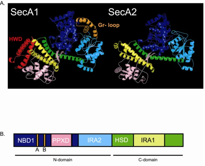
Finally, we performed a comparison of the sequences and structures of SecA1 and SecA2. Sequence alignments show a high degree of similarity (~50%) between SecA1 and SecA2, even though SecA2 is 20 kDa smaller than SecA1. The crystal structure of M. tuberculosis SecA1 is solved (59), but the structure of M. tuberculosis SecA2 has not been determined. Using a computer-generated homology model of SecA2, built on to the crystal structure of SecA1, we identified potential structural differences between these two proteins. While strikingly similar across most of the structure, two regions present in SecA1 are notably absent in SecA2. We believe this model will be a useful tool in designing future experiments to characterize the accessory SecA2 protein of mycobacteria.
Materials and Methods
used to grow E. coli cultures. When needed, kanamycin or hygromycin was added at 40 µg/ml or 150 µg/ml, respectively. The identity of all vectors used in this study was confirmed by DNA sequencing (UNC-CH automated DNA sequencing facility and Eton Biosciences).
Macrophage infections. Bone marrow macrophages were elicited from femurs of C57BL/6 mice, as described previously (37, 40), and 2.5 x 10⁵ macrophages were seeded into wells of
8-well-chamber slides 24 h prior to infection. The M. tuberculosis strains were grown to mid-exponential phase, washed with phosphate-buffered saline containing 0.05% (w/v) Tween 80, diluted in tissue culture medium (Dulbecco’s modified Eagle’s medium supplemented with 10% heat-inactivated fetal calf serum, 2 mM glutamine, and 1X nonessential amino acids [Gibco]) and added to the macrophage monolayer to achieve a multiplicity of infection of 1.0. Macrophage monolayers were infected with M. tuberculosis strains for 4 h at 37°C in 5% CO2. On days 0, 1, and 5 post-infection, the contents of
triplicate wells for each infection were washed to remove extracellular bacilli and then lysed with 0.05% (w/v) Tween 80. The resulting lysates were diluted and plated on Middlebrook 7H10 agar to enumerate intracellular bacteria during the course of infection.
Homology model of M. tuberculosis SecA2. To generate a structural model of SecA2, we
as a raw sequence alignment generated using ClustalW, we determined where several insertions and deletions in SecA2 should be placed. This was achieved by comparing the sequence alignments versus the local environment of the SecA1 amino acids in the crystal structure. By examining the side chains of the amino acids that flank an insertion or deletion, we decided whether the position of a given insertion/deletion made structural sense. We then generated the homology model using Insight II (Accelrys, Inc.). To assess the validity of the model, we used the Profiles3D/verify function in Insight II to determine self-compatibility scores.
Subcellular fractionation. Cell envelope and soluble fractions were prepared by differential ultracentrifugation as described previously (24). Briefly, 100 ml cultures of M. smegmatis grown in Mueller Hinton were harvested by centrifugation at 3,000 x g. Cell pellets were resuspended in 4 ml of breaking buffer (PBS, 0.6 µg/ml each of DNase and RNase, and a cocktail of protease inhibitors (2 µg/ml each of aprotinin, E-64, leupeptin, and pepstatin A and 100 µg/ml Pefabloc SC) and then lysed by five passages in a French pressure cell. Unbroken cells were pelleted at 3,000 x g for 20 min to generate a clarified whole cell lysate, which was centrifuged at 100,000 x g for 2 h to separate the cell envelope (pellet) fraction from the soluble fraction (supernatant). The cell envelope fraction was washed once and then resuspended in PBS.
quantitative immunoblots, whole-cell lysates of exponential phase M. tuberculosis cultures grown in Middlebrook 7H9 medium were prepared following fixation in an equal volume of 10% (w/v) formalin for 1 h. Fixed cells were pelleted by centrifugation, resuspended in extraction buffer, and lysed by bead beating. For quantitative SecA1 and SecA2 Western blot analysis, 100 µg of formalin-fixed whole-cell lysates were electrophoresed on a 12% sodium dodecyl sulfate (SDS)-polyacrylamide gel alongside known amounts of purified SecA1 or SecA2 protein (29). Proteins were transferred to nitrocellulose membranes and probed with rabbit polyclonal anti-SecA1 (1:50,000 dilution) or anti-SecA2 (1:20,000 dilution) antibody. The secondary antibody was goat anti-rabbit conjugated to alkaline phosphatase (1:20,000 dilution), and detection of the fluorescence from ECF (Amersham/GE Healthcare) was done with a PhosphorImager (Molecular Dynamics). The signal intensity values from six independently prepared whole-cell lysates were quantified by comparison with standard curve values to determine the number of moles of SecA1 and SecA2 per mg of cellular protein.
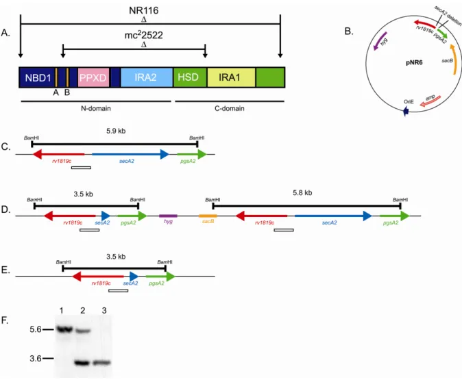
secA2 suicide plasmid pNR6. Sequences immediately upstream and downstream of secA2
were PCR amplified from M. smegmatis genomic DNA using primers GTCGACCGACAGGTTCCAGCCGTAGAA-3’ /
GTCGACCACGGCGTCAGTTGTGCCTCG-3’ and GTCGACTAGGCCCCAGCCATTAGGTTC-3’ /
5’-TGATATCGAGCACCTCCCAGCCCCATTC-3’, respectively. SalI restriction sites were added to the ends of the upstream PCR product which was then cloned into the vector pCC1 using the Copy Control PCR cloning kit (Epicentre) to generate pNR3. The 901 bp
vector pCR2.1 (Invitrogen) to generate pNR4. A 1087 bp SalI fragment of pNR3 was ligated into SalI-cut pNR4. The resulting plasmid, pNR5, contained a 2385 bp deletion of secA2 leaving only three codons. A 1737 bp MscI-EcoRV fragment containing the secA2 deletion was cut from pNR5 and cloned into the EcoRV site of the counterselectable suicide plasmid pMP62, yielding vector pNR6.
Two step allelic exchange. The ∆secA2 mutant strain NR116 was constructed by two step allelic exchange as described previously (2, 50). Wild-type M. smegmatis strain mc2155 was electroporated with pNR6, and hygromycin-resistant transformants were selected on 7H10 plates. Transformants were screened by Southern blot to identify a single-crossover
integration of pNR6 at the chromosomal secA2 locus in strain SCO5. To resolve the single crossover strain, a saturated culture (7H9 0.2% glucose, 0.5% glycerol, 0.1% Tween 80, hygromycin 50 µg/ml) of SCO5 was diluted 1:100 into 7H9 without hygromycin and grown overnight at 37˚C. This culture was plated onto 7H10 supplemented with 4.5% (w/v) sucrose to select against the sacB marker encoded on the backbone of the suicide vector. Sucrose-resistant clones were screened for hygromycin sensitivity. Then sucrose-Sucrose-resistant,
Azide assay. 200 µl of saturated (OD600nm = 2.0) M. smegmatis culture was mixed with 3.5
ml of molten 7H9 top agar, and then poured onto a 7H10 bottom agar plate. After the top agar cooled, sterile 6 mm filter discs were placed onto the surface. 10 µl of 0.15 M sodium azide was then added to the disc. The plates were inverted and incubated for 2 days at 37˚C. After incubation, any resulting zone of inhibition on the plate was measured. Each strain was tested in triplicate, and untreated plates were included as a control.
Agar plate growth assays. All M. smegmatis strains were grown in 7H9 medium at 37˚C prior to plating. Serial dilutions of each strain were made in 7H9 and then plated onto the appropriate agar medium. All plates were incubated at 37˚C until colonies were visible.
Results
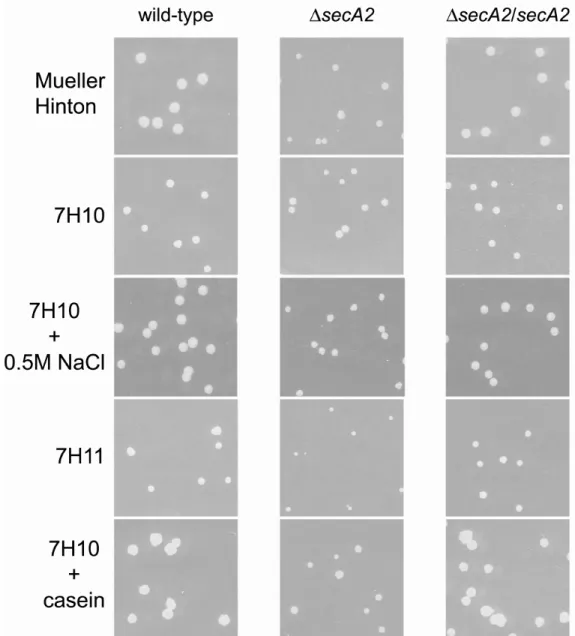
M. smegmatis is a valid model for studying the M. tuberculosis SecA2 system. M. tuberculosis is difficult to manipulate experimentally because it is extremely slow growing and requires containment in a BSL3 laboratory. As a result, the fast growing nonpathogen M. smegmatis is often used as a model mycobacterium (54). Throughout this work, we take advantage of M. smegmatis as a convenient tool to study SecA2-mediated protein export in mycobacteria. It is already known that expression of M. tuberculosis SecA2 can complement the rich agar growth defect and azide hypersensitivity phenotypes of the M. smegmatis ∆secA2 mutant (3). In order to validate using M. smegmatis as a model to study M.
M. smegmatis secA2 complements the macrophage growth defect of a M.
tuberculosis ∆∆∆∆secA2 mutant. Unlike the wild-type M. tuberculosis H37Rv strain which can
infect and grow inside macrophages over a period of several days, the ∆secA2 mutant is defective in intracellular growth (37). A ∆secA2 mutant of M. tuberculosis is attenuated for growth in macrophages and in mice (4, 37). Introduction of a wild-type copy of M.
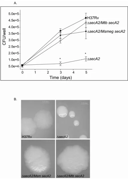
tuberculosis secA2 at the attB locus complements the mutant phenotype. Here we tested if the M. smegmatis secA2 gene can also complement the M. tuberculosis ∆secA2 mutant phenotype. Murine bone marrow-derived macrophages were infected with wild-type M. tuberculosis (H37Rv), ∆secA2 mutant mc23112 (∆secA2), the ∆secA2 mutant complemented with wild-type M. tuberculosis secA2 (∆secA2/Mtb secA2), or the ∆secA2 mutant expressing wild-type M. smegmatis secA2 (∆secA2/Msm secA2) (Figure 2.1A). As shown previously (37), H37Rv and the ∆secA2 mutant complemented with M. tuberculosis secA2 grew similarly in macrophages over a 5-day period of infection while the ∆secA2 mutant failed to grow in these macrophages. Introduction of M. smegmatis secA2 in single copy at the chromosomal attB site in the mutant also fully restored the ability of the ∆secA2 mutant to grow in macrophages. Western blot analysis confirmed that the ∆secA2 mutant strains carrying either M. tuberculosis or M. smegmatis secA2 expressed the corresponding SecA2 protein at similar levels (data not shown). These results indicated that M. smegmatis SecA2 was able to promote growth of M. tuberculosis in macrophages, just like M. tuberculosis SecA2.
Expression of M. smegmatissecA2 complements the smooth colony morphology of the M. tuberculosis ∆ ∆ ∆ ∆secA2 mutant. The colony morphology of the M. tuberculosis
shaped colonies of wild-type M. tuberculosis, colonies of the ∆secA2 mutant are round and smooth with a glossy appearance. This phenotype could indicate a difference in composition of the cell envelope between the two strains. Notably, this difference is only detected when the bacteria are grown on media containing Tween 80. When grown on 7H10 plates without Tween 80, wild-type and ∆secA2 mutant colonies are indistinguishable. The rough colony morphology can be restored in the ∆secA2 mutant by expression of a wild-type copy of M. tuberculosis secA2.
To further test the ability of M. smegmatis secA2 to complement the M. tuberculosis ∆secA2 mutant, we grew the bacterial strains described in Figure 2.1A on 7H10 plates supplemented with 0.05% Tween 80. After 3 weeks of growth, plates were removed from incubation and photographed. As shown in Figure 2.1B, the ∆secA2 mutant is noticeably smoother than H37Rv. Expression of M. tuberculosis secA2 is able to restore the rough colony morphology to the ∆secA2 mutant. As expected, when M. smegmatis secA2 is expressed in the ∆secA2 mutant, the colonies appear rough (Figure 2.1B). This provides further indication that M. smegmatis secA2 is able to function normally in M. tuberculosis. Together, these data showed that M. smegmatis SecA2 can substitute for M. tuberculosis SecA2 during growth in macrophages and under standard laboratory conditions. We believe our findings show functional conservation between M. tuberculosis SecA2 and M. smegmatis SecA2; this provides strong justification for using M. smegmatis as a model to understand how SecA2 functions in protein export.
Construction and characterization of a complete in-frame unmarked deletion of
this mutant spans the ATP binding site of SecA2 (Figure 2.2A). Several in vitro phenotypes are reported for this mutant, including a growth defect on rich agar plates, hypersensitivity to sodium azide, and an export defect of the lipoproteins Msmeg1704 and Msmeg1712 (3, 24). Expression of a wild-type secA2 allele in trans complements these phenotypes, showing that the phenotypes are attributable to the deletion in secA2.
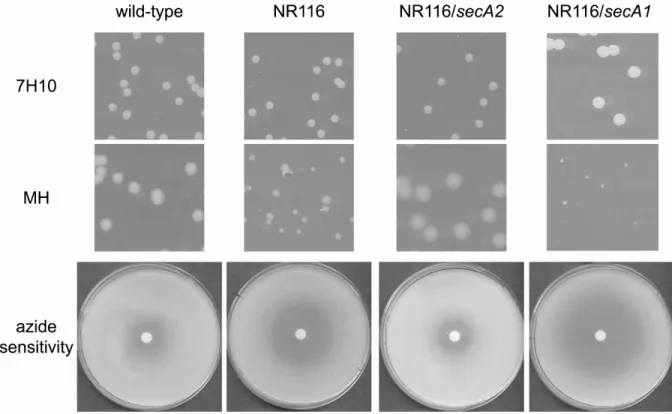
Another phenotype observed in earlier studies is that while overexpression of SecA1 has no effect on wild-type M. smegmatis, overexpression of SecA1 in mc22522 exacerbates the rich agar growth defect of the ∆secA2 mutant (3). This synthetic phenotype suggests a relationship between SecA1 and SecA2 or their respective export pathways. The ∆secA2 deletion allele in mc22522 still encodes a protein of 366 amino acids, although no truncated SecA2 protein is observed by immunoblot (data not shown). In other bacteria, SecA is capable of forming dimers (12, 14, 31, 46). Thus, it is possible that the SecA proteins of mycobacteria also form dimers, perhaps even SecA1/SecA2 heterodimers. We considered the possibility that a truncated SecA2 protein might have dominant effects through
interfering with SecA1-protein complexes and thereby produce the observed phenotype. To eliminate this potentially complicating factor, we constructed a new M. smegmatis ∆secA2 mutant in which the secA2 gene is fully deleted and only three codons remain. This strain, NR116, was then used for the remainder of this thesis work as an improved M. smegmatis ∆secA2 mutant.
(sacB). Hygromycin-resistant transformants were selected on 7H10 plates and then analyzed by Southern blot to confirm recombination of the deletion allele into the secA2 chromosomal locus, yielding the single-crossover strain SCO5 (Figure 2.2D). Upon counterselection for sucrose resistance, the integrated suicide vector will undergo a second recombination event, either yielding a ∆secA2 deletion mutant (Figure 2.2E) or regenerating the starting wild-type secA2 allele (Figure 2.2C) without the intervening suicide vector backbone. Sucrose resistant colonies were screened for hygromycin sensitivity, which reports on loss of the vector
backbone. SucR HygS clones were analyzed by PCR and Southern blot until we identified NR116 as a ∆secA2 deletion mutant (Figure 2.2F).
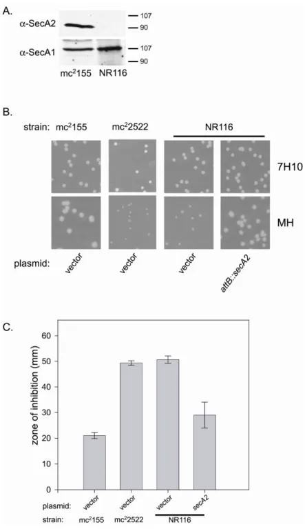
After constructing NR116, immunoblot analysis of whole cell lysates confirmed that SecA2 is no longer present in this new ∆secA2 mutant. As expected, deletion of SecA2 in NR116 had no effect on expression of SecA1 (Figure 2.3A). NR116 was then tested for all the previously reported phenotypes of mc22522 (3, 24). When grown on rich Mueller Hinton agar, NR116 colonies were smaller than colonies of wild-type M. smegmatis. The phenotype of NR116 was indistinguishable from that of the original ∆secA2 mutant mc22522 on rich agar (Figure 2.3B). As reported previously, neither secA2 mutant exhibited a mutant phenotype when growing on minimal 7H10 agar media (3). We also tested NR116 for sensitivity to sodium azide. Sodium azide is known to target ATPases. In bacteria, the major target of azide is SecA (23). NR116 was more sensitive to azide than wild-type M.