ROLE OF NANOMATERIAL PHYSICOCHEMICAL PROPERTIES ON FATE AND TOXICITY IN BACTERIA AND PLANTS
Danielle Slomberg
A dissertation submitted to the faculty of the University of North Carolina at Chapel Hill in the partial fulfillment of the requirements for the degree of Doctor of Philosophy in the
Department of Chemistry (Analytical Chemistry).
Chapel Hill 2013
Approved by:
ABSTRACT
Danielle Slomberg: Role of Nanomaterial Physicochemical Properties on Fate and Toxicity in Bacteria and Plants
(Under the direction of Mark H. Schoenfisch)
Nanomaterials, defined as having at least one dimension <100 nm, are ubiquitous in nature. However, engineered nanomaterials have gained increasing attention for use in drug-delivery applications and consumer goods. Examination of nanomaterial toxicity, both beneficial (e.g., drug delivery to bacterial pathogens) and detrimental (e.g., death of terrestrial plants), thus warranted. Herein, I present the evaluation of nitric oxide-releasing nanomaterial toxicity to bacteria and silica particle toxicity to plants as a function of nanomaterial physicochemical properties.
Nanomaterial toxicity toward planktonic (i.e., free-floating) Pseudomonas aeruginosa and Staphylococcus aureus bacteria was evaluated as a function of scaffold
weight due to rapid association between the cationic scaffolds and negatively-charged bacterial cell membranes.
Given the importance of nanomaterial physicochemical properties in planktonic bacterial killing, the NO-releasing scaffolds were also evaluated against clinically-relevant bacterial biofilms. Similar to planktonic studies, smaller particle sizes proved more efficient in delivering NO throughout the biofilm. Particles with rod-like shape also eradicated biofilms more effectively. The role of NO-releasing dendrimer and chitosan oligosaccharide hydrophobicity was prominent in scaffold diffusion through the biofilm and subsequent NO delivery, with scaffolds modified with hydrophobic functionalities generally exhibiting better bacterial association. Lastly, biofilm eradication was more effective for NO-releasing dendrimers exhibiting sustained NO-release compared to delivery of NO via an initial burst.
Phytotoxicity and uptake of silica nanoparticles was evaluated for the plant Arabidopsis thaliana as a function of particle size, surface composition, and shape (i.e.,
ACKNOWLEDGEMENTS
I would first like to thank my advisor, Dr. Mark Schoenfisch, for providing guidance and very practical knowledge and training that will last throughout the years.
I thank Dr. Alexis Carpenter, Dr. Bin Sun, and Dr. Yuan Lu for allowing me to assist in their NO-releasing nanomaterial projects early on. I thank Angela Broadnax for helping to synthesize particles for phytotoxicity experiments. I thank Dr. Wallace Ambrose for assistance with sample sectioning and thoughtful discussion. I also thank Dr. Neal Kramarcy and Dr. Michael Chua for assistance with confocal microscopy.
To the past and present Schoenfisch lab members: I thank you for making work an enjoyable and inspiring place to be. To my non-chemistry friends: I thank you for reminding me to keep balance in my life.
I thank my Mom, Dad, Christine, and Jim, as well as my siblings Allen, Ashley, and Zack for always supporting me through thick and thin…no matter what crazy ideas I
TABLE OF CONTENTS
LIST OF TABLES ... xii
LIST OF FIGURES ... xiv
LIST OF ABBREVIATIONS AND SYMBOLS ...xx
CHAPTER 1. RECENT ADVANCES IN EVALUATION OF NANOMATERIAL TOXICITY TOWARD BACTERIA AND PLANTS ...1
1.1 Overview of nanomaterial toxicity ...1
1.1.1 Engineered nanomaterials ...2
1.1.2 Nanomaterial-cell interactions ...3
1.1.3 Evaluation of nanomaterial toxicity ...4
1.2 Nanomaterial toxicity toward bacteria ...5
1.2.1 Bacteria in clinical settings ...5
1.2.2 Current bacteria eradication strategies ...8
1.2.3 Nitric oxide as an antibacterial agent ...8
1.2.4 Nitric oxide donors ...10
1.2.5 Tuning properties of nitric oxide-releasing nanomaterials ...20
1.3 Nanomaterial toxicity toward plants ...22
1.3.2 Nanomaterial physicochemical properties in
phytotoxicity ...24
1.3.3 Silica nanoparticle phytotoxicity ...26
1.4 Summary of dissertation research ...26
REFERENCES ...29
CHAPTER 2. ROLE OF NITRIC OXIDE-RELEASING SCAFFOLD PROPERTIES ON ANTIBACTERIAL EFFICACY AGAINST PLANKTONIC BACTERIA ...41
2.1 Introduction ...41
2.1.1 Nitric oxide-releasing silica particles...42
2.1.2 Nitric oxide-releasing dendrimers ...43
2.1.3 Nitric oxide-releasing chitosan oligosaccharides...44
2.2 Material and Methods ...45
2.2.1 Synthesis of nitric oxide-releasing silica particles ...47
2.2.2 Synthesis of nitric oxide-releasing PPI dendrimers ...48
2.2.3 Synthesis of nitric oxide-releasing chitosan oligosaccharides ...49
2.2.4 Fluorescently-labeled scaffolds ...50
2.2.5 Scaffold characterization and nitric oxide release ...51
2.2.6 Planktonic bactericidal assays...52
2.2.7 Confocal microscopy ...53
2.2.8 In vitro cytotoxicity...56
2.3 Results and Discussion ...57
2.3.2. Nitric oxide-releasing dendrimers ...64
2.3.3 Nitric oxide-releasing chitosan oligosaccharides...68
REFERENCES ...73
CHAPTER 3. ROLE OF NITRIC OXIDE-RELEASING SCAFFOLD PROPERTIES ON ANTIBACTERIAL EFFICACY AGAINST BIOFILM-BASED BACTERIA ...78
3.1 Introduction ...78
3.1.1 Nitric oxide-releasing silica particles...80
3.1.2 Nitric oxide-releasing dendrimers ...80
3.1.3 Nitric oxide-releasing chitosan oligosaccharides...81
3.2 Materials and Methods ...81
3.2.1 Synthesis of nitric oxide-releasing silica particles ...83
3.2.2 Synthesis of nitric oxide-releasing amphiphilic dendrimers...86
3.2.3 Synthesis of nitric oxide-releasing chitosan oligosaccharides. ...86
3.2.4 Fluorescently-labeled scaffolds ...87
3.2.5 Scaffold characterization and nitric oxide release ...88
3.2.6 Planktonic bactericidal assays...89
3.2.7 Bacterial biofilm assays ...90
3.2.8 Confocal microscopy ...92
3.2.9 In vitro cytotoxicity...93
3.3.1 Nitric oxide-releasing silica particles...93
3.3.2. Nitric oxide-releasing amphiphilic dendrimers ...110
3.3.3 Nitric oxide-releasing chitosan oligosaccharides...117
REFERENCES ...121
CHAPTER 4. ROLE OF DENDRIMER NITRIC OXIDE-RELEASE KINETICS IN ERADICATION OF PLANKTONIC BACTERIA AND BIOFILMS ...127
4.1 Introduction ...127
4.2 Materials and Methods ...130
4.2.1 Synthesis and characterization of secondary amine-functionalized PAMAM dendrimers...131
4.2.2 N-diazeniumdiolation of secondary amine-functionalized dendrimers ...132
4.2.3 Nitric oxide-release measurements. ...133
4.2.4 Planktonic bactericidal assays...133
4.2.5 Biofilm bacterial assays. ...134
4.2.6 Confocal microscopy. ...135
4.2.7 In vitro cytotoxicity...136
4.3 Results and Discussion ...137
4.3.1 Bactericidal efficacy against planktonic bacteria ...139
4.3.2 Bactericidal efficacy against biofilm bacteria...141
4.3.3 Confocal microscopy. ...144
4.3.5 Cytotoxicity of PAMAM dendrimers as a function
of nitric oxide-release kinetics. ...151
4.4 Conclusions ...153
REFERENCES ...155
CHAPTER 5. SIZE- AND SHAPE-DEPENDENT SILICA NANOPARTICLE PHYTOTOXICITY AND UPTAKE IN ARABIDOPSIS THALIANA ...161
5.1 Introduction ...161
5.2 Materials and methods ...163
5.2.1 Silica nanoparticle synthesis and characterization ...164
5.2.2 Plant growth ...166
5.2.3 Transmission electron microscopy ...167
5.2.4 Silicon elemental analysis ...167
5.3 Results and discussion ...168
5.3.1 Size-dependent phytotoxicity and uptake ...168
5.3.2 Shape-dependent phytotoxicity and uptake ...196
5.4 Conclusions ...200
REFERENCES ...203
CHAPTER 6. SUMMARY AND FUTURE DIRECTIONS ...207
6.1 Summary ...207
6.2 Future Directions ...212
6.2.2 Effects of nitric oxide against additional
clinically-relevant bacterial strains ...214 6.2.3 Effects of nitric oxide against polymicrobial
biofilms ...216 6.2.4 Phytotoxicity as a function of plant species and
LIST OF TABLES
Table 3.1. Particle size as determined by transmission electron microscopy (TEM) and total micromoles of NO released per mg of particle as measured by the Griess assay. Size measurements are n≥20 and total NO release is n≥3
syntheses ...95 Table 3.2. Size and aspect ratio of silica nanorods as determined by
scanning electron microscopy (SEM) and total micromoles NO released per mg of particle as measured by the Griess assay. Size measurements are n≥50 and total NO release is
n≥3 syntheses. . ...96
Table 3.3. Determination of planktonic and biofilm MBCs and bactericidal NO doses for NO-releasing 14, 50, and 200 nm
silica particles against P. aeruginosa and S. aureus biofilms. . ...98 Table 3.4. Determination of planktonic and biofilm MBCs and
bactericidal NO doses for NO-releasing AR1, AR4, and AR8
silica particles against P. aeruginosa and S. aureus biofilms. ...99 Table 4.1. Nitric oxide release properties of
N-diazeniumdiolate-modified PAMAM dendrimers as measured by NOA. All
values are n≥3 measurements. ...138
Table 4.2. Determination of planktonic and biofilm MBCs and bactericidal NO doses for NO-releasing PAMAM dendrimers
against P. aeruginosa and S. aureus bacteria. ...140 Table 5.1. Size and charge characterization of SiNPs via TEM, DLS,
and zeta potential. Measurements were made in Hoagland’s #2 nutrient solution (400 mg L-1) with SiNP concentration of 250 ppm unless noted. Measurement of 14 nm SiNPs made
in ethanol.a ...171 Table 5.2. Carbon and hydrogen weight percents for unmodified and
Table 5.3. Variation in pH for exposure groups where pH was not adjusted over 6 weeks. Blank exposure groups were grown in
a solution of Hoagland’s #2 Basal Salt Mixture (400 mg L-1). ...177 Table 5.4. Silicon determination in roots, rosette, and stems for 250 ppm
exposure group after 6 weeks using ICP-OES. Results are reported in mg Si per kg plant tissue. Error represents variation
between three instrument integrations. ...191
Table 5.5. Silicon determination in roots, rosette, and stems for 1000 ppm exposure group after 6 weeks using ICP-OES. Results are reported in mg Si per kg plant tissue. Error represents variation
between three instrument integrations. ...192
Table 5.6. Si determination in roots, rosette, and stem for 250 ppm exposure group after 6 weeks using ICP-OES. Results are
normalized for the nanoparticle volume. . ...193
Table 5.7. Si determination in roots, rosette, and stem for 1000 ppm exposure group after 6 weeks using ICP-OES. Results are
normalized for the nanoparticle volume. ...194
Table 5.8. Silicon determination in roots, rosette, and stems for 250 ppm exposure to AR1 and AR3 particles after 6 weeks using ICP-OES. Results are reported in mg Si per kg plant tissue. Error
LIST OF FIGURES
Figure 1.1. Stages of bacterial adhesion to a substrate and subsequent
biofilm formation ...6
Figure 1.2. N-diazeniumdiolate (A) formation and (B) proton-initiated
decomposition ...12
Figure 1.3. Representative formation of silica particles via (A)
hydrolysis and (B) condensation under basic conditions ...14
Figure 1.4. Formation of secondary amine sites on dendrimers via Michael addition or ring-opening reaction and subsequent
N-diazeniumdiolation...18
Figure 1.5. Formation of secondary amine-modified chitosan
oligosaccharides and subsequent N-diazeniumdiolation ...21
Figure 1.6. Transformation and key processes of engineered
nanoparticles in soil ...23
Figure 2.1. Confocal microscopy images of RITC-modified (A) 50, (B) 100, and (C) 200 nm silica nanoparticle (10 μg/mL) association with planktonic P. aeruginosa after 10 min
incubation. Scale bar is 5 μm. ...60 Figure 2.2. Confocal images of 50 nm RITC-modified NO-releasing
silica particle association with planktonic P. aeruginosa. Images were acquired (A) 0 (addition of particles), (B) 2.4, (C), 6.4, (D) 19.5, and (E) 39 min after addition of
nanoparticles at 10 μg/mL. Scale bar is 10 μm. . ...61
Figure 2.4. Confocal microscopy images of P. aeruginosa cells exposed to the same NO dosage (10 μmol/L) via incubation with NO-releasing G2-and G5-PPI-SO (8.7 and 10 μg/mL, respectively) and 50 nm AHAP3/TEOS nanoparticles (22 μg/mL). Intracellular NO is indicated by the appearance of DAF-2 green fluorescence, while PI red fluorescence indicates compromised membranes (cell death). Images were acquired (A) 30, (B) 46, (C) 60, (D) 64, (E) 86, and (F) 94
min after dendrimer/nanoparticle addition. . ...66 Figure 2.5. Confocal microscopy images of RITC-modified control and
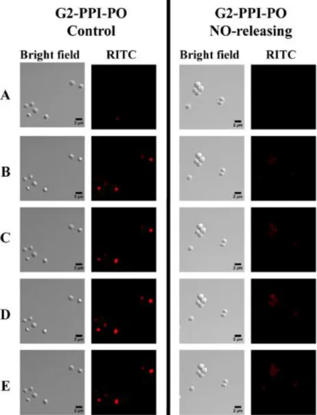
NO-releasing G2 PPI-PO dendrimer (400 μg/mL) association with S. aureus cells. Images were acquired (A) 4, (B) 12, (C)
18, (D) 30, and (E) 45 min following dendrimer addition. . ...67 Figure 2.6. Synthesis of secondary amine-and
N-diazeniumdiolate-functionalized chitosan oligosaccharide derivatives. A) Chitosan 1 and Chitosan 2) and subsequent N-diazeniumdiolate formation of the resulting materials (Chitosan 1/NO and Chitosan 2/NO); B) Modification of secondary amine-functionalized chitosan oligosaccharides
with PEG (Chitosan 3 and Chitosan 3/NO). . ...69 Figure 2.7. Confocal microscopy images of RITC-modified Chitosan
2/NO-5k; (A) 24 min, (B) 28 min, (C) 42 min and Chitosan 3/NO-5k; (D) 82 min, (E) 86 min, (F) 110 min, (H) 120 min association with P. aeruginosa cells (150 μg/ mL). Overlay images of P. aeruginosa cells incubated with (G) Chitosan
2/NO-5k at 44 min and (H) Chitosan 3/NO-5k at 120 min. . ...71 Figure 3.1. Fluorescent images of P. aeruginosa biofilm exposed to the
same particle concentration (1 mg/mL) and NO dosage (~250 μmol/L) of NO-releasing (A) 14, (B) 50, or (C) 150 nm particles for 30 or 60 min. DAF-2 green fluorescence indicates increased intracellular NO and PI red fluorescence
indicates compromised cell membranes (i.e., cell death). . ...103 Figure 3.2. Fluorescent images of P. aeruginosa biofilm exposed to the
μmol/L) of NO-releasing (A) AR1, (B) AR4, or (C) AR8 particles for 15 or 60 min. DAF-2 green fluorescence indicates increased intracellular NO and PI red fluorescence
indicates compromised cell membranes (i.e., cell death). ...105 Figure 3.3. Fluorescent images of RITC-modified (A) 14 and (B) 150 nm
control particle (0.1 mg/mL) diffusion in P. aeruginosa biofilm 30 min after particle addition. Green Syto 9 fluorescence shows biofilm cells. Increased RITC red fluorescence indicates more efficient particle diffusion within
biofilm. . ...107 Figure 3.4. Cytotoxicity of NO-releasing (white) and control (gray) silica
particles against L929 mouse fibroblasts at MBC concentrations required for biofilm killing listed in Tables 3
and 4; (A) P. aeruginosa and (B) S. aureus. ...109 Figure 3.5. Confocal microscopy images of P. aeruginosa biofilms
incubated with (A) PE-37-NO, (B) ED-NO, (C) G1-PE 73-NO, and (D) G3-G1-PE 73-NO RITC-labeled dendrimers for 1 h (50 μg/mL). Increased red fluorescence indicates more efficient dendrimer–bacteria association and improved
diffusion. Scale bar is 300 μm. . ...114
Figure 3.6. Confocal microscopy images of intracellular DAF-2 fluorescence in P. aeruginosa biofilms incubated with (A) G1-and (B) G3-PE 73-NO for 1 h (20 μg/mL) Green DAF-2 fluorescence indicates increased intracellular NO levels.
Scale bar is 50 µm ...116 Figure 3.7. Confocal fluorescence images of RITC-labeled chitosan
oligosaccharide association with P. aeruginosa biofilms: A) Chitosan 2/NO-5k, B) Chitosan 3/NO-5k, C) Chitosan 2-10k) and images of Syto 9 labeled biofilms incubated with D) Chitosan 2/NO-5k, E) Chitosan 3/NO-5k and F) Chitosan 2/NO-10k. Scale bar is 40 μm. The Syto 9 (green) fluorescence shows biofilm cells. Increased RITC (red) fluorescence indicates more efficient chitosan diffusion
Figure 4.1. Determination of bacterial viability for (A) P. aeruginosa and (B) S. aureus biofilms exposed to control (i.e., non-NO-releasing) PAMAM dendrimers at 5.0 mg/mL. Viability is
similar for the four control scaffolds at the highest MBC. .. ...143 Figure 4.2. Fluorescent images of RITC-modified (A) PO, (B) 1:1, (C)
1:7, and (D) ACN control PAMAM dendrimers (200 μg/mL) diffusion in P. aeruginosa biofilm 1 h after dendrimer addition. Green Syto 9 fluorescence shows biofilm cells. Similar RITC red fluorescence indicates similar association
and diffusion within biofilm for each scaffold. . ...145 Figure 4.3. Fluorescent images of P. aeruginosa biofilm exposed to the
same dendrimer concentration (200 μg/mL) of NO-releasing (A) PO, (B) 1:1, (C) 1:7, or (D) ACN dendrimers for 1 or 4 h. DAF-2 green fluorescence indicates increased intracellular NO and PI red fluorescence indicates compromised cell
membranes (i.e., cell death). . ...147 Figure 4.4. Cytotoxicity of NO-releasing (white) and control (gray)
PAMAM dendrimers against L929 mouse fibroblasts at MBC concentrations required for killing of planktonic (A) P.
aeruginosa and (B) S. aureus as listed in Table 4.2. . ...149
Figure 4.5. Cytotoxicity of NO-releasing (white) and control (gray) PAMAM dendrimers against L929 mouse fibroblasts at MBC concentrations required for (A) P. aeruginosa and (B)
S. aureus biofilm killing as listed in Table 4.2. . ...150
Figure 4.6. Cytotoxicity of (A) NO-releasing and (B) control PAMAM dendrimers against L929 mouse fibroblasts at concentrations of 0.2 (dashed), 1.0 (light gray), 3.0 (dark gray), and 5.0
Figure 5.1. TEM of SiNPs with sizes of 14.8 ± 2 nm (A), 51.4 ± 5 nm (B), and 211.5 ± 24 nm (C). Scale bar is 100 nm (A) or 200
nm (B and C)...170 Figure 5.2. Growth data for plants harvested at 3 weeks with (A, C) pH
5.8 and (B, D) pH unadjusted after exposure to 250 ppm (white), 1000 ppm (light gray), or calcined (dark gray) SiNPs. Values are normalized to plants grown in blank solution. *Significant difference at 95% relative to blank. Of note, stems were not developed by 3 week harvest for
measurement. . ...174 Figure 5.3. Growth data for plants harvested at 6 weeks with (A, C, E)
pH 5.8 and (B, D, F) pH unadjusted after exposure to 250 ppm (white), 1000 ppm (light gray), or calcined (dark gray) SiNPs. Values are normalized to plants grown in blank
solution. *Significant difference at 95% relative to blank. . ...175 Figure 5.4. Growth after 6 weeks in (A) blank nutrient solution with no
pH adjustment and (B) after exposure to 250 ppm of 200 nm
SiNPs with no pH adjustment. Seed holder diameter is 2 cm.. ...176 Figure 5.5. Plants grown for 6 weeks in (A) pH 5.8 nutrient solution and
(B) nutrient solution adjusted to pH 8. Scale bar is 15 cm. . ...179 Figure 5.6. Growth of plants exposed to 1000 ppm SiNPs after 6 weeks
showing development with (A) no pH adjustment; (B) pH 5.8; and (C) pH unadjusted with calcined SiNPs. Scale bar is
15 cm. ...182 Figure 5.7. Growth of plants after 6 week exposure to 1000 ppm calcined
SiNPs with growth medium maintained at pH 8. Scale bar is
15 cm. ...183 Figure 5.8. Transmission electron microscopy images of roots from (A)
blank solution and from 1000 ppm exposure after 6 weeks with SiNP sizes of (B) 14 nm; 14 kx magnification; (C) 50 nm; 29 kx; and (D) 200 nm; 14 kx. Arrows point to SiNPs in
each image. ...186 Figure 5.9. Transmission electron microscopy image of roots from 1000
Figure 5.10. Scanning electron micrographs of (A) AR1 and (B) AR3
silica particles. Scale bar is 500 nm (A) or 2 μm (B). . ...197
Figure 5.11.Transmission electron micrographs of root cells after exposure to 250 ppm (A) AR1 or (B) AR3 particles. Scale bar is 0.2 μm. Arrow
LIST OF ABBREVIATIONS AND SYMBOLS
% percentage
[…] concentration
[NO]max maximum NO flux
~ approximately
°C degree(s) Celsius
µg microgram(s)
µL microliter(s)
µmol micromole
AEAI
N-(2-aminoethyl)-3-amino-isobutyl-dimethyl methoxysilane
AHAP N-(6-aminohexyl)aminopropyltrimethoxysilane
Ar argon
atm atmosphere(s)
ATTC American Type Culture Collection
BET Brunauer-Emmett-Teller
CDC Centers for Disease Control and Prevention CFU colony forming units
cm centimeter(s)
CO2 carbon dioxide
d day(s)
DAF-2 4,5-diaminofluorescein
DAF-2 DA 4,5-diaminofluorescein diacetate DLS dynamic light scattering
DMF N,N-dimethylformamide
E. coli Escherichia coli
e.g. for example
ED 1,2-epoxy-9-decene
EPS exopolysaccharide
et al. and others
etc. and so forth
EtOH ethanol
g gravitational force
h hour(s)
H2O water
HCl hydrochloric acid
i.e. that is
ICP-OES
inductively coupled plasma-optical emission spectroscopy
kg kilogram
KOH potassium hydroxide
m meter(s)
M molar
MAP 3-methylaminopropyltrimethoxysilane MBC 3-methylaminopropyltrimethoxysilane
MeOH methanol
mg milligram(s)
min minute(s)
mL milliliter(s)
mm millimeter(s)
mM millimolar
mmol millimole(s)
mol mole
mol% percent of total moles
N2 nitrogen
N2O3 nitrogen
NaCl sodium chloride
NaOH sodium hydroxide
NaOMe sodium methoxide
NH4OH ammonium hydroxide
nm nanometer(s)
nmol nanomole(s)
NO nitric oxide analyzer
NO2 nitrogen dioxide
NO2- nitrite
NOA nitric oxide analyzer
O2 oxygen
OH- hydroxide ion
ONOO- peroxynitrite
P.
aeruginosa Pseudomonas aeruginosa
PAMAM poly(amidoamine)
PBS phosphate buffered saline, pH 7.4 PDI polydispersity index
PEG poly(ethylene glycol)
PI propidium iodide
pmol picomole(s)
PO propylene oxide
ppb parts per billion PPI poly(propylene imine) ppm parts per million
PROLI/NO N-diazeniumdiolate-modified L-proline RSNO S-nitrosothiol
S. aureus Staphylococcus aureus
S.
epidermidis Staphylococcus epidermidis
SEM scanning electron microscope/microscopy
SO styrene oxide
t time
t1/2 half-life
TEM transmission electron microscope/microscopy TEOS tetraethoxysilane
THF tetrahydrofuran
tmax time to max NO flux TMOS tetramethoxysilane
TSA tryptic soy agar
TSB tryptic soy broth
v/v volume/volume
w/w weight/weight
CHAPTER 1: RECENT ADVANCES IN EVALUATION OF NANOMATERIAL TOXICITY TOWARD BACTERIA AND PLANTS
In 1959, physicist and Nobel laureate, Richard Feynman, challenged the scientific
community to think small, noting that “[a]toms on a small scale behave like nothing on a
large scale.”1 Feynman also suggested that “[the] problems of chemistry and biology
[could] be greatly helped” if the ability “to do things on an atomic level, [was] ultimately
developed.” Inspired by these challenges, the field of nanotechnology has rapidly
developed as researchers work to create materials with new molecular organization,
properties, and functions relative to the bulk material.2 However, evaluation of the
toxicity of these engineered nanomaterials toward biological systems has only begun in
recent years. In this introductory chapter, nanomaterial toxicity, both beneficial (e.g.,
drug delivery to bacterial pathogens) and detrimental (e.g., death of terrestrial plants),
will be discussed as a function of the material’s physicochemical properties.
1.1 Overview of nanomaterial toxicity
1.1.1 Engineered nanomaterials
Since the advent of nanotechnology, a wide variety of nanomaterials have been
developed for a range of applications including drug delivery, biotechnology, water
decontamination, and communication technologies.3, 4 These engineered nanomaterials
for distinct applications include metal (e.g., Fe and Ag) and metal oxide nanoparticles
(e.g., TiO2 and SiO2), dendrimers, carbon nanotubes, and quantum dots among others.6 As the quantities and types of ENMs increase and consumers begin to use
nanomaterial-containing products with greater frequency, the need for an improved understanding of
nanomaterial physicochemical behavior and prevention of unintended biological and
environmental consequences will also rise.7 Humans and a myriad of other organisms will be exposed to ENMs through intended (e.g., common therapeutic use) or incidental
routes (e.g., release into atmosphere, rivers, soil, etc). Thus, proactive measures are
necessary to fully evaluate both the beneficial and detrimental implications associated
with ENMs.
The behavior of ENMs, and ultimately their toxicity, is influenced by several
physicochemical properties, such as increased surface area to volume ratio,8 size, shape, exterior functionality,9 and drug-release kinetics.10 Each of these characteristics is significant in dictating how a nanomaterial will interact with cells and the surrounding
environment. Generally, nanomaterials exhibit increased reactivity and toxicity compared
to their bulk counterparts.8 Midander et al. reported increased DNA damage in human lung cells exposed to nano-copper compared to micron-sized copper particles.11 However, in some cases there is no difference in toxicity between nanomaterials and bulk
formulations. Heinlaan et al. noted no significant differences in ecotoxicity to select
bacteria and crustaceans exposed to ZnO, TiO2, and CuO nanoparticles and their bulk
dissolved Zn and not nanoparticle effects.13 Evaluation of nanomaterial toxicity relative to the corresponding bulk material on a case by case basis is thus warranted.
1.1.2 Nanomaterial-cell interactions
Nanomaterial-cell interactions will greatly depend on nanomaterial size, shape,
surface characteristics, and drug-release properties (if any). Generally, improved
penetration of cell membranes is observed for smaller nanoparticles. Association of
nanomaterials with the cell membrane may also be governed by nanomaterial shape.
Nanomaterial surface charge is important in that the cell membrane is negatively-charged
and will associate more readily with positively-charged materials. In addition, more
hydrophobic ENMs are better able to penetrate the lipophilic membrane.
After association with the cell, nanomaterials can cause DNA damage, degrade
the membrane, or interrupt key processes for cellular function. The mechanism of action
varies with nanomaterial composition, size, and shape. For example, Yang et al. studied
the cytotoxicity, oxidative stress, and genotoxicity as a function of ENM composition
(i.e., carbon nanotubes and silica and ZnO nanoparticles), size, and shape.14 Particle composition played the primary role in cytotoxicity, and genotoxicity was significantly
influenced by ENM shape. However, no significant toxicity was attributed to size. The
effects of nanomaterial physicochemical properties on their interactions with cells is
clear, thus researchers are currently focused on further developing methodology to
understand and evaluate these interactions and any resulting toxicity.
Given the importance of assessing ENM parameters in toxicity, a variety of in vitro and in vivo assays are currently used for screening risk. In vitro systems are ideal in that they are cost effective and generate rapid, reproducible results.15 Common methods for screening new ENMs include the LDH (cell membrane integrity) and MTT assays (mitochondrial function), as well as assays for measuring cell-generated reactive oxygen species and immunochemistry markers (apoptosis and necrosis).15, 16 Due to the variety of physiological environments and applications in which ENMs may be encountered, these in vitro assays are tested against numerous cell types including phagocytic, neural, hepatic, epithelial, endothelial, and red blood cells.16 Despite the toxicity data generated with in vitro tests, determinations of safety need to be made based on the final fate of ENMs in biological systems.15 Thus, tests to evaluate ENM absorption, distribution, transformation, and excretion in vivo are ultimately warranted. Researchers are now focused on a predictive toxicological approach in which high-throughput screening is used in vitro and in vivo to determine ENM structure-activity relationships and hazard risk.17-19
1.2 Nanomaterial toxicity toward bacteria
1.2.1 Bacteria in clinical settings
Although bacteria are ubiquitous in aquatic and soil-based ecosystems, their prevalence in clinical settings has become particularly problematic, with hospital-acquired infections being the fourth-leading cause of death in the United States.22, 23 Open wounds24, 25 and implanted medical devices such as prosthetic heart valves,23 orthopedic implants, and catheters are frequent sites of microbial infection despite aseptic procedures and instrument sterilization.23, 26, 27 With >99% of all bacteria existing in a biofilm state, these infections are increasingly difficult to treat.28 Bacterial biofilms have shown decreased susceptibility to antibacterials compared to their planktonic (i.e., free-floating) counterparts.28-31 Consequently, infected medical implants often necessitate device removal,32 and hospital-acquired infections can result in sepsis,33 and even death. Current research is focused on developing nanomaterial-based drugs to address these problems. Furthermore, understanding the clinical significance of nanomaterial toxicity to bacteria will aid in the design of future antibacterial and anti-biofilm agents.
Irreversible Attachment Reversible
Attachment Maturation I Maturation II Dispersal
reversibly attach via gravitational, electrostatic, and van der Waals forces. Short range
(i.e., hydrogen boding, dipole-dipole, ionic, and hydrophobic) interactions also influence
the degree of bacterial adhesion. After a few hours, the bacteria begin to secrete
extracellular polymeric substances (EPS) and form cell-to-cell bridges, thus irreversibly
attaching to the surface.35, 36 Maturation of the biofilm continues as bacterial cells grow, divide, and form microcolonies (clusters of bacteria).37 The biofilm formation process continues over hours to days depending on several abiotic (e.g., nutrient conditions and
pH) and biotic (e.g., quorum-sensing) factors. Following maturation, detachment of
biofilm bacteria from microcolonies may occur via regulated dispersal mechanisms to
release planktonic cells that can further colonize surfaces.34, 38
Given the frequency and complexity of bacterial surface colonization, and the rate
at which bacterial biofilms are increasingly difficult to treat, research into antibacterial
resistance mechanisms has continued to garner attention. The traditional antibacterial
resistance mechanisms attributed to planktonic bacteria include efflux pumps, modifying
enzymes, and target mutations, but these mechanisms do not always explain the
resistance of biofilm-embedded bacteria.39 Genetic mutation of the biofilm bacteria is not likely the main cause of resistance since biofilm cells will once again exhibit
susceptibility to antibacterial agents upon dispersal.40, 41 Three proposed mechanisms for the increased resistance of biofilms are slow antibacterial penetration beyond surface
layers of the biofilm, differentiation of cells into a resistant phenotype, and reduced
As a result, complete eradication of biofilms using common antibacterials has been met with increasing difficulty and is often not feasible.
1.2.2 Current bacteria eradication strategies
Ideally, hospital-acquired infections would be controlled by eliminating initial bacteria attachment to a surface and thus preventing biofilm formation by adherent cells.34 However, superhydrophobic,42 heparin-coated,43 and antibacterial-doped substrates44, 45 have met varied success in eliminating bacterial adhesion and reducing clinical infection. These strategies are also often not amenable to implanted sensor applications, which can hinder their utility. As such, additional bacteria eradication strategies have been implemented for cases in which surface colonization cannot be prevented. Bacterial biofilm formation has been minimized through interference of iron metabolism,46, 47 enhancement of macrophage phagocytosis of the bacteria,48 or disruption of quorum-signaling.23, 34 Another biofilm control strategy has focused on targeting the EPS layer. Hatch and Schiller observed enhanced diffusion of the antibiotics, gentamicin and tobramycin, after the enzyme, alginate lyase, degraded the EPS layer of P. aeruginosa biofilms.49 Lastly, several researchers are working toward the development of new antibacterial agents, that will be effective against current and emerging multi-drug resistant bacteria strains.50-53 Successful new antibacterial agents will need to not only prevent initial adhesion or eradicate established biofilms, but must do so without fostering resistance or harming healthy host cells.
Nitric oxide (NO) is an endogenously-produced, diatomic free radical that participates in several concentration-dependent processes within the body.54-56 At low concentrations (~pM–nM), NO mediates vasodilation, angiogenesis, and neurotransmission.57-59 However, at higher concentrations (~µM), NO can serve as a potent antibacterial agent, inducing oxidative and nitrosative stresses that damage the bacteria membrane.60-62 In the presence of bacterial pathogens, macrophages utilize inducible nitric oxide synthase (iNOS) to generate NO. Once formed, NO can react with oxygen or other reactive oxygen intermediates (e.g., superoxide) to yield antibacterial byproducts (e.g., peroxynitrite, nitrogen dioxide, and dinitrogen trioxide).60 These reactive species can then induce oxidative stress, resulting in lipid peroxidation, tyrosine nitrosation, and oxidative DNA cleavage. Concurrently, thiol nitrosation, deamination of cellular proteins, and nitrosamine formation occur via nitrosative stress. Through disruption of bacterial components and disabling of crucial cell functions, NO and its reactive byproducts have proven effective as antibacterial agents against both Gram-positive and Gram-negative species.63, 64 Additionally, NO is a known anti-biofilm agent, capable of eradicating a variety of microbial strains including P. aeruginosa, E. coli, S. aureus, S. epidermidis, and C. albicans.62 The utility of NO as an antibacterial and anti-biofilm agent is demonstrated by its broad-spectrum and multi-mechanistic killing, thereby reducing the chance for bacteria resistance to treatment.
antioxidant enzymes, including superoxide dismutase, catalase, and peroxidase.65 Superoxide dismutase converts superoxide formed from the reaction of NO and oxygen into hydrogen peroxide. Catalase and peroxidase then convert the produced hydrogen peroxide into water. Using these enzymatic pathways, mammalian cells are able to turn superoxide and hydrogen peroxide into water, thus reducing NO-induced toxicity. Of note, some bacterial cells (e.g., S. aureus) produce lower levels of antioxidant enzymes and can adapt to oxidative stress via NO-mediated cytoprotection; however, bacteria are generally unable to mitigate the toxic effects of NO and its reactive byproducts as readily as mammalian cells.66
In addition to NO produced endogenously by immune cells, exogenous gaseous NO has proven effective in eradicating bacterial infections.67 However, control over the dose and location of delivery is challenging due to the highly reactive nature of gaseous NO. While administration of gaseous NO is appropriate for topical and pulmonary treatments, the use of NO in other clinical applications necessitates greater control of dosage and delivery.68 As such, compounds (NO donors) that store NO and release it upon an appropriate trigger (e.g., pH change, light, heat) have been developed.56
1.2.4 Nitric oxide donors
under physiological conditions, and varied NO-release kinetics.69 The N-diazeniumdiolate moieties are formed on amine sites of the donor compound upon
reaction with high pressures (~10 atm) of gaseous NO in the presence of a strong base
(Figure 1.2 A).70 In the more generally accepted mechanism, the amine reacts with an NO dimer (N2O2) to form the N-diazeniumdiolate. Release of NO then occurs via proton-intitiated decomposition, in which the N-diazeniumdiolate decomposes under aqueous conditions to yield 2 moles of NO and the parent secondary amine (Figure 1.2 B).
Overall, the N-diazeniumdiolate functionality is stable in the absence of a proton source
and at low temperature, making it an appropriate NO donor for a variety of
clinically-relevant applications.
N-diazeniumdiolate NO donors have been formed on small molecules such as proline (PROLI/NO) and diethylenetriamine (DETA/NO) for treatment of cardiovascular
disease and respiratory distress.55, 71 However, low molecular weight (LMW) donors diffuse rapidly, exhibit short circulation times, and often necessitate higher doses of NO
to elicit an effect.56 Side effects including hypotension, headaches, and possible tolerance have been noted during clinical use.72 Given these shortcomings, macromolecular scaffolds can instead be utilized to obtain controlled and localized NO delivery, facilitate
targeting, and decrease potential for tolerance.56, 61
Relative to LMW donors, macromolecular NO-releasing scaffolds have numerous
advantages including increased NO payload, targeted and localized delivery, and tunable
A
B
control over the concentration, location, timing, and duration of NO release can decrease the potential for tolerance and unwanted toxic side effects. As such, several types of NO-releasing protein, organic, inorganic, and hybrid polymer macromolecular scaffolds have been designed.73 An ideal NO-releasing macromolecular scaffold would be functionalized with multiple NO donors and/or targeting ligands to achieve localized release with the desired payload while controlling toxicity. The NO-releasing macromolecular scaffolds presented in this work include silica nanoparticles, dendrimers, and chitosan oligosaccharides. These NO-release vehicles allow for the tuning of size, shape, hydrophobicity, and NO-release kinetics to evaluate the effects of nanomaterial physicochemical properties on bactericidal efficacy to planktonic bacteria and biofilms.
A
B
Silica particles can be synthesized via the Stӧber method through the hydrolysis and condensation of a tetraalkoxysilane in a solution of water, an alcohol solvent (e.g., ethanol), and a base catalyst.76 Monodispersity of the synthesized particles is dependent on the rates of nucleation and growth during the reaction. Conditions favoring simultaneous formation of particle nuclei, with similar subsequent growth result in particles with narrow distributions in size. Silica particles synthesized via the Stӧber method are generally ≥100 nm as the conditions favor particle growth over nucleation. Alternatively, silica particles with sizes on the low end of the nanoparticle regime (i.e., <100 nm) can be synthesized by a reverse microemulsion approach in which particles are confined to micelles of a pre-determined size as they form.77 Tuning of particle size is critical in biomedical applications since effective drug delivery is generally inversely proportional to particle size.
In addition to size, the aspect ratio of silica particles can be altered to evaluate drug delivery efficiency and nanoparticle–cell interactions as a function of shape. Rod-like particles of various compositions (e.g., poly(ethylene glycol), silica, and poly(lactide-co-glycotide)) have demonstrated increased mammalian cell internalization and circulation time compared to spherical particles, thus improving their capacity for drug delivery.79-83 Silica nanorods of varied aspect ratio (i.e., 1–8) have been synthesized via a surfactant-templated synthesis, where the nanorods are grown along micelles. Control over the shape and geometry of the micelle ultimately impacts the final shape of the silica nanorod. Particle aspect ratio has been tuned by altering the type of surfactant, reaction temperature, ammonia concentration, or solution volume. Despite previous studies on mammalian cell interactions and uptake, there is a lack of understanding on how nanoparticle shape influences bactericidal efficacy against planktonic and biofilm-based bacteria.
To fully explore the effects of NO-releasing nanomaterial characteristics on toxicity toward bacteria, dendrimers have been evaluated as a second type of delivery scaffold. Dendrimers are highly-branched, nano-scale macromolecules.86 Bonds emanate from a central core at the interior of the dendrimer, forming a hyper-branched, multivalent structure. A wide range of dendritic scaffolds have been used in drug delivery,87 gene transfection,88, 89 tissue engineering,90 and even antibacterial applications. For example, quaternary ammonium-modified dendrimers have demonstrated efficacy in bacterial killing, but their inherent positive charge resulted in significant toxicity to mammalian cells.91 Masking this charge with poly(ethylene glycol) (PEG) helped mitigate the cytotoxicity while still providing potent antibacterial efficacy. Poly(amidoamine) (PAMAM) dendrimers and partially PEG-modified dendrimers were also proven effective against planktonic bacteria, with complete eradication of P. aeruginosa and S. aureus at µg/mL concentrations.92
In drug delivery applications, important dendrimer parameters to evaluate are size (i.e., generation), surface functionalization, and cytotoxicity. Unlike silica particles that range from ~10–1000 nm, dendrimers can be reproducibly synthesized with sizes <10 nm. Of note, as dendrimer generation is increased, the number of surface groups at the exterior also increases, likely enhancing biocidal action. The type of functional group bound to the dendrimer scaffold has also been shown to influence NO-release properties. For example, NO-releasing PAMAM dendrimers functionalized with varied carbon chain lengths (i.e., C1–C7) exhibited half-lives of 2.5–86 min, with the longer chain resulting in extended NO release.94
and thus offer a better approach for designing NO-releasing scaffolds with improved conversion efficiency and total storage.
Similar to NO-releasing silica and dendrimer scaffolds, the physicochemical properties (e.g., molecular weight and exterior functionality) of chitosan oligosaccharides can be tailored to influence NO-release kinetics and bactericidal action. The molecular weight (MW) of chitosan oligosaccharides can be controlled through the oxidative degradation of chitosan using hydrogen peroxide. Hydrogen peroxide concentration, (0.5–3.5%), reaction temperature (≥80 °C), and reaction time (~2.5–12 h) have all been shown to influence the degree of chitosan degradation and the resulting chitosan oligosaccharide MW.101 Additionally, a variety of functional groups can be introduced to the chitosan oligosaccharides via covalent modification. For example, secondary amine functionality can be imparted to the chitosan oligosaccharides via a cationic ring opening reaction with 2-methyl aziridine (MAz), after which PEG modification can be achieved via the Michael addition.
1.2.5 Tuning properties of nitric oxide-releasing nanomaterials
Figure 1.5 Formation of secondary amine-modified chitosan oligosaccharides and subsequent N-diazeniumdiolation.
A
B
Chitosan 1 x=1 Chitosan 2 x=2
Chitosan 1/NO x=1 Chitosan 2/NO x=2
Chitosan 3 x=2 Chitosan 3/NO x=2
A
B
Chitosan 1 x=1 Chitosan 2 x=2
Chitosan 1/NO x=1 Chitosan 2/NO x=2
Chitosan 3 x=2 Chitosan 3/NO x=2
A
B
Chitosan 1 x=1 Chitosan 2 x=2
Chitosan 1/NO x=1 Chitosan 2/NO x=2
Chitosan 3 x=2 Chitosan 3/NO x=2
H+ H2O
number of their N-diazeniumdiolates at the particle surface, likely promoting faster NO release and bacterial killing. Smaller nanoparticles are also likely to exhibit improved bactericidal efficacy due to more rapid diffusion to the bacterial cell surface. Particles with high aspect ratios (i.e., rod-like) may also enhance the efficiency of NO delivery given the potential for an increased surface area of interaction between the nanoparticle and bacterial membrane. Increased bactericidal efficacy of nanomaterials with more hydrophobic character will likely be observed due to improved association with the lipid-containing bacterial membrane; however, cytotoxicity may present a challenge. Lastly, evaluating scaffold NO-release kinetics will be crucial to understanding the desired release profile (i.e., short burst of NO versus low sustained levels) for eradication of planktonic bacteria versus biofilms.
1.3 Nanomaterial toxicity toward plants
1.3.1 Phytotoxicity and uptake of nanomaterials
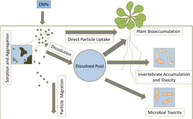
Figure 1.6 Transformation and key processes of engineered nanoparticles in soil (modified from Klaine et al).10
ENPs
Part
ic
le
Mi
gr
ati
on
Direct Particle Uptake
Microbial Toxicity Invertebrate Accumulation
and Toxicity Plant Bioaccumulation
Sorp
ti
on
and
Ag
gr
eg
ati
on
nanoparticles tend to be transient in the environment, disappearing by either dissolution or aggregation, some ENPs have been shown to persist due to stabilization by surfactants or organic matter. A better understanding of particle fate, behavior, and potential toxicity in the environment is thus warranted. As such, researchers are evaluating ENP mobility, fate, and bioavailability in the environment as a function of size, shape, and surface charge.10, 102 Prior studies have assessed the toxicity of ENPs toward mammalian cells, bacteria, aquatic invertebrates, and other terrestrial organisms. However, plant toxicity (i.e., phytotoxicity) and potential uptake due to nanoparticle exposure have received less attention to date, especially with regard to ENP physicochemical properties.107, 108 Of note, ENP–plant interactions may not always result in toxicity, as some nanoparticles (e.g., mesoporous silica) have been used for target-specific delivery of proteins, nucleotides, or other chemicals for plant biotechnology applications.109
Previous work with plants has evaluated the toxicity of silica (SiO2), zinc oxide (ZnO), nickel hydroxide (Ni(OH)2), copper (Cu), cerium oxide (CeO2), titanium dioxide (TiO2), iron oxide (Fe3O4), gold (Au), silver (Ag), and iron (Fe) nanoparticles, as well as CdSe/ZnS quantum dots and carbon nanotubes to Arabidopsis thaliana,105, 110 rye grass,111, 112 mesquite,113 and select edible plant species including wheat and mung bean,106 alfalfa, tomato, corn, and cucumber.114, 115 Although current research has made some progress in determining the effects of nanomaterials on terrestrial plants, systematic studies are still lacking in the literature.
Some researchers have started to evaluate phytotoxicity as a function of nanomaterial physicochemical properties such as size, shape, and surface charge. Nanoparticle size has been the most readily examined parameter, with increased phytotoxicity generally observed in correlation with decreased particle size. Lee et al. noted that ZnO nanoparticles (~44 nm) were more toxic to Arabidopsis thaliana than micron-sized counterparts.105 Similarly, smaller (~3.5 nm) Au NPs were more toxic to tobacco plants and exhibited greater uptake compared to larger (~18 nm) particles.116 After 7 d exposure to silver NPs (20 and 100 nm), size-dependent NP toxicity was observed in L. minor.117 However, no significant difference in toxicity was noted between the 20 and 100 nm particles after a 14 d exposure.
1.3.3 Silica nanoparticle phytotoxicity
Silica nanoparticles (SiNPs) have received special interest in toxicology studies due to their prominence in cosmetic and biomedical applications.121-123 In one study, 12.5 and 27.0 nm SiNPs (20.0 and 28.8 mg L-1, respectively) were shown to reduce growth of green alga by 20% after 72 h.124 Phytotoxicity assays with Cucurbita pepo (zucchini), however, showed no significant difference in germination percent, root elongation, or biomass after exposure to 1000 mg L-1 bulk silicon powder and SiNPs (< 100 nm) for 5– 14 d.125 Lee and coworkers found that 42.8 nm SiNPs promoted Arabidopsis thaliana root elongation at a low concentration (400 mg L-1), but resulted in toxicity at higher concentrations (≥ 2000 mg L-1).105 While prior work on the phytotoxicity of SiNPs to higher plants has established a strong foundation, lack of toxicity examination as a function of increased SiNP size range (i.e., < 42.8 nm and > 100 nm), shape (i.e., rod-like versus spherical), duration of exposure, and surface composition necessitated further investigation. Additionally, visualization of SiNPs in the plant cells and determination of uptake location (i.e., roots, rosette, and stem) had not been previously observed.
1.4 Summary of dissertation research
1. determine the bactericidal efficacy of NO-releasing silica, dendrimer, and chitosan scaffolds against planktonic and biofilm bacteria as a function of size, shape, and surface exterior functionality;
2. evaluate the mechanisms of antibacterial activity for these scaffolds using confocal microscopy; and,
3. determine the phytotoxicity and uptake of silica nanoparticles for Arabidopsis thaliana as a function of the physical properties of this scaffold.
REFERENCES
1. Feynman, R. P., There's plenty of room at the bottom. Caltech Engineering and Science 1960, pp 22-36.
2. Alvarez, P., Nanotechnology in the Environment: The good, the bad, and the ugly. Journal of Environmental Engineering 2006, 132, 1233.
3. Roco, M. C.; Mirkin, C. A.; Hersam, M. C., Nanotechnology research directions for societal needs in 2020. New York, 2011.
4. Cañas, J. E.; Long, M.; Nations, S.; Vadan, R.; Dai, L.; Luo, M.; Ambikapathi, R.; Lee, E. H.; Olszyk, D., Effects of functionalized and nonfunctionalized single-walled carbon nanotubes on root elongation of select crop species. Environ. Toxicol. Chem. 2008, 27, 1922-1931.
5. Handy, R.; Kammer, F.; Lead, J.; Hassellov, M.; Owen, R.; Crane, M., The ecotoxicology and chemistry of manufactured nanoparticles. Ecotoxicol. 2008, 17, 287-314.
6. Wiesner, M. R.; Lowry, G. V.; Alvarez, P.; Dionysiou, D.; Biswas, P., Assessing the risks of manufactured nanomaterials. Environ. Sci. Technol. 2006, 40, 4336-4345.
7. Colvin, V. L., The potential environmental impact of engineered nanomaterials. Nat. Biotechnol. 2003, 21, 1166-1170.
8. Rotello, V., Nanoparticles: Building Blocks for Nanotechnology. Springer: New York, 2004.
9. Kim, S. T.; Saha, K.; Kim, C.; Rotello, V. M., The role of surface functionality in determining nanoparticle cytotoxicity. Accounts of Chemical Research 2013, 46, 681-691.
10. Klaine, S. J.; Alvarez, P. J. J.; Batley, G. E.; Fernandes, T. F.; Handy, R. D.; Lyon, D. Y.; Mahendra, S.; McLaughlin, M. J.; Lead, J. R., Nanomaterials in the environment: Behavior, fate, bioavailability, and effects. Environ. Toxicol. Chem. 2008, 27, 1825-1851.
12. Heinlaan, M.; Ivask, A.; Blinova, I.; Dubourguier, H.-C.; Kahru, A., Toxicity of nanosized and bulk ZnO, CuO and TiO2 to bacteria Vibrio fischeri and crustaceans Daphnia magna and Thamnocephalus platyurus. Chemosphere 2008, 71, 1308-1316.
13. Franklin, N. M.; Rogers, N. J.; Apte, S. C.; Batley, G. E.; Gadd, G. E.; Casey, P. S., Comparative toxicity of nanoparticulate ZnO, Bulk ZnO, and ZnCl2 to a freshwater microalga (Pseudokirchneriella subcapitata): The importance of particle solubility. Environ. Sci. Technol. 2007, 41, 8484-8490.
14. Yang, H.; Liu, C.; Yang, D.; Zhang, H.; Xi, Z., Comparative study of cytotoxicity, oxidative stress and genotoxicity induced by four typical nanomaterials: the role of particle size, shape and composition. J. Appl. Toxicol. 2009, 29, 69-78.
15. Sharifi, S.; Behzadi, S.; Laurent, S.; Forrest, M. L.; Stroeve, P.; Mahmoudi, M., Toxicity of nanomaterials. Chem. Soc. Rev. 2012, 41, 2323-2343.
16. Jones, C. F.; Grainger, D. W., In vitro assessments of nanomaterial toxicity. Adv. Drug Deliv. Rev. 2009, 61, 438-456.
17. Nel, A.; Xia, T.; Meng, H.; Wang, X.; Lin, S.; Ji, Z.; Zhang, H., Nanomaterial toxicity testing in the 21st century: use of a predictive toxicological approach and high-throughput screening. Accounts of Chemical Research 2012, 46, 607-621. 18. Sun, B.; Li, R.; Wang, X.; Xia, T., Predictive toxicological paradigm and high
throughput approach for toxicity screening of engineered nanomaterials. Int. J. Biomed. Nanosci. Nanotechnol. 2013, 3, 4-18.
19. Nel, A.; Zhao, Y.; Mädler, L., Environmental health and safety considerations for nanotechnology. Accounts of Chemical Research 2013, 46, 605-606.
20. Gagner, J. E.; Shrivastava, S.; Qian, X.; Dordick, J. S.; Siegel, R. W., Engineering Nanomaterials for biomedical applications requires understanding the nano-bio interface: A perspective. J. Phys. Chem. Lett. 2012, 3, 3149-3158.
21. Ray, P. C.; Singh, A. K.; Senapati, D.; Fan, Z.; Yu, H., Toxicity and environmental risks of nanomaterials: An Update. Bio-Nanotechnology, Blackwell Publishing Ltd.: 2013; 733-748.
23. Bryers, J. D., Medical biofilms. Biotechnol. Bioeng. 2008, 100, 1-18.
24. D'Avignon, L. C.; Hogan, B. K.; Murray, C. K.; Loo, F. L.; Hospenthal, D. R.; Cancio, L. C.; Kim, S. H.; Renz, E. M.; Barillo, D.; Holcomb, J. B.; Wade, C. E.; Wolf, S. E., Contribution of bacterial and viral infections to attributable mortality in patients with severe burns: An autopsy series. Burns 2010, 36, 773-779.
25. Goswami, A. P.; Goswami, N.; Patel, T.; Tripathi, C.; Trivedi, H., Antibiotic sensitivity profile of bacterial pathogens in postoperative wound infections at a tertiary care hospital in Gujarat, India. J. Pharmacol. Pharmacother. 2011, 2, 158-164.
26. Nablo, B. J.; Prichard, H. L.; Butler, R. D.; Klitzman, B.; Schoenfisch, M. H., Inhibition of implant-associated infections via nitric oxide release. Biomaterials 2005, 26, 6984-6990.
27. Rutala, W. A.; Weber, D. J., Infection control: the role of disinfection and sterilization. Journal of Hospital Infection 1999, 43, Supplement 1, S43-S55. 28. Smith, A. W., Biofilms and antibiotic therapy: Is there a role for combating
bacterial resistance by the use of novel drug delivery systems? Adv. Drug Deliv. Rev. 2005, 57, 1539-1550.
29. Anderl, J. N.; Franklin, M. J.; Stewart, P. S., Role of antibiotic penetration limitation in Klebsiella pneumoniae biofilm resistance to ampicillin and ciprofloxacin. J. Antimicrob. Chemother. 2000, 44, 1818-1824.
30. Nickel, J. C.; Ruseska, I.; Wright, J. B.; Costerton, J. W., Tobramycin resistance of Pseudomonas aeruginosa cells growing as a biofilm on urinary catheter material. J. Antimicrob. Chemother. 1985, 27, 619-624.
31. Cerca, N.; Martins, S.; Cerca, F.; Jefferson, K. K.; Pier, G. B.; Oliveira, R.; Azeredo, J., Comparative assessment of antibiotic susceptibility of coagulase-negative staphylococci in biofilm versus planktonic culture as assessed by bacterial enumeration or rapid XTT colorimetry. Journal of Antimicrobial Chemotherapy 2005, 56, 331-336
32. Mack, D.; Rohde, H.; Harris, L. G.; Davies, A. P.; Hortskotte, M. A.; Knobloch, J. K., Biofilm formation in medical-device related infection. Int. J. Artif. Organs 2006, 29, 343-359.
Epidemiology of sepsis and infection in ICU patients from an international multicentre cohort study. Intensive Care Medicine 2002, 28, 108-121.
34. Lindsay, D.; von Holy, A., Bacterial biofilms within the clinical setting: what healthcare professionals should know. J. Hosp. Infect. 2006, 64, 313-325.
35. Donlan, R. M., Biofilms: microbial life on surfaces. Emerging Infectious Diseases 2002, 8, 881-90.
36. Hall-Stoodley, L.; Costerton, J. W.; Stoodley, P., Bacterial biofilms: from the natural environment to infectious diseases. Nat. Rev. Microbiol. 2004, 2, 95-108. 37. Costerton, J. W.; Stewart, P. S.; Greenberg, E. P., Bacterial biofilms: A common
cause of persistent infections. Science 1999, 284, 1318-1322.
38. Parsek, M. R.; Fuqua, C., Biofilms 2003: Emerging themes and challenges in studies of surface-associated microbial life. Journal of bacteriology 2004, 186, 4427-4440.
39. Stewart, P. S.; William Costerton, J., Antibiotic resistance of bacteria in biofilms. The Lancet 2001, 358, 135-138.
40. Anwar, H.; van Biesen, T.; Dasgupta, M.; Lam, K.; Costerton, J. W., Interaction of biofilm bacteria with antibiotics in a novel in vitro chemostat system. Antimicrob. Agents Chemother. 1989, 33, 1824-1826.
41. Barraud, N.; Hassett, D. J.; Hwang, S.-H.; Rice, S. A.; Kjelleberg, S.; Webb, J. S., Involvement of nitric oxide in biofilm dispersal of Pseudomonas aeruginosa. J. Bacteriol. 2006, 188, 7344-7353.
42. Fadeeva, E.; Truong, V. K.; Stiesch, M.; Chichkov, B. N.; Crawford, R. J.; Wang, J.; Ivanova, E. P., Bacterial retention on superhydrophobic titanium surfaces fabricated by femtosecond laser ablation. Langmuir 2011, 27, 3012-3019.
43. Appelgren, P.; Ransjӧ, U.; Bindslev, L.; Larm, O., Does surface heparinisation reduce bacterial colonisation of central venous catheters? The Lancet 1995, 345, 130.
44. Chilukuri, D. M.; Shah, J. C., Local delivery of vancomycin for the prophylaxis of prosthetic device-related infections. Pharm. Res. 2005, 22, 563-572.
46. Narasimhan, J.; Antholine, W. E.; Chitambar, C. R., Effect of gallium on the tyrosyl radical of the iron-dependent M2 subunit of ribonucleotide reductase. Biochem. Pharmacol. 1992, 44, 2403-2408.
47. Singh, P., Iron sequestration by human lactoferrin stimulates P. aeruginosa surface motility and blocks biofilm formation. Biometals 2004, 17, 267-270. 48. Krishnamurthy, V. M.; Quinton, L. J.; Estroff, L. A.; Metallo, S. J.; Isaacs, J. M.;
Mizgerd, J. P.; Whitesides, G. M., Promotion of opsonization by antibodies and phagocytosis of Gram-positive bacteria by a bifunctional polyacrylamide. Biomaterials 2006, 27, 3663-3674.
49. Hatch, R. A.; Schiller, N. L., Alginate lyase promotes diffusion of aminoglycosides through the extracellular polysaccharide of mucoid Pseudomonas aeruginosa. Antimicrob. Agents Chemother. 1998, 42, 974-977. 50. Saravanakumari, P.; Mani, K., Structural characterization of a novel xylolipid
biosurfactant from Lactococcus lactis and analysis of antibacterial activity against multi-drug resistant pathogens. Bioresource Technol. 2010, 101, 8851-8854. 51. Norrby, S. R.; Nord, C. E.; Finch, R., Lack of development of new antimicrobial
drugs: a potential serious threat to public health. The Lancet Infectious Diseases 2005, 5, 115-119.
52. Khan, R.; Islam, B.; Akram, M.; Shakil, S.; Ahmad, A. A.; Ali, S. M.; Siddiqui, M.; Khan, A., Antimicrobial activity of five herbal extracts against multi drug resistant (MDR) strains of bacteria and fungus of clinical origin. Molecules 2009, 14, 586-597.
53. Malka, E.; Perelshtein, I.; Lipovsky, A.; Shalom, Y.; Naparstek, L.; Perkas, N.; Patick, T.; Lubart, R.; Nitzan, Y.; Banin, E.; Gedanken, A., Eradication of multi-drug resistant bacteria by a novel Zn-doped CuO nanocomposite. Small 2013, doi: 10.1002/smll.201301081.
54. Ignarro, L. J.; Buga, G. M.; Wood, K. S.; Byrns, R. E.; Chaudhuri, G., Endothelium-derived relaxing factor produced and released from artery and vein is nitric oxide. Proc. Natl. Acad. Sci. 1987, 84, 9265-9269.
56. Carpenter, A. W.; Schoenfisch, M. H., Nitric oxide release: Part II. Therapeutic applications. Chem. Soc. Rev. 2012, 41, 3742-3752.
57. Ferrari, C. K. B.; Franca, E. L.; Honorio-Franca, A. C., Nitric oxide, health and disease. J. Appl. Biomed. 2009, 7, 163-173.
58. Hill, B. G.; Dranka, B. P.; Bailey, S. M.; Lancaster, J. R.; Darley-Usmar, V. M., What part of NO don't you understand? Some answers to the cardinal questions in nitric oxide biology. J. Biol. Chem. 2010, 285, 19699-19704.
59. Ziche, M.; Morbidelli, L.; Masini, E.; Amerini, S.; Granger, H.; Maggi, C.; et al.; Geppetti, P.; Ledda, F., Nitric oxide mediates angiogenesis in vivo and endothelial cell growth and migration in vitro promoted by substance P. Journal of Clinical Investigation 1994, 94, 2036-2044.
60. Fang, F. C., Perspectives series: host/pathogen interactions. Mechanisms of nitric oxide-related antimicrobial activity. J. Clin. Invest. 1997, 99, 2818-2825.
61. Hetrick, E. M.; Shin, J. H.; Stasko, N. A.; Johnson, C. B.; Wespe, D. A.; Holmuhamedov, E.; Schoenfisch, M. H., Bactericidal efficacy of nitric oxide-releasing silica nanoparticles. ACS Nano 2008, 2, 235-246.
62. Hetrick, E. M.; Shin, J. H.; Paul, H. S.; Schoenfisch, M. H., Anti-biofilm efficacy of nitric oxide-releasing silica nanoparticles. Biomaterials 2009, 30, 2782-2789.
63. De Groote, M. A.; Fang, F. C., NO inhibitions: antimicrobial properties of nitric oxide. Clinical Infectious Diseases 1995, 21, S162-S165.
64. Privett, B. J.; Deupree, S. M.; Backlund, C. J.; Rao, K. S.; Johnson, C. B.; Coneski, P. N.; Schoenfisch, M. H., Synergy of nitric oxide and silver sulfadiazine against gram-negative, gram-positive, and antibiotic-resistant pathogens. Mol. Pharm. 2010, 7, 2289-2296.
65. Weydert, C. J.; Cullen, J. J., Measurement of superoxide dismutase, catalase and glutathione peroxidase in cultured cells and tissue. Nature protocols 2009, 5, 51-66.
66. Gusarov, I.; Nudler, E., NO-mediated cytoprotection: Instant adaptation to oxidative stress in bacteria. Proc. Natl. Acad. Sci. 2005, 102, 13855-13860.
68. Eroy-Reveles, A. A.; Mascharak, P. K., Nitric oxide-donating materials and their potential in pharmacological applications for site-specific nitric oxide delivery. Future Medicinal Chemistry 2009, 1, 1497-1507.
69. Wang, P. G.; Xian, M.; Tang, X.; Wu, X.; Wen, Z.; Cai, T.; Janczuk, A. J., Nitric oxide donors: chemical activities and biological applications. Chem. Rev. 2002, 102, 1091-1134.
70. Hrabie, J. A.; Keefer, L. K., Chemistry of the nitric oxide-releasing diazeniumdiolate ("nitrosohydroxylamine") functional group and its oxygen-substituted derivatives. Chem. Rev. 2002, 102, 1135-1154.
71. Lam, C.-F.; van Heerden, P. V.; Blott, J.; Roberts, B.; Ilett, K. F., The selective pulmonary vasodilatory effect of inhaled DETA/NO, a novel nitric oxide donor, in ARDS–a pilot human trial. J. Crit. Care 2004, 19, 48-53.
72. Yamamoto, T.; Kakar, N. R.; Vina, E. R.; Johnson, P. E.; Bing, R. J., The effect of aspirin and two nitric oxide donors on the infarcted heart in situ. Life Sciences 2000, 67, 839-846.
73. Riccio, D. A.; Schoenfisch, M. H., Nitric oxide release: Part I. Macromolecular scaffolds. Chem. Soc. Rev. 2012, 41, 3731-3741.
74. Lee, J. E.; Lee, N.; Kim, T.; Kim, J.; Hyeon, T., Multifunctional mesoporous silica nanocomposite nanoparticles for theranostic applications. Accounts of Chemical Research 2011, 44, 893-902.
75. Brinker, C. J.; Scherer, G. W., Sol-gel science: the physics and chemistry of sol-gel processing. Elsevier: 1990.
76. Stӧber, W.; Fink, A.; Bohn, E., Controlled growth of monodisperse silica spheres in micron size range. J. Colloid Interf. Sci. 1968, 26, 62-69.
77. Carpenter, A. W.; Slomberg, D.L.; Rao, K.S.; Schoenfisch, M.H., Influence of scaffold size on bactericidal activity of nitric oxide-releasing silica nanoparticles. ACS Nano 2011, 5, 7235-7244.
78. Bogush, G. H.; Tracy, M. A.; Zukoski Iv, C. F., Preparation of monodisperse silica particles: Control of size and mass fraction. J. Non-Cryst. Solids 1988, 104, 95-106.