Control of
gdhR
Expression in
Neisseria
gonorrhoeae
via Autoregulation and a
Master Repressor (MtrR) of a Drug Efflux
Pump Operon
Corinne E. Rouquette-Loughlin,a,bYaramah M. Zalucki,a,b*Vijaya L. Dhulipala,a,b Jacqueline T. Balthazar,a,bRaúl G. Doyle,d*Robert A. Nicholas,dAfrin A. Begum,e Erica L. Raterman,eAnn E. Jerse,eWilliam M. Shafera,b,c
Laboratories of Bacterial Pathogenesis, VA Medical Center, Decatur, Georgia, USAa; Department of
Microbiology and Immunology, Emory University School of Medicine, Atlanta, Georgia, USAb; Emory Antibiotic Resistance Center, Emory University School of Medicine, Atlanta, Georgia, USAc; Departments of Pharmacology and Microbiology and Immunology, University of North Carolina, Chapel Hill, North Carolina, USAd;
Department of Microbiology and Immunology, F. Edward Hébert School of Medicine, Uniformed Services University, Bethesda, Maryland, USAe
ABSTRACT The MtrCDE efflux pump ofNeisseria gonorrhoeae contributes to gono-coccal resistance to a number of antibiotics used previously or currently in treat-ment of gonorrhea, as well as to host-derived antimicrobials that participate in in-nate defense. Overexpression of the MtrCDE efflux pump increases gonococcal survival and fitness during experimental lower genital tract infection of female mice. Transcription of mtrCDEcan be repressed by the DNA-binding protein MtrR, which also acts as a global regulator of genes involved in important metabolic, physiologic, or regulatory processes. Here, we investigated whether a gene downstream of mtrCDE, previously annotatedgdhRinNeisseria meningitidis, is a target for regulation by MtrR. In meningococci, GdhR serves as a regulator of genes involved in glucose catabolism, amino acid transport, and biosynthesis, including gdhA, which encodes an L-glutamate dehydrogenase and is located next to gdhR but is transcriptionally divergent. We report here that in N. gonorrhoeae, expression ofgdhR is subject to autoregulation by GdhR and direct repression by MtrR. Importantly, loss of GdhR sig-nificantly increased gonococcal fitness compared to a complemented mutant strain during experimental murine infection. Interestingly, loss of GdhR did not influence expression ofgdhA, as reported for meningococci. This variance is most likely due to differences in promoter localization and utilization between gonococci and menin-gococci. We propose that transcriptional control of gonococcal genes through the action of MtrR and GdhR contributes to fitness ofN. gonorrhoeaeduring infection.
IMPORTANCE The pathogenic Neisseria species are strict human pathogens that can cause a sexually transmitted infection (N. gonorrhoeae) or meningitis or fulmi-nant septicemia (N. meningitidis). Although they share considerable genetic informa-tion, little attention has been directed to comparing transcriptional regulatory sys-tems that modulate expression of their conserved genes. We hypothesized that transcriptional regulatory differences exist between these two pathogens, and we used the gdh locus as a model to test this idea. For this purpose, we studied two conserved genes (gdhR andgdhA) within the locus. Despite general conservation of the gdh locus in gonococci and meningococci, differences exist in noncoding se-quences that correspond to promoter elements or potential sites for interacting with DNA-binding proteins, such as GdhR and MtrR. Our results indicate that implications drawn from studying regulation of conserved genes in one pathogen are not neces-sarily translatable to a genetically related pathogen.
Received17 March 2017Accepted23 March 2017 Published11 April 2017
CitationRouquette-Loughlin CE, Zalucki YM, Dhulipala VL, Balthazar JT, Doyle RG, Nicholas RA, Begum AA, Raterman EL, Jerse AE, Shafer WM. 2017. Control ofgdhRexpression in
Neisseria gonorrhoeaevia autoregulation and a master repressor (MtrR) of a drug efflux pump operon. mBio 8:e00449-17.https://doi.org/10 .1128/mBio.00449-17.
EditorMichael S. Gilmore, Harvard Medical School
Copyright© 2017 Rouquette-Loughlin et al. This is an open-access article distributed under the terms of theCreative Commons Attribution 4.0 International license.
Address correspondence to William M. Shafer, [email protected].
*Present address: Yaramah M. Zalucki, Institute for Glycomics, Griffith University, Gold Coast Campus, Gold Coast, QLD, Australia; Raúl G. Doyle, Agile Sciences, Raleigh, North Carolina, USA.
This article is a direct contribution from a Fellow of the American Academy of Microbiology. External solicited reviewers: Michael Apicella, University of Iowa; Xavier Nassif, Universite Paris Descartes Faculte de medecine Necker/INSERM U1002.
crossm
KEYWORDS gonococci, transcription, efflux pumps, physiology
N
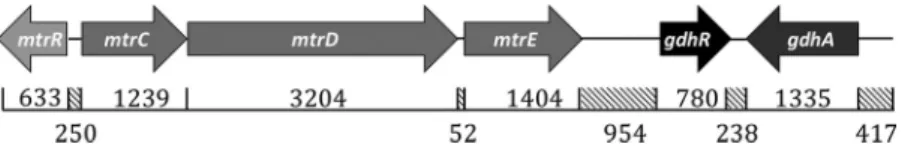
eisseria gonorrhoeaeis the etiologic agent of the sexually transmitted infection (STI) termed gonorrhea, which is the second most prevalent bacterial STI in the United States and had a worldwide incidence of an estimated 78 million infections in 2012 (1). The capacity of gonococci to develop resistance to antibiotics is now of great concern with the recent emergence of strains resistant to current and past frontline antibiotics (2–5). With respect to the clinical efficacy of antibiotic treatment regimens, evidence has been presented that overproduction of the gonococcal MtrCDE efflux pump due to cis- or trans-acting mutations that elevate transcription of mtrCDEcan contribute to clinically relevant levels of antibiotic resistance (6–10).The mtrR gene, which encodes the master repressor (MtrR) of the mtrCDEefflux pump operon (8–10), is located immediately upstream of themtrCDEoperon (Fig. 1). ThemtrRandmtrCDEgenes are oriented away from each other and have overlapping promoters. Transcription of mtrCDEis repressed when MtrR is bound to themtrCDE promoter, which overlaps themtrRpromoter (7, 8). Point mutations in the MtrR-binding site (8, 10), a single base pair deletion within a 13-bp inverted repeat sequence in the mtrR promoter (7), a point mutation that creates a new promoter (9), or missense mutations that cause radical amino acid replacements within the helix-turn-helix DNA-binding motif of MtrR can increase mtrCDEexpression and antimicrobial resis-tance (8, 10). Such elevated expression ofmtrCDEalso increased gonococcal fitnessin vivo when assessed by use of an experimental female murine lower genital tract infection model (11). In addition to regulatingmtrCDE, MtrR serves as a global regulator of gonococcal genes (12) and directly or indirectly activates or represses at least 65 genes outside themtrCDElocus. Included in these so-called “off-target” genes are those that are involved in the stress response (rpoH), amino acid synthesis (glnAandglnE), peptidoglycan biosynthesis (ponA), and regulation of gene expression (farR); the reg-ulatory properties of MtrR have been summarized elsewhere (2, 13).
Our analysis of the whole-genome sequence of strain FA1090 (http://www.genome .ou.edu) revealed an open reading frame (termed NGO 1360) positioned 943 bp downstream of themtrCDEoperon that encodes a transcriptional regulator previously annotated GdhR inNeisseria meningitidis(Fig. 1). GdhR belongs to the bacterial GntR family of proteins, which serve as gene regulators and contain a highly conserved N-terminal DNA-binding domain and a variable C-terminal domain involved in effector binding and oligomerization (14). InN. meningitidis, which causes often deadly men-ingitis or fulminant septicemia (15), GdhR regulates the expression of a number of genes, some of which are involved in metabolism (16, 17). Meningococcal GdhR has been reported to activate gdhA, which encodes an NADP-specificL-glutamate dehy-drogenase (16). Given the prominent role of MtrR in modulating gonococcal resistance to antimicrobials, its control of genes involved in metabolism, its influence on the fitness of gonococci in an experimental infection model, and its proximity togdhR, we tested the capacity of MtrR to regulate expression ofgdhRinN. gonorrhoeae, as well as the ability of GdhR to regulate genes in the gdhlocus (gdhR andgdhA). Our results suggest that gonococcal and meningococcal GdhRs have distinct regulatory properties that are driven by differences in promoter utilization of regulated genes and emphasize
the importance of bacterial species-specific studies for examining regulatory properties of a common DNA-binding protein.
RESULTS
The gdh locus in N. gonorrhoeae. Similar to N. meningitidis, the gdh locus in N. gonorrhoeaeFA19 is positioned 954 bp downstream from themtrlocus (Fig. 1) and contains two open reading frames, gdhR, which encodes a GntR-like DNA-binding protein, andgdhA, which encodesL-glutamate dehydrogenase. The gonococcal GdhR and GdhA proteins are 97% and 98% identical, respectively (data not shown) to the equivalent proteins described for meningococci (16). The end of gdhAis positioned 238 bp from the end ofgdhRand is transcribed in the opposite direction; transcription ofgdhAhas been reported to be activated by GdhR in meningococci (16).
Bioinformatic analysis (http://www.ncbi.nlm.nih.gov) revealed that the 200 bp up-stream ofgdhRin five gonococcal strains (FA19, FA1090, MS11, FA6140, and F89) were identical, except for a C-to-T change in FA1090 29 bp upstream of the translation start codon but after the transcription start site (TSS) (see below). In these same gonococcal strains, 100% identity was noted for the 500-bp sequence upstream ofgdhA(data not shown). When the same regions fromN. meningitidisstrain MC58 in the corresponding upstream regions of gdhR and gdhA were used in a BLAST search against whole-genome sequences, two other meningococcal isolates (LNP21362 and H44/76) were found to have identical sequences, while 10 others showed 99% identity (data not shown). Thus, our use of gonococcal strain FA19 for comparison to meningococcal strain MC58 is suitable for determining differences in regulation of thegdh locus in these pathogens. Although the DNA sequences of the gdh loci in gonococci and meningococci are very similar, important differences exist, especially in the location of promoters and potentialcis-acting regulatory sequences (see Fig. 3 for the CE insertion in the meningococcalgdhRand see Fig. 6 for thegdhApromoters, respectively). We hypothesized that the differences in these sequences between gonococci and menin-gococci could impact GdhR-mediated regulation of gene expression in gonococci and influence gonococcal biology. Accordingly, we sought to identify a phenotype that is linked to GdhR production in gonococci and then to examine regulation of gdhR expression and the capacity of GdhR to control model genes.
Loss of GdhR impactsin vivofitness of gonococci independently of the mtr
locus.Given the close location ofgdhRto themtrlocus (Fig. 1), we determined whether expression of GdhR influences transcription ofmtrCDEand resistance of gonococci to antimicrobials recognized by the MtrCDE efflux pump (2, 6–12). For this purpose, we constructed agdhRnull mutant as well as a complemented strain. We found that the wild-type parent (FA19), thegdhR::kanmutant, and the complemented strain displayed identical levels of susceptibility to antimicrobials (erythromycin [Erm] MIC, 0.25g/ml; penicillin MIC, 0.015 g/ml; Triton X-100 MIC, 100 g/ml), and these MICs varied according to the levels of the MtrCDE efflux pump (6, 7, 11, 18). Moreover, results from quantitative reverse transcription-PCR (qRT-PCR) experiments indicated that expression ofmtrC(the first gene in themtrCDEoperon [Fig. 1]) was not impacted by loss of GdhR (data not shown).
suggested that GdhR may be a negative regulator of in vivofitness; therefore, we sought to determine howgdhRis regulated in gonococci and if GdhR controls expres-sion of model genes (gdhRandgdhA) previously studied in meningococci (16).
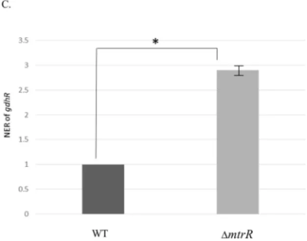
MtrR is a direct repressor ofgdhRexpression.MtrR exerts transcriptional repres-sion on themtrCDEoperon by binding to a promoter located upstream ofmtrC(8, 19). Given the close proximity ofgdhRto themtrlocus in both gonococci and meningo-cocci, we asked if MtrR regulatesgdhRexpression in gonococci. AlthoughgdhRwas not previously assigned to be an MtrR-regulated gene in an earlier transcriptional profiling study that employed gonococcal RNA prepared from mid-logarithmic-phase cultures (12), we reexamined MtrR control of gdhR for two reasons. First, the presence of a putative MtrR-binding site upstream of thegdhRgene (Fig. 3A) suggested such control is possible. This MtrR-binding site (boxed in Fig. 3A) was 60% homologous to the MtrR-binding site on themtrCpromoter region (Fig. 3B). Second, the work of Mercante et al. (20) showed that a different transcriptional factor (MpeR) expressed in gonococci displays growth phase-dependent regulons.
Results from qRT-PCR experiments indicated that deletion ofmtrR increasesgdhR transcription, supporting the hypothesis that MtrR controlsgdhRexpression in gono-cocci by functioning as a repressor of this gene in the late-logarithmic phase of growth (Fig. 3C). Using primer extension (PE) analysis (see Fig. S2), we identified threegdhR TSSs, positioned 73, 72, and 70 nucleotides upstream of the start of translation ofgdhR. These TSSs allowed us to identify a promoter element (5=-TAGAAT-3= for the ⫺10 hexamer and 5=-TTGACG-3= for the⫺35 hexamer) 81 bp upstream of the ATG trans-lational start codon (Fig. 3A). Importantly, the putative MtrR-binding site overlapped the⫺10 hexamer sequence of the predictedgdhRpromoter. Based on this promoter mapping and the identification of a predicted MtrR-binding site within the putative gdhR promoter, we tested if MtrR bound in a specific manner upstream of thegdhR coding sequence, and we used an electrophoretic mobility shift assay (EMSA) for this purpose. We found that 2 g of MBP-MtrR was sufficient to completely shift a 32P-labeled probe, termed R3/R2, that consisted of 393 bp of sequence upstream of the
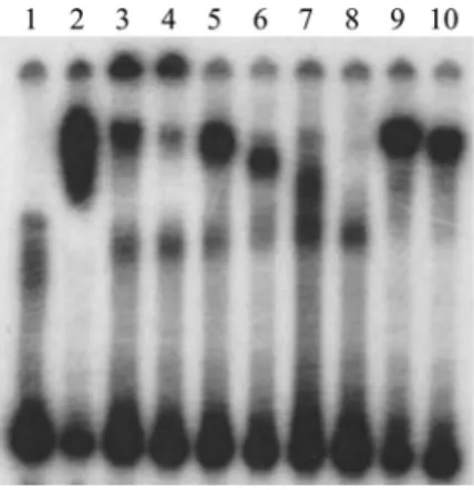
gdhRtranslational start (Fig. 4, lane 2). In order to better localize the MtrR-binding site(s) within this region, we performed a competitive EMSA with nonradioactive fragments of the R3/R2 probe used in the aforementioned EMSA. Binding competition assays showed that a smaller probe encompassing thegdhR promoter and its downstream region (probe R4/R2 [Fig. 3A]) competed with MtrR binding to the labeled R3/R2 probe
(Fig. 4, lanes 3 and 4), while a fragment located upstream of the promoter (R3/R5 [Fig. 3A]) did not (Fig. 4, lanes 5 and 6). Accordingly, we propose that MtrR represses gdhRexpression by binding within the promoter sequence that contains the predicted MtrR-binding site.
GdhR regulation of model genes in gonococci.We selected two GdhR genes for study:gdhRandgdhA, which constitute thegdhlocus (Fig. 1). We chose these 2 genes to test ifgdhRis subject to autoregulation by its gene product and becausegdhAhas been reported to be a GdhR-activated gene in meningococci (16) and is positioned near gdhRin both pathogens.
Pagliarulo et al. (16) suggested that the meningococcalgdhRtranscript originates in a Correia element (CE) (21) located upstream of thegdhRgene. An examination of 23 publicly available gonococcal genome sequences revealed that this CE is absent in the gdhR promoter region (data not shown). However, a putative GntR-like binding site (5=-TGTCATTA-3=) was identified between the⫺10 and the⫺35 sites of the gonococcal gdhR promoter overlapping the predicted MtrR-binding site (Fig. 3A, underlined in green). In order to investigate autoregulation ofgdhR, qRT-PCR analysis of total RNA prepared from mid- and late-log-phase cultures of strains FA19 and FA19gdhR::kanwas performed. The expression ofgdhRwas increased by 4-fold at mid-log phase and by a little more than 2-fold at late-log phase in the GdhR-negative mutant compared to the parental strain (Fig. 5A). We hypothesize that insertion of a CE upstream of gdhRin meningococci, but not in gonococci, results in the utilization of distinctgdhRpromoters FIG 4 Competitive EMSAs. The MtrR-binding site located on fragment R4/R2 has the highest affinity for the MtrR protein. Lanes: 1, probe R3/R2*alone; 2, probe R3/R2*plus 2g of MtrR; 3, probe R3/R2*plus 2g of MtrR plus 50⫻unlabeled R3/R2; 4, probe R3/R2*plus 2g of MtrR plus 100⫻unlabeled R3/R2; 5, probe R3/R2*plus 2g of MtrR plus 50⫻unlabeled R3/R5; 6, probe R3/R2*plus 2g of MtrR plus 100⫻unlabeled R3/R5; 7, probe R3/R2*plus 2g of MtrR plus 50⫻unlabeled R4/R2; 8, probe R3/R2*plus 2g of MtrR plus 100⫻unlabeled R4/R2; 9, probe R3/R2*plus 2g of MtrR plus 50⫻rnpB; 10, probe R3/R2*plus 2g of MtrR plus 100⫻rnpB. An asterisk indicates a radioactive probe. The location of the differentgdhRprobes are shown in Fig. 3A.
by these two related pathogens. Therefore, competing mechanisms ofgdhRregulation by MtrR and GdhR itself may not occur in meningococci. In this respect, it is important to note that most meningococci encode an MtrR protein that contains loss-of-function mutations inmtrR (22, 23), while nearly 80% of gonococci encode a wild-type MtrR (reviewed in reference 2).
Previous work indicated that expression ofgdhAin meningococci is directed by two promoters, only one of which is regulated by GdhR (16). The GdhR-activated promoter ofgdhAin meningococci has a putative GdhR-binding site (5=-TGTCAACA-3=) upstream of the⫺35 hexamer, based on similarity to the known GntR-binding site (5= -TGTcaACA-3=; the lowercase letters refer to nucleotides that differ from consensus GntR-binding site) in other bacteria (14); this site is also located in the corresponding gonococcal sequence (underlined in Fig. 6). In order to investigate whether GdhR binds to this site in gonococci, as was shown previously in meningococci, we performed EMSA compe-tition analysis using purified gonococcal His-tagged GdhR protein. The results showed that gonococcal GdhR binds specifically to a DNA fragment encompassing the GntR-binding site present upstream ofgdhAin gonococci (Fig. 7).
FIG 6 Alignment ofgdhApromoters from gonococcal strain FA19 (top) and meningococcal strain MC58 (bottom). The TSSs determined by primer extension experiments for strain FA19 identified in this study and that of MC58 as reported by Pagliarulo et al. (16) are represented in blue, green, and red, with their respective putative promoter elements. The consensus binding sequence for the GntR protein is underlined. ThegdhAtranslation start site is represented in purple.
In meningococci, the presence of anothergdhATSS was detected 207 bp upstream of the ATG translational start codon (represented in blue in Fig. 6). However, a GdhR-binding site was not identified within or near this distal promoter in meningo-cocci. Interestingly, we did not detect a TSS 207 bp upstream of the ATG in gonomeningo-cocci. This could be due to the presence of a mutation which changes the⫺10 hexamer from 5=-TAATTA-3= in meningococcal strain MC58 to 5=-TAACTA-3= in gonococcal strain FA19. To determine if gonococci have an additional promoter(s) forgdhAtranscription, we used PE analysis to identify transcription start sites. The results suggested the presence of two promoters (Fig. 6). We identified a TSS located 8 bp downstream from a⫺10 hexamer that constitutes the homolog of the above-mentioned meningococcal promoter (shown in red). We also identified three TSSs located upstream of a noncon-sensus⫺10 hexamer (5=-ATTTGT-3=) that is spaced 17 nucleotides from a weak⫺35 hexamer (5=-ATATGG-3=) (represented in green in Fig. 6). Importantly, this putative promoter has the previously identified GdhR-binding site (underlined sequence in Fig. 6) between its⫺10 and⫺35 hexamer sequences. This second gonococcal pro-moter was not identified in meningococci. Based on the location of the two putative gdhA promoters in gonococci, GdhR could impact expression of gdhA from both promoters through interaction with the identified GdhR-binding site.
Taken together, our promoter mapping studies suggest that differences exist re-gardinggdhAtranscription in gonococci and meningococci and that a GdhR-binding site influencesgdhAtranscription in gonococci. In order to assess promoter utilization in gonococci and any influence of GdhR ongdhAtranscription, we performed qRT-PCR analysis on RNA prepared from strain FA19 and its isogenicgdhR::kanmutant at mid-and late-logarithmic phases of growth. Unlike its influence ongdhRexpression, loss of GdhR did not impact gdhA expression in either mid-log- or late-log-phase cultures (Fig. 5B). One explanation for why we did not observe changes ingdhAexpression is that GdhR binds upstream of the most proximal promoter (represented in red in Fig. 6) and inhibits the binding of the RNA polymerase to the secondgdhRpromoter (repre-sented in green in Fig. 6), but it does not interfere with transcription from the proximal promoter, just as in meningococci. Consequently, when GdhR is present, the most proximal promoter (represented in red in Fig. 6) is the primary promoter used for transcription ofgdhA. When GdhR is absent, the most distal promoter (green in Fig. 6) becomes the primary promoter forgdhRtranscription.
DISCUSSION
Our interest in gdhR was spurred by its close location to the mtr locus, which encodes the tripartite RND-type efflux pump MtrC-MtrD-MtrE and a transcriptional repressor (Fig. 1). We hypothesized that GdhR and MtrR might have cross-regulatory activities on themtr andgdh loci, respectively. While we did not find evidence that GdhR regulatesmtrCDEor antimicrobial resistance, we did find that its loss significantly increased fitness of gonococci during an experimental infection of the lower genital tract of female mice. This experimental model of infection has been used by us to show the importance of the MtrCDE pump for gonococcal survivalin vivoand that gradients of fitness can be observed (11, 24), depending on the presence of distinct cis- or trans-acting mutations that influencemtrCDEexpression (2).
instance, GdhR autoregulation ofgdhRmay be unique to gonococci, because the presence of a CE in this region in meningococci, but not gonococci, likely influences promoter utilization and GdhR binding.
GdhR has been previously studied in meningococci for its capacity to regulate genes involved in metabolism, but heretofore it has not been investigated for its regulatory properties in gonococci. Although GdhR has been reported to activate expression of gdhAandgltT, which encodes anL-glutamate transporter that appeared to be essential for full virulence in a rodent model of invasive meningococcal disease (25), its capacity to autoregulate its own gene or be controlled by trans-acting factors has not been elucidated in either pathogen. The work presented here illustrates that although two genetically related pathogens can encode the same transcription factor (e.g., GdhR) and have conserved target genes (e.g.,gdhA), gene regulatory principles that have evolved for one pathogen may not necessarily apply to the related pathogen. Thus, although the GdhRs in meningococci and gonococci are identical, regulation of one target,gdhA, is distinct. WhilegdhAis a GdhR-activated gene in meningococci, based on quantitative analysis of levels of mRNA transcripts, our work failed to reveal differences in gdhA transcript levels in isogenic GdhR-positive and -negative gonococci. This does not mean that GdhR cannot activate gdhA in gonococci. We draw this conclusion because PE analysis suggested the presence of two promoters in gonococci that could direct transcription and be differentially impacted (activated or repressed) by GdhR, thereby giving the impression of lack ofgdhAregulation. We propose that differences in the DNA sequence in thegdh locus in gonococci versus meningococci result in distinct promoter utilization and regulation.
Additional studies are needed to define the GdhR regulon in gonococci in order to understand the role of this DNA-binding protein in controlling genes important for me-tabolism andin vivofitness ofN. gonorrhoeae. In this respect, the increased fitness of the gdhRmutant observed on days 3 and 5 (Fig. 2) corresponds to the time inflammation is detected in the mouse model (26). With the protocol we use, proinflammatory cytokines and chemokines begin to increase on day 3 and peak on day 5, along with a peak polymorphonuclear leukocyte influx on day 5; expression of antimicrobial peptides also peaks on day 5 (A. E. Jerse et al., unpublished data). Thus, it is possible thatgdhRmay downregulate genes important in the invasion of innate defenses. Depression or induction of genes that are important in growth and metabolism could also contribute to the increased fitness observed with thegdhRmutant. With these possibilities in mind, which form the basis for future studies, our results emphasize that pathogen-specific regulatory actions of a common DNA-binding protein likely exist even between closely related bacteria (e.g., gonococci versus meningococci) and that differences in gene control, which could be influenced bycis-regulatory elements, may have consequences for the overall biology of members in same genus.
MATERIALS AND METHODS
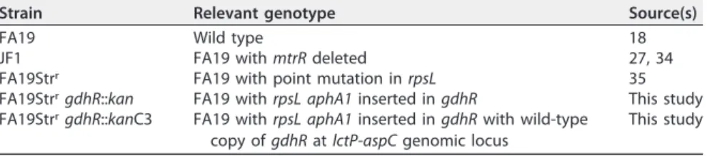
Gonococcal strains, growth conditions, and determination of susceptibility to antimicrobial agents.Strains used in this study are presented in Table 1. Gonococcal strains were grown overnight at 37°C under 5% (vol/vol) CO2on GC agar containing defined supplements I and II (27). Determination of susceptibility of test strains to antibiotics was performed by the agar dilution method, and results were reported as the MIC (18). Antibiotics were purchased from Sigma Chemical Co. (St. Louis, MO). Esche-richia colistrains were grown overnight at 37°C on LB agar.
TABLE 1 Strains ofNeisseria gonorrhoeaeemployed in this study
Strain Relevant genotype Source(s)
FA19 Wild type 18
JF1 FA19 withmtrRdeleted 27, 34
FA19Strr FA19 with point mutation inrpsL 35
FA19StrrgdhR::kan FA19 withrpsL aphA1inserted ingdhR This study
FA19StrrgdhR::kanC3 FA19 withrpsL aphA1inserted ingdhRwith wild-type
copy ofgdhRatlctP-aspCgenomic locus
Construction of thegdhR-negative mutant and its complemented strain.The plasmid construct used to insertionally inactivate thegdhRgene was created in pUC18us, which is pUC18 containing the 10-bp uptake sequence preceding the HindIII site in the polylinker. Overlap extension PCR was used to amplify thegdhRgene containing an internal XbaI site by using the upstream primer 5=GepR-new-Bam (5=-AGAGGATCCTAGAAACTGGTAAGGCCTCAGA-3=) and midstream reverse primer 3=GepR-mid-XbaI (5= -CTTCCTCAAACTTTTCTAGACAAAACCGAATCCGC-3=) to amplify the first half of the gene and the mid-stream forward primer 5=GepR-mid-XbaI (5=-GCGGATTCGGTTTTGTCTAGAAAAGTTTGAGGAAG-3=) and the downstream reverse primer 3=-gepR-EcoRI (5=-AGAGAATTCATACCTCCCAATCCTGCAC-3=) to amplify the second half of the gene. The two midstream primers are complementary to one another, and so in the second round of PCR, aliquots of each amplification product were used as the template with the forward upstream and reverse downstream primers to generate thegdhRgene containing an XbaI site in the middle of the gene. This modifiedgdhRconstruct was digested with EcoRI and BamHI and cloned into similarly digested pUC18us in which the existing XbaI site was destroyed by cutting with XbaI, filling in the 5=-overhangs with Klenow fragment, and religation. To create thegdhRinactivation construct, a blunt-ended kanamycin (Kan) resistance cassette derived from pLG338 (28) was ligated into the pUC18us-gdhRplasmid at the filled-in XbaI site in the middle of thegdhRgene. This construct was linearized by digestion with EcoRI and used to transform FA19Strr(FA19 containing therpsLallele from FA1090 that confers resistance to streptomycin [Str]), with transformants selected on GC agar plates containing 50g/ml Kan and verified by colony PCR and sequencing.
The pGCC3 vector (29) was used to complement FA19StrrgdhR::kanbecause it allows the integration of a wild-type copy ofgdhRunder its own promoter at the transcriptionally silent intergenic region betweenlctPandaspC. pgntR3pac1 (5=-GATCTTAATTAAGCCGATTGCCGTGTAGTTTT-3=) and pme1gepR4 (5=-GATCGTTTAAACCCAGACCGTCTGAAC-3=) were used to amplify thegdhRgene. The resulting PCR product was cloned into the pGCC3 vector. The pGCC3gdhRconstruct was verified by sequencing and then transformed into FA19StrrgdhR::kan. FA19StrrgdhR::kanC3 transformants were selected on GC agar plates supplemented with 1g/ml of Erm and verified by colony PCR.
Competitive murine infection.Mixed bacterial inocula containing similar numbers of the two strains being tested were prepared by harvesting test strains from GC agar plates grown for 18 to 21 h and suspending the bacteria in 4 to 5 ml of 1⫻phosphate-buffered saline (PBS). The suspensions were passed through a 1.2-m filter to remove bacterial aggregates and, using previously determined standard values for each strain relating readings of the optical density at 600 nm (OD600) to CFU counts, the bacterial suspensions were diluted to ~5⫻107CFU/ml before being mixed in a 1:1 ratio (actual ratios were determined by plating as described below). Female NCI BALB/c mice (6 to 8 weeks old; Charles River, Inc.) in the diestrus stage or anestrus were injected subcutaneously with 0.5 mg of Premarin (Pfizer) on days⫺2, 0, and⫹2. On day 0, the mice were inoculated vaginally with 20l of the mixed suspension (~1⫻106to 2⫻106CFU/mouse). Mice were also treated with Str, vancomycin, and trimethoprim as described elsewhere (30) to suppress the overgrowth of commensal flora that occurs under the influence of estrogen. The vaginas were gently swabbed with a PBS-moistened sterile swab on days⫹1,⫹3, and
⫹5 postinfection, and the swab material was suspended in 1 ml of PBS. Serial dilutions were performed in GC broth with 0.05% saponin, and dilutions were plated on GC agar with 100g/ml of Str for determination of total CFU, GC agar with 100g/ml of Str and 50g/ml Kan for determination of FA19StrrgdhR::kanCFU, or 1g/ml Erm for FA19StrrgdhR::kanC3 CFU. Plates were incubated overnight at 37°C under 7% (vol/vol) CO2, and colonies were counted after 24 to 48 h. The number of CFU recovered on GC-Str plus Kan or GC-Str plus Em agar plates was subtracted from the number of CFU recovered on GC-Str agar to determine the number of wild-type/parent CFU recovered. The CI was calculated according to the following equation: [(CFUMutant/CFUWt)Output]/[(CFUMutant/CFUWt)Input]. A value of 20 CFU (limit of detection) was used to calculate the CI for cultures from which CFU from one of the two strains were not recovered. Competitive infections were repeated, and the data were combined to test reproducibility and increase the statistical power. The Kruskall-Wallis test with Dunn’s multiple-comparisons test (GraphPad Prism) was used to compare the differences in the CIs for mice inoculated with each mixture.
Animal experiments were conducted in the laboratory animal facility at USUHS, which is fully accredited by the Association for Assessment and Accreditation of Laboratory Animal Care, under a protocol approved by the USUHS Institutional Animal Care and Use Committee.
Mapping transcriptional start sites by primer extension analysis.Total RNA from strain FA19 was prepared at the late-logarithmic phase of growth in GC broth as described above, using the method of Baker and Yanofsky (31). Primer extension experiments were performed as described previously (7) with 6g of total RNA with primers PEgntR (5=-CCAGTTTCATCACTCCTCCT-3=) or PEgdhA (5=-TTTGAGGTTGG CAAACAGGG-3=). The AMV reverse transcriptase primer extension system from Promega (Madison, WI) was used as described by the manufacturer. The TSSs were determined via electrophoresis of the extension products on a 6% (wt/vol) DNA sequencing acrylamide gel adjacent to reference sequencing reaction mixtures.
Purification of the GdhR protein.Construction of pET15bgdhRwas done by amplifying thegdhR open reading frame using the primers gdhR_F (5=-GATCGCCATATGAAACTGGTAAGGCCTCAG-3=) and gdhR_R (5=-GCGGATCCTCATACCTCCCAATCCTG-3=). The resulting PCR product along with the pET15b vector were digested with NdeI and BamHI, ligated overnight, and transformed intoE. coliDH5␣. The pET15bgdhRconstruct was confirmed by sequencing with vector-specific primers T7F (5=-TTAATACGAC TCACTATAGG-3=) and T7R (5=-GCTAGTTATTGCTCAGCGG-3=).
For protein expression, pET15bgdhRwas transformed intoE. coliBL21(DE3) cells. Cultures (5 ml) of BL21(DE3)-pET15bgdhRcells were grown overnight at 30°C and added to 500 ml of LB broth the next morning. The culture was grown at 30°C until mid-log phase and then induced with 0.3 mM
isopropyl--D-thiogalactopyranoside and grown overnight at 30°C. Cells were harvested and resuspended in 20 ml of 10 mM Tris (pH 7.5), 200 mM NaCl, and then EDTA-free protease inhibitor was added to the bacterial suspension. The cells were lysed by use of a French press cell as described elsewhere (32), membranes and unbroken cells were removed by centrifugation at 100,000⫻g, and the supernatant was collected and filtered. GdhR-His was purified over a 2-ml nickel-nitrilotriacetic acid (Ni⫹2-NTA) column. After flowing the supernatant over the Ni⫹2-NTA column, the resin was washed successively with buffer containing 20 mM and 50 mM imidazole to remove contaminants and weakly bound proteins, and GdhR-His was eluted successively with buffer containing 100 and 200 mM imidazole. The fractions containing GdhR-His were concentrated and the imidazole-containing buffer was removed by dialysis into storage buffer (10 mM Tris-HCl [pH 7.5], 200 mM NaCl, and 1 mM EDTA). Dithiothreitol and glycerol were added to final concentrations of 1 mM and 10%, respectively. To verify the stability and purity of the GdhR and MtrR fusion proteins, we subjected 1 g of purified proteins to sodium dodecyl sulfate-polyacrylamide gel electrophoresis (SDS-PAGE) using a 12% (wt/vol) polyacrylamide gel (33), and then stained the resolved proteins with Coomassie brilliant blue (CBB). Each protein preparation contained a single CBB-staining band; the respective proteins migrated in the SDS-PAGE gel with a molecular mass consistent with their fusion protein status (32.0 kDa for GdhR-His and 65 kDa for MtrR-MBP [data not shown]).
EMSA.DNA probes encompassing thegdhRor thegdhApromoter regions that were used in the EMSAs were amplified by PCR from FA19 genomic DNA using the upstream primer R3 (5=-CGCCGATTG CCGTGTAGTTTT-3=) or R4 (5=-TGCCGTTGACGGCGGGAACGG-3=) and the downstream primer R2 (5=-GTT TCATCACTCCTCCTTTAT-3=) or R5 (5=-CCGTTCCCGCCGTCAACGGCA-3=) forgdhR(relative to the direction of transcription) and P1958F (5=-GTTGTTGGCAATTTCAGCCCTT-3=) and P1358R (5=-CGTCATTCGGATACTC CTTTT-3=) forgdhA. When making radioactive probes, the indicated PCR products were labeled with [32P]dATP using T4 polynucleotide kinase (New England Biolabs). The labeled DNA fragments were incubated with 2g of MtrR-MBP, purified as described previously (8, 22), or with 1g of GdhR-His, in 30l of reaction buffer at room temperature. For the competition assays, the same nonlabeled probe or a nonlabeled PCR product along with rnpBF1 (5=-CGGGACGGGCAGACAGTCGC-3=) and rnpBR1 (5=-GGA CAGGCGGTAAGCCGGGTTC-3=) primers were added to the reaction mixture. Samples were subjected to electrophoresis in a 6% native polyacrylamide gel at 4°C, followed by autoradiography.
SUPPLEMENTAL MATERIAL
Supplemental material for this article may be found athttps://doi.org/10.1128/mBio .00449-17.
FIG S1,TIF file, 10.6 MB
FIG S2,TIF file, 2.8 MB.
ACKNOWLEDGMENTS
The contents of this article are solely the responsibility of the authors and do not necessarily reflect the official views of the National Institutes of Health, the U.S. Department of Veterans Affairs, or the U.S. Government.
We have no competing interests to declare.
We thank V. Stringer for technical assistance and Cara Olsen for help with statistical analysis of the results from the competitive mouse infection studies.
REFERENCES
1. Newman L, Rowley J, Vander Hoorn S, Wijesooriya NS, Unemo M, Low N, Stevens G, Gottlieb S, Kiarie J, Temmerman M. 2015. Global estimates of the prevalence and incidence of four curable sexually transmitted infec-tions in 2012 based on systematic review and global reporting. PLoS One 10:e0143304.https://doi.org/10.1371/journal.pone.0143304. 2. Unemo M, Shafer WM. 2014. Importance of multidrug efflux pumps in
the antimicrobial resistance property ofNeisseria gonorrhoeae. Clin Mi-crobiol Rev 27:587– 613.https://doi.org/10.1128/CMR.00010-14. 3. Fifer H, Natarajan U, Jones L, Alexander S, Hughes G, Golparian D,
Unemo M. 2016. Failure of dual antimicrobial therapy in treatment of gonorrhea. N Engl J Med 374:2504 –2506. https://doi.org/10.1056/ NEJMc1512757.
4. Golparian D, Brilene T, Laaring Y, Viktorova E, Johansson E, Domeika M, Unemo M. 2014. First antimicrobial resistance data and genetic charac-teristics ofNeisseria gonorrhoeaeisolates from Estonia, 2009 –2013. New Microbes New Infect 2:150 –153.https://doi.org/10.1002/nmi2.57. 5. Unemo M, del Rio C, Shafer WM. 2016. Antimicrobial resistance
ex-pressed byNeisseria gonorrhoeae: a major global public health problem in the 21st century. Microbiol Spectr 4:EI10-0009-2015.https://doi.org/ 10.1128/microbiolspec.EI10-0009-2015.
6. Veal WL, Nicholas RA, Shafer WM. 2002. Overexpression of the MtrC-MtrD-MtrE efflux pump due to anmtrRmutation is required for chro-mosomally mediated penicillin resistance inNeisseria gonorrhoeae. J Bacteriol 184:5619 –5624. https://doi.org/10.1128/JB.184.20.5619-5624 .2002.
7. Hagman KE, Shafer WM. 1995. Transcriptional control of themtrefflux system ofNeisseria gonorrhoeae. J Bacteriol 177:4162– 4165.https://doi .org/10.1128/jb.177.14.4162-4165.1995.
8. Lucas CE, Balthazar JT, Hagman KE, Shafer WM. 1997. The MtrR repressor binds the DNA sequence between themtrRandmtrCgenes ofNeisseria gonorrhoeae. J Bacteriol 179:4123– 4128.https://doi.org/10.1128/jb.179 .13.4123-4128.1997.
9. Ohneck EA, Zalucki YM, Johnson PJ, Dhulipala V, Golparian D, Unemo M, Jerse AE, Shafer WM. 2011. A novel mechanism of high-level, broad-spectrum antibiotic resistance caused by a single base pair change in Neisseria gonorrhoeae. mBio 2:e00187-11.https://doi.org/10.1128/mBio .00187-11.
10. Shafer WM, Balthazar JT, Hagman KE, Morse SA. 1995. Missense mutations that alter the DNA-binding domain of the MtrR protein occur frequently in rectal isolates ofNeisseria gonorrhoeaethat are resistant to faecal lipids. Microbiology 141:907–911.https://doi.org/10.1099/13500872-141-4-907. 11. Warner DM, Folster JP, Shafer WM, Jerse AE. 2007. Regulation of the
MtrC-MtrD-MtrE efflux-pump system modulates the in vivo fitness of Neisseria gonorrhoeae. J Infect Dis 196:1804 –1812.https://doi.org/10 .1086/522964.
12. Folster JP, Johnson PJ, Jackson L, Dhulipali V, Dyer DW, Shafer WM. 2009. MtrR modulatesrpoHexpression and levels of antimicrobial resistance in Neisseria gonorrhoeae. J Bacteriol 191:287–297.https://doi.org/10.1128/ JB.01165-08.
13. Unemo M, Nicholas RA, Jerse AE, Davies C, Shafer WM. 2014. Molecular mechanisms of antibiotic resistance expressed by the pathogenic Neis-seria, p 161–192.InDavies J, Kahler C (ed), Pathogenic Neisseria: genom-ics, molecular biology and disease intervention. Caister Academic Press, Haverhill, United Kingdom.
14. Rigali S, Derouaux A, Giannotta F, Dusart J. 2002. Subdivision of the helix-turn-helix GntR family of bacterial regulators in the FadR, HutC, MocR, and YtrA subfamilies. J Biol Chem 277:12507–12515.https://doi .org/10.1074/jbc.M110968200.
15. Stephens DS, Greenwood B, Brandtzaeg P. 2007. Epidemic meningitis, meningococcaemia, andNeisseria meningitidis. Lancet 369:2196 –2210.
https://doi.org/10.1016/S0140-6736(07)61016-2.
16. Pagliarulo C, Salvatore P, De Vitis LR, Colicchio R, Monaco C, Tredici M, Talà A, Bardaro M, Lavitola A, Bruni CB, Alifano P. 2004. Regulation and differential expression ofgdhAencoding NADP-specific glutamate de-hydrogenase in Neisseria meningitidis clinical isolates. Mol Microbiol 51:1757–1772.https://doi.org/10.1111/j.1365-2958.2003.03947.x. 17. Monaco C, Talà A, Spinosa MR, Progida C, De Nitto E, Gaballo A, Bruni CB,
Bucci C, Alifano P. 2006. Identification of a meningococcalL-glutamate ABC transporter operon essential for growth in low-sodium environments. Infect Immun 74:1725–1740.https://doi.org/10.1128/IAI.74.3.1725-1740.2006.
18. Sarubbi FA, Jr., Blackman E, Sparling PF. 1974. Genetic mapping of linked antibiotic resistance loci in Neisseria gonorrhoeae. J Bacteriol 120: 1284 –1292.
19. Hoffmann KM, Williams D, Shafer WM, Brennan RG. 2005. Characteriza-tion of the multiple transferable resistance repressor, MtrR, from Neis-seria gonorrhoeae. J Bacteriol 187:5008 –5012.https://doi.org/10.1128/JB .187.14.5008-5012.2005.
20. Mercante AD, Jackson L, Johnson PJ, Stringer VA, Dyer DW, Shafer WM. 2012. MpeR regulates themtrefflux locus inNeisseria gonorrhoeaeand modulates antimicrobial resistance by an iron-responsive mechanism. Antimicrob Agents Chemother 56:1491–1501. doi:10.112/AAC.06112-11. 21. Correia FF, Inouye S, Inouye M. 1988. A family of small repeated ele-ments with some transposon-like properties in the genome ofNeisseria gonorrhoeae. J Biol Chem 263:12194 –12198.
22. Rouquette-Loughlin CE, Balthazar JT, Hill SA, Shafer WM. 2004. Modula-tion of themtrCDE-encoded efflux pump gene complex ofNeisseria meningitidisdue to a Correia element insertion sequence. Mol Microbiol 54:731–741.https://doi.org/10.1111/j.1365-2958.2004.04299.x. 23. Enríquez R, Abad R, Chanto G, Corso A, Cruces R, Gabastou JM, Gorla MC,
Maldonado A, Moreno J, Muros-Le Rouzic E, Sorhouet C, Vázquez JA. 2010. Deletion of the Correia element in themtr gene complex of Neisseria meningitidis. J Med Microbiol 59:1055–1060.https://doi.org/10 .1099/jmm.0.021220-0.
24. Warner DM, Shafer WM, Jerse AE. 2008. Clinically relevant mutations that cause derepression of theNeisseria gonorrhoeaeMtrC-MtrD-MtrE efflux pump system confer different levels of antimicrobial resistance andin vivofitness. Mol Microbiol 70:462– 478.https://doi.org/10.1111/j.1365 -2958.2008.06424.x.
25. Colicchio R, Ricci S, Lamberti F, Pagliarulo C, Pagliuca C, Braione V, Braccini T, Talà A, Montanaro D, Tripodi S, Cintorino M, Troncone G, Bucci C, Pozzi G, Bruni CB, Alifano P, Salvatore P. 2009. The meningo-coccal ABC-TypeL-glutamate transporter GltT is necessary for the de-velopment of experimental meningitis in mice. Infect Immun 77: 3578 –3587.https://doi.org/10.1128/IAI.01424-08.
26. Packiam M, Yedery RD, Begum AA, Carlson RW, Ganguly J, Sempowski GD, Ventevogel MS, Shafer WM, Jerse AE. 2014. Phosphoethanolamine decoration ofNeisseria gonorrhoeaelipid A plays a dual immunostimu-latory and protective role during experimental genital tract infection. Infect Immun 82:2170 –2179.https://doi.org/10.1128/IAI.01504-14. 27. Hagman KE, Pan W, Spratt BG, Balthazar JT, Judd RC, Shafer WM. 1995.
Resistance of Neisseria gonorrhoeae to antimicrobial agents is modu-lated by themtrRCDEeffluc system. Microbiology 141:611– 622. 28. Stoker NG, Fairweather NF, Spratt BG. 1982. Versatile low-copy-number
plasmid vectors for cloning inEscherichia coli. Gene 18:335–341.https:// doi.org/10.1016/0378-1119(82)90172-X.
29. Skaar EP, Lecuyer B, Lenich AG, Lazio MP, Perkins-Balding D, Seifert HS, Karls AC. 2005. Analysis of the Piv recombinase-related gene family of Neisseria gonorrhoeae. J Bacteriol 187:1276 –1286. https://doi.org/10 .1128/JB.187.4.1276-1286.2005.
30. Jerse AE, Wu H, Packiam M, Vonck RA, Begum AA, Garvin LE. 2011. Estradiol-treated female mice as surrogate hosts forNeisseria gonor-rhoeaegenital tract infections. Front Microbiol 2:107.https://doi.org/10 .3389/fmicb.2011.00107.
31. Baker RF, Yanofsky C. 1968. Direction ofin vivodegradation of a mes-senger RNA. Nature 219:26 –29.https://doi.org/10.1038/219026a0. 32. Zalucki YM, Dhulipala V, Shafer WM. 2012. Dueling regulatory properties
of a transcriptional activator (MtrA) and repressor (MtrR) that control efflux pump gene expression inNeisseria gonorrhoeae. mBio 3:e00446 -12.https://doi.org/10.1128/mBio.00446-12.
33. Laemmli UK. 1970. Cleavage of structural proteins during the assembly of the head of bacteriophage T4. Nature 227:680 – 685.https://doi.org/ 10.1038/227680a0.
34. Folster JP, Shafer WM. 2005. Regulation ofmtrFexpression inNeisseria gonorrhoeaeand its role in high-level antimicrobial resistance. J Bacteriol 187:3713–3720.https://doi.org/10.1128/JB.187.11.3713-3720.2005. 35. Jerse AE, Sharma ND, Simms AN, Crow ET, Snyder LA, Shafer WM. 2003.