Chapter Outline
5.1 Membrane Structure 5.2 Fluidity of Membranes 5.3 Overview of Membrane Transport 5.4 Transport Proteins 5.5 Intercellular Channels 5.6 Exocytosis and Endocytosis 5.7 Cell JunctionsAssess and Discuss The plasma membrane separates the
internal contents of a cell from its exter-nal environment. With such a role, you might imagine that the plasma membrane would be thick and rigid. Remarkably, the opposite is true. All cel-lular membranes, including the plasma membrane, are thin (typically 5–10 nm) and even somewhat fluid. It would take 5,000–10,000 membranes stacked on top of each other to equal the thickness of the page you are reading! Despite their thinness, membranes are impressively dynamic structures that effectively main-tain the separation between a cell and its surroundings.
Cellular membranes, also known as biological membranes, are an essential characteristic of all living cells. Membranes provide an interface to carry out many vital cellular activities (Table 5.1). In this chapter, we will begin by considering the components that provide the structure and fluid properties of membranes and then explore how they are synthesized. Next, we will examine one of a mem-brane’s primary functions— membrane transport. Biological membranes regulate
the traffic of substances into and out of the cell and its organelles. As you will learn, this occurs via diffusion, transport proteins, intercellular channels, exocyto-sis, and endocytosis. Finally, we will con-sider how the membranes of adjacent cells are held together by proteins that form cell junctions.
Membrane Structure, Transport,
and Cell Junctions
5
A model for the structure of aquaporin. This pro-tein, found in the plasma membrane of many cell types, such as red blood cells and plant cells, forms a channel that allows the rapid movement of water molecules across the membrane.
Table 5.1
Important
Functions
of Biological
Membranes
FunctionSelective uptake and export of ions and molecules
Cell compartmentalization Protein sorting
Anchoring of the cytoskeleton Production of energy intermediates such as ATP
Cell signaling
Cell and nuclear division
Adhesion of cells to each other and to the ECM
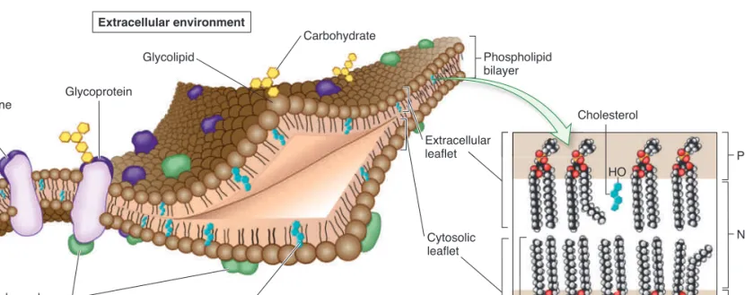
hydrophilic (water-loving), or polar region. The hydrophobic tails of the lipids, referred to as fatty acyl tails, are found in the interior of the membrane, and the hydrophilic heads are on the surface. Biological membranes also contain proteins, and most membranes have carbohydrates attached to lipids and proteins. Overall, the membrane is considered a mosaic of lipid, protein, and carbohydrate molecules. The membrane structure illustrated in Figure 5.1 is referred to as the fluid-mosaicmodel, originally proposed by S. Jonathan Singer and Garth Nicolson in 1972. As discussed later, the membrane exhibits properties that resemble a fluid because lipids and proteins can move relative to each other within the membrane.
Half of a phospholipid bilayer is termed a leaflet. Each leaf-let faces a different region. For example, the plasma membrane contains a leaflet that faces the cytosol (cytosolic leaflet) and a leaflet that faces the extracellular environment (extracellular leaflet) (see Figure 5.1). With regard to lipid composition, the two leaflets are highly asymmetric, with certain types of lipids more abundant in one leaflet than the other. A striking asymme-try occurs with glycolipids—lipids with carbohydrate attached. These are found primarily in the extracellular leaflet. The carbo-hydrate portion of a glycolipid protrudes into the extracellular environment. The types of carbohydrates on the surfaces of cells often allow cells to recognize each other, a phenomenon called cell surface recognition.
Proteins Associate with Membranes
in Three Different Ways
Although the phospholipid bilayer forms the basic foundation of biological membranes, the protein component carries out many key functions. Some of these functions were considered in
5.1
Membrane Structure
Learning Outcomes:
1. Describe the fluid-mosaic model of membrane structure.
2. Identify the three different types of membrane proteins.
3. Explain the technique of freeze fracture electron microscopy.
An important biological principle is that structure determines function. Throughout this chapter, we will see how the structure of cellular membranes enables them to compartmentalize the cell while selectively importing and exporting vital substances. The two primary components of membranes are phospholipids, which form the basic matrix of a membrane, and proteins, which are embedded in the membrane or loosely attached to its surface. A third component is carbohydrates, which may be attached to membrane lipids and proteins. In this section, we will be mainly concerned with the organization of these components to form a biological membrane and how they are important in the overall function of membranes.
Biological Membranes Are a Mosaic of Lipids,
Proteins, and Carbohydrates
Figure 5.1 shows the biochemical organization of a membrane,
which is similar in composition among all living organisms. The framework of the membrane is the phospholipid bilayer, which consists of two layers of phospholipids. Recall from Chapter 3 that phospholipids are amphipathic molecules (see Figure 3.9). They have a hydrophobic (water-fearing), or nonpolar region, and a
Cholesterol C Extracellular environment Cytosol Integral membrane protein Glycoprotein Glycolipid Carbohydrate Extracellular leaflet Phospholipid bilayer Cytosolic leaflet Peripheral membrane proteins Cholesterol (found only in animal cells) Polar Nonpolar Polar HO Phospholipid
Figure 5.1
Fluid-mosaic model of membrane structure. The basic framework of a plasma membrane is a phospholipid bilayer. Proteins may span the membrane and may be bound on the surface to other proteins or to lipids. Proteins and lipids that have covalently bound carbohydrates are called glycoproteins and glycolipids, respectively. The inset shows nine phospholipids and one cholesterol molecule (blue) in a bilayer, and it emphasizes the two leaflets and the polar and nonpolar regions of the bilayer.bro32271_ch05_087-110.indd 88
MEMBRANE STRUCTURE, TRANSPORT, AND CELL JUNCTIONS 89
3. Peripheral membrane proteins are proteins that are
noncovalently bound to regions of transmembrane membrane proteins that project out from the membrane or are bound to the polar head groups of phospholipids (see Figure 5.1). Peripheral membrane proteins are typically bound to the membrane by hydrogen bonds or ionic bonds (or both).
Membrane Structure Can Be Viewed
with an Electron Microscope
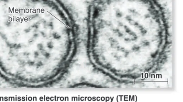
Electron microscopy, discussed in Chapter 4, is a valuable tool for probing membrane structure and function. In transmission elec-tron microscopy (TEM), a biological sample is thin-sectioned and stained with heavy-metal dyes such as osmium tetroxide. This compound binds tightly to the polar head groups of phospholip-ids, but it does not bind well to the fatty acyl tails. As shown in
Figure 5.3a, membranes stained with osmium tetroxide resemble
a railroad track. Two thin dark lines, which are the stained polar head groups, are separated by a uniform light space about 2 nm thick. This railroad track morphology is seen when cell mem-branes are subjected to electron microscopy.
A specialized form of TEM, freeze fracture electron micros-copy (FFEM), is used to analyze the interiors of phospholipid bilayers. Russell Steere invented this method in 1957. In FFEM, a sample is frozen in liquid nitrogen and split with a knife (Figure 5.3b). The knife does not actually cut through the bilayer, but it fractures the frozen sample. Due to the weakness of the central membrane region, the leaflets separate into a P face (the protoplasmic face that was next to the cytosol) and the E face (the extracellular face). Most transmembrane proteins do not break in half. They remain embedded within one of the leaflets, usu-ally in the P face. The samples, which are under a vacuum, are then sprayed with a heavy metal such as platinum, which coats the sample and reveals architectural features within each leaflet. For example, when viewed under TEM, membrane proteins are visible as bumps that provide significant three-dimensional detail about their form and shape.
5.1
Reviewing the Concepts
• A plasma membrane separates a cell from its surroundings. Biological membranes provide interfaces for carrying out vital cellular functions (Table 5.1).
• The accepted model of membranes is the fluid-mosaic model, and its basic framework is the phospholipid bilayer. Cellular membranes also contain proteins, and most membranes have attached carbohydrates (Figure 5.1).
• The three main types of membrane proteins are transmembrane proteins, lipid-anchored proteins, and peripheral membrane proteins. Transmembrane proteins and lipid-anchored proteins are classified as integral membrane proteins (Figure 5.2). • Electron microscopy is a valuable tool for studying membrane
structure and function. Freeze fracture electron microscopy (FFEM) is used to analyze the interiors of phospholipid bilayers (Figure 5.3).
Chapter 4. Later in this chapter, we will explore how membrane proteins are involved in transporting ions and molecules across membranes and in the adhesion of cells to each other. In later chapters, we will examine how membrane proteins are respon-sible for other functions, including ATP synthesis (Chapter 6), photosynthesis (Chapter 7), and cell signaling (Chapter 8).
The three types of membrane proteins are transmembrane proteins, lipid-anchored proteins, and peripheral membrane pro-teins. Transmembrane proteins and lipid-anchored proteins are called integral membrane proteins, because a portion of them is integrated into the hydrophobic region of the membrane. Each of the three types has a different way of associating with a mem-brane (Figure 5.2).
1. Transmembrane proteins span or traverse the membrane
from one leaflet to the other. Transmembrane segments within these proteins have stretches of nonpolar amino acids that are inserted into the hydrophobic interior of the phospholipid bilayer, making it possible for the entire protein to traverse the membrane. In most transmembrane proteins, each transmembrane segment is folded into an
a helix structure. Such a segment is stable in a membrane because the nonpolar amino acids interact favorably with the hydrophobic fatty acyl tails of the lipid molecules.
2. Lipid-anchored proteins are proteins that associate with a membrane because they have a lipid molecule that is covalently attached to an amino acid side chain within the protein. The fatty acyl tails are inserted into the hydrophobic portion of the membrane, thereby keeping the protein firmly attached to the membrane.
Transmembrane protein Lipid Peripheral membrane protein Transmembrane helix 1 7 2 6 3 4 5 Extracellular environment Cytosol Lipid-anchored protein
Figure 5.2
Types of membrane proteins. Integral membrane proteins are of two types: transmembrane proteins and lipid-anchored proteins. Peripheral membrane proteins are noncovalently bound to the hydrophilic regions of integral membrane proteins or to the polar head groups of lipids. Inset: The protein bacteriorhodopsin contains seven transmembrane segments, depicted as cylinders, in an a helix structure. Bacteriorhodopsin is found in halophilic (salt-loving) archaea.5.1
Testing Your Knowledge
1. Which of the following is not a characteristic of a biological membrane?
a. contains a bilayer of phospholipids
b. contains proteins that are inserted into the membrane
c. contains a high percentage of water molecules
d. has carbohydrates attached to lipids and proteins
e. has asymmetric leaflets
2. A membrane protein that is noncovalently attached to a protein or a lipid is a
a. transmembrane protein.
b. lipid-anchored protein.
c. peripheral membrane protein.
d. glycoprotein.
e. all of the above.
5.2
Fluidity of Membranes
Learning Outcomes:
1. Describe the fluidity of membranes.
2. Predict how fluidity will be affected by changes in lipid composition.
Let’s now turn our attention to the dynamic properties of mem-branes. Although a membrane provides a critical interface between a cell and its environment, it is not a solid, rigid struc-ture. Rather, biological membranes exhibit properties of fluidity,
which means that individual molecules remain in close associa-tion yet have the ability to readily move within the membrane. In this section, we will examine the fluid properties of biological membranes.
Membranes Are Semifluid
Though membranes are often described as fluid, it is more appro-priate to say they are semifluid. In a fluid substance, molecules can move in three dimensions. By comparison, most phospho-lipids can rotate freely around their long axes and move laterally within the membrane leaflet (Figure 5.4a). This type of motion is considered two-dimensional, which means it occurs within the plane of the membrane. Because rotational and lateral move-ments keep the fatty acyl tails within the hydrophobic interior, such movements are energetically favorable. At 378C, a typical lipid molecule exchanges places with its neighbors about 107 times per second, and it can move several micrometers per sec-ond. At this rate, a lipid can traverse the length of a bacterial cell (approximately 1 μm) in only 1 second and the length of a typical animal cell in 10 to 20 seconds.
In contrast to rotational and lateral movements, the “flip-flop” of lipids from one leaflet to the opposite leaflet does not occur spontaneously. Flip-flop is energetically unfavorable because the hydrophilic polar head of a phospholipid has to travel through the hydrophobic interior of the membrane. How are lipids moved
Membrane bilayer
(b) Freeze fracture electron microscopy (FFEM)
Transmembrane protein Lipid bilayer Direction of fracture P face exposed P face E face E face exposed E face P face Cytosolic leaflet Extracellular leaflet 40 nm
(a) Transmission electron microscopy (TEM)
Membrane bilayer
10 nm 10 nm
10 nm
Figure 5.3
Electron micrographs of a biological membrane. (a) In the standard form of TEM, a membrane appears as two dark parallel lines. These lines are the lipid head groups, which stain darkly with osmium tetroxide. The fatty acyl tails do not stain well and appear as a light region sandwiched between the dark lines. (b) In the technique of freeze fracture electron microscopy (FFEM), a sample is frozen in liquid nitrogen and fractured. The sample is then coated with metal and viewed under the electron microscope.Concept Check:
If a heavy metal labeled the hydrophobic tails rather than the polar head groups (as osmium tetroxide does), do you think you would see a bilayer (that is, a railroad track) under TEM?bro32271_ch05_087-110.indd 90
MEMBRANE STRUCTURE, TRANSPORT, AND CELL JUNCTIONS 91
• Presence of cholesterol: Cholesterol, which is found in
animal cells, tends to stabilize membranes because it is a short, rigid molecule (see inset to Figure 5.1). Its effects depend on temperature. At higher temperatures, such as those observed in mammals that maintain a constant body temperature, cholesterol makes the membrane less fluid. At lower temperatures, such as icy water, cholesterol has the opposite effect. It makes the membrane more fluid and prevents it from freezing. Plant cell membranes contain phytosterols that resemble cholesterol in their chemical structure.
An optimal level of bilayer fluidity is essential for normal cell function, growth, and division. If a membrane is too fluid, which may occur at higher temperatures, it can become leaky. However, if a membrane becomes too solid, which may occur at lower tem-peratures, the functioning of membrane proteins will be inhib-ited. How can organisms cope with changes in temperature? The cells of many species adapt to changes in temperature by alter-ing the lipid composition of their membranes. For example, when the water temperature drops, the cells of certain fish will incor-porate more cholesterol in their membranes, making the mem-brane more fluid. If a plant cell is exposed to high temperatures for many hours or days, it will alter its lipid composition to have longer fatty acyl tails and fewer double bonds, which will make the membrane less fluid.
Many Transmembrane Proteins Can Rotate and Move
Laterally, but Some Are Restricted in Their Movement
Like lipids, many transmembrane proteins may rotate and later-ally move throughout the plane of a membrane. Because trans-membrane proteins are larger than lipids, they move within the membrane at a much slower rate. Flip-flop of transmembrane from one leaflet to the other? The transport of lipids betweenleaf-lets requires the action of the enzyme flippase, which requires energy input in the form of ATP (Figure 5.4b).
Although most lipids diffuse rotationally and laterally within the plane of the lipid bilayer, researchers have discovered that cer-tain types of lipids in animal cells tend to strongly associate with each other to form structures called lipid rafts. As the term raft suggests, a lipid raft is a group of lipids that float together as a unit within a larger sea of lipids. Lipid rafts have a lipid composition that differs from the surrounding membrane. For example, they usually have a high amount of cholesterol. In addition, lipid rafts may contain unique sets of lipid-anchored proteins and trans-membrane proteins. The functional importance of lipid rafts is the subject of a large amount of current research. Lipid rafts may play an important role in endocytosis (discussed later in this chapter) and cell signaling.
Lipid Composition Affects Membrane Fluidity
The biochemical properties of phospholipids affect the fluidity of the phospholipid bilayer. These include the following:
• Length of the fatty acyl tail: Shorter acyl tails are less likely
to interact with each other, which makes the membrane more fluid. The tails typically range from 14 to 24 carbon atoms, with 18 to 20 carbons being the most common.
• Presence of double bonds: Double bonds make a membrane
more fluid. When a double bond is present, the lipid is said to be unsaturated with respect to the number of hydrogens that are bound to the carbon atoms (refer back to Figure 3.8). A double bond creates a kink in the fatty acyl tail (shown in the inset to Figure 5.1), making it more difficult for neighboring tails to interact and making the bilayer more fluid.
(a) Spontaneous lipid movements (b) Lipid movement via flippase
Lateral movement
Rotational movement
Flip-flop Flippase
ATP ADP Pi
Figure 5.4
Semifluidity of the lipid bilayer. (a) Spontaneous movements in the bilayer. Lipids can rotate (that is, move 360°) and move laterally (for example, from left to right in the plane of the bilayer). (b) Flip-flop does not happen spontaneously, because the polar head group would have to pass through the hydrophobic region of the bilayer. Instead, the enzyme flippase uses ATP to flip phospholipids from one leaflet to the other.microscope. If the cells were maintained at 08C, a temperature that greatly inhibits lateral movement, the fluorescence was seen on only one side of the fused cell. However, if the cells were incu-bated for several hours at 378C and then cooled to 08C, the fluo-rescence was distributed throughout the plasma membrane of the fused cell. This occurred because the higher temperature allowed the lateral movement of the H-2 protein throughout the fused cell.
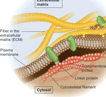
Unlike the example shown in Figure 5.5, not all transmem-brane proteins are capable of rotational and lateral movement. Depending on the cell type, 10–70% of membrane proteins may be restricted in their movement. Transmembrane proteins may be bound to components of the cytoskeleton, which restricts the proteins from moving (Figure 5.6), or may be attached to molecules that are outside the cell, such as the interconnected network of proteins that forms the extracellular matrix (ECM) of animal cells (see Chapter 4, Figure 4.27).
5.2
Reviewing the Concepts
• Bilayer semifluidity is essential for normal cell function, growth, and division. Lipids and many proteins can move rotationally and laterally, but the flip-flop of lipids from one leaflet to the opposite does not occur spontaneously. Some membrane pro-teins are restricted in their movements (Figures 5.4, 5.5, 5.6). • The chemical properties of phospholipids—such as tail length
and the presence of double bonds—and the amount of choles-terol affect the fluidity of membranes.
proteins does not occur, because the proteins also contain hydro-philic regions that project out from the phospholipid bilayer, and it would be energetically unfavorable for the hydrophilic regions of membrane proteins to pass through the hydrophobic portion of the phospholipid bilayer.
In 1970, Larry Frye and Michael Edidin conducted an experi-ment that verified the lateral moveexperi-ment of transmembrane pro-teins (Figure 5.5). Mouse and human cells were mixed together and exposed to agents that caused them to fuse with each other to produce mouse-human cell hybrids. Some cells were cooled to 08C, while others were incubated at 378C before being cooled. Both sets of cells were then exposed to fluorescent antibodies that became specifically bound to a mouse transmembrane protein called H-2. The fluorescent label was observed with a fluorescence
Biology Principle
Biology Is an Experimental Science
This experiment verifi ed that membrane proteins can diff use laterally within the plane of the lipid bilayers.
H-2 mouse protein
Mouse cell Human cell
Add agents that cause mouse cell and human cell to fuse. 1 2 Fluorescent dye Antibody H-2 Fuse cells
Lower the temperature to 0C and add a fluorescently labeled antibody that recognizes the mouse H-2 protein in the plasma membrane. Observe with a fluorescence microscope. H-2 protein is unable to move laterally and remains on one side of the fused cell.
Incubate cell at 37C, then cool to 0C and add a fluorescently labeled antibody that recognizes the mouse H-2 protein in the plasma membrane. Observe with a fluorescence microscope. Due to lateral movement at 37C, the mouse H-2 protein is distributed throughout the fused cell surface.
Figure 5.5
A method to measure the lateral movement of membrane proteins.Concept Check:
Explain why the H-2 proteins are found on only one side of the cell when the cells were incubated at 08C.Cytoskeletal filament Linker protein Transmembrane protein Fiber in the extracellular matrix (ECM) Plasma membrane Cytosol Extracellular matrix
Figure 5.6
Attachment of transmembrane proteins to the cytoskeleton and ECM of an animal cell. Some transmembrane proteins have regions that extend into the cytosol and are anchored to large cytoskeletal filaments via linker proteins. Being bound to these large filaments restricts the movement of these proteins. Similarly, some transmembrane proteins are bound to large, immobile fibers in the ECM, which restricts their movements.bro32271_ch05_087-110.indd 92
MEMBRANE STRUCTURE, TRANSPORT, AND CELL JUNCTIONS 93
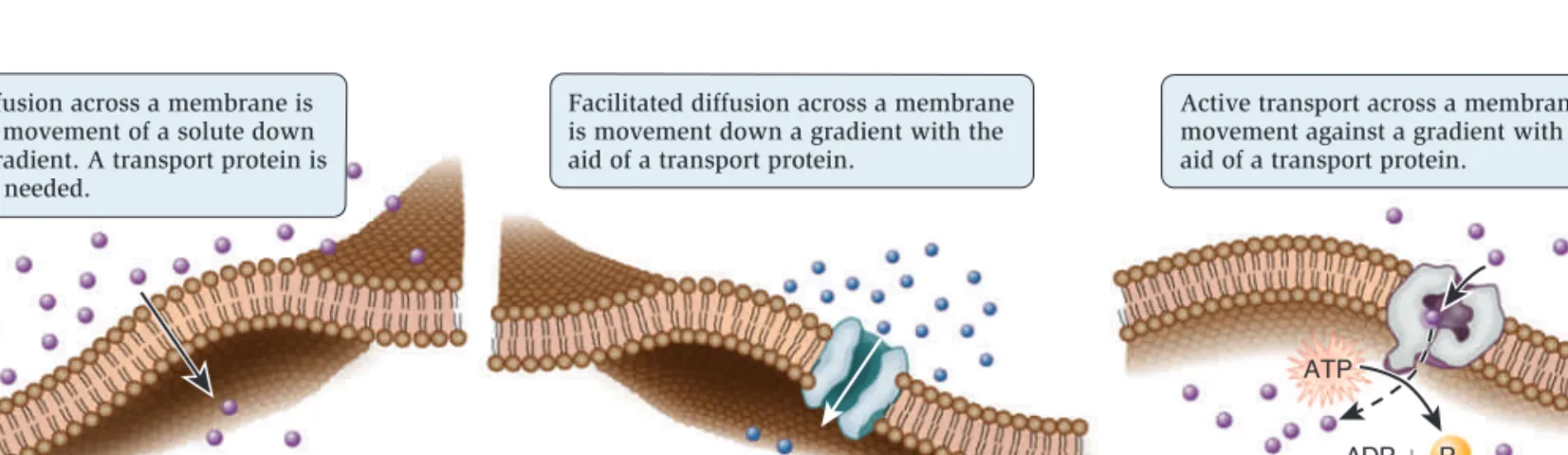
Substances can move directly across a membrane in three general ways (Figure 5.7).
• Diffusion occurs when a substance moves from a region of high concentration to a region of lower concentration. Some substances can move directly through a biological membrane via diffusion.
• In facilitated diffusion, a transport protein provides a passageway for a substance to diffuse across a membrane. Diffusion and facilitated diffusion are examples of
passive transport—the transport of a substance across a membrane that does not require an input of energy.
• A third mode of transport, called active transport, moves a substance from an area of low concentration to one of higher concentration with the aid of a transport protein. Active transport requires an input of energy from a source such as ATP.
In this section, we will begin with a discussion of how the phospholipid bilayer presents a barrier to the movement of ions and molecules across membranes. We will then consider the concept of gradients across membranes and how such gradients affect the movement of water.
The Phospholipid Bilayer Is a Barrier to the Diffusion
of Hydrophilic Solutes
Because of their hydrophobic interiors, phospholipid bilayers are a barrier to the movement of ions and hydrophilic molecules. Such ions and molecules are called solutes; they are dissolved in water, which is a solvent. The rate of diffusion across a phos-pholipid bilayer depends on the chemistry of the solute and its concentration. Three factors greatly affect the ability of solutes to diffuse through a phospholipid bilayer.
• Size: small substances diffuse faster than larger ones. • Polarity: nonpolar substances diffuse faster than polar
ones.
• Charge: noncharged substances diffuse faster than charged
ones.
5.2
Testing Your Knowledge
1. Which of the following lipid movements would not occur spon-taneously in a membrane?
a. lateral movements (side-to-side)
b. rotational movements
c. flip-flop from one leaflet to the other
d. both a and c
e. all of the above
2. Which of the following changes would make a membrane more fluid?
a. decrease the temperature
b. increase the percentage of lipids with short tails
c. decrease the percentage of lipids with double bonds in their tails
d. increase the percentage of glycoproteins
e. increase the percentage of glycolipids
5.3
Overview of Membrane Transport
Learning Outcomes:
1. Describe the concepts of diffusion, facilitated diffusion, passive transport, gradients, and active transport.
2. Explain the process of osmosis and how it affects cell structure.
We now turn to one of the key functions of membranes, membrane transport—the movement of ions and molecules across bio-logical membranes. All cells contain a plasma membrane that exhibits selective permeability, allowing the passage of some ions and molecules but not others. As a protective envelope, its structure ensures that essential molecules such as glucose and amino acids enter the cell, metabolic intermediates remain in the cell, and waste products exit. The selective permeability of the plasma membrane allows the cell to maintain a favorable internal environment.
(b) Facilitated diffusion—passive transport (c) Active transport (a) Diffusion—passive transport
Diffusion across a membrane is the movement of a solute down a gradient. A transport protein is not needed.
Facilitated diffusion across a membrane is movement down a gradient with the aid of a transport protein.
Active transport across a membrane is movement against a gradient with the aid of a transport protein.
ATP
ADP Pi
are a universal feature of all living cells. For example, immediately after you eat a meal containing carbohydrates, a higher concen-tration of glucose is found outside your cells than inside; this is an example of a chemical gradient (Figure 5.9a).
Gradients involving ions have two components: electrical and chemical. An electrochemicalgradient is a dual gradient with both electrical and chemical components (Figure 5.9b). It occurs with solutes that have a net positive or negative charge. For exam-ple, let’s consider a gradient involving Na⫹. An electrical gradient could exist in which the amount of net positive charge outside a cell is greater than inside. In Figure 5.9b, an electrical gradient is due to differences in the amounts of different types of ions across the membrane, including sodium, potassium, and chloride (Na⫹, K⫹, and Cl⫺). At the same time, a chemical gradient—a difference in Na⫹ concentration across the membrane—could exist in which the concentration of Na⫹ outside is greater than inside. Thus, the
Figure 5.8 compares the relative permeabilities of various solutes
through an artificial phospholipid bilayer that does not contain any proteins or carbohydrates. Gases, such as O2 and CO2, and a few small, uncharged molecules, such as ethanol, can readily diffuse across the bilayer. However, the permeability of ions and larger polar molecules, such as sugars, is relatively low, and the permeability of macromolecules, such as proteins and polysac-charides, is even lower.
Cells Maintain Gradients Across Their Membranes
A hallmark of living cells is their ability to maintain a relatively constant internal environment that is distinctively different from their external environment. Solute gradients are formed across the plasma membrane and across organellar membranes. When we speak of a transmembranegradient, or concentrationgradient,
we mean the concentration of a solute is higher on one side of a membrane than the other. Transmembrane gradients of solutes
High permeability Moderate permeability Low permeability Very low permeability Gases Very small, uncharged molecules CO2 N2 O2 Ethanol Water Urea H2O H2NCONH2 Polar organic molecules Sugars Ions Charged polar molecules and macro-molecules Na⫹, K⫹, Mg2⫹, Ca2⫹, Cl⫺ Amino acids ATP Proteins Polysaccharides Nucleic acids
(DNA and RNA) Artificial bilayer
Figure 5.8
Relative permeability of an artificial phospholipid bilayer to a variety of solutes. Solutes that easily penetrate are shown with a straight arrow that passes through the bilayer. The dotted line indicates solutes that have moderate permeability. The remaining solutes shown at the bottom are relatively impermeable.BioConnections:
Which amino acid, described in Chapter 3 (see Figure 3.11), would you expect to cross an artificial membrane more quickly, leucine or lysine?Concept Check:
Which molecule would you expect to pass through a phospholipid bilayer more quickly, methanol (CH3OH) or methane (CH4)?(a) Chemical gradient for glucose—a higher glucose concentration outside the cell
Plasma membrane Plasma membrane Kⴙ Naⴙ Clⴚ
(b) Electrochemical gradient for Na+—more positive charges outside
the cell and a higher Na+ concentration outside the cell
Glucose ⴚ ⴚ ⴚ ⴚ ⴚ ⴚ ⴚ ⴚ ⴚ ⴚ ⴚ ⴚ ⴚ ⴚ ⴚ ⴚ ⴙ ⴙ ⴙ ⴙ ⴙ ⴙ ⴙ ⴙ ⴙ ⴙ ⴙ ⴙ ⴙ ⴙ ⴙ ⴙ ⴙ ⴙ ⴙ ⴙ ⴙ ⴙ ⴙ ⴙ ⴙ ⴙ ⴙ ⴙ ⴙ
Figure 5.9
Gradients across cell membranes.BioConnections:
Look ahead to Figure 32.8. What types of ion gradients are important for the conduction of an action potential across the plasma membrane of a neuron?bro32274_ch05_087-110.indd 94
MEMBRANE STRUCTURE, TRANSPORT, AND CELL JUNCTIONS 95
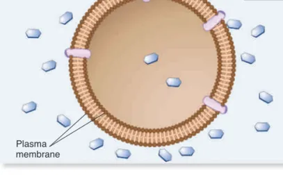
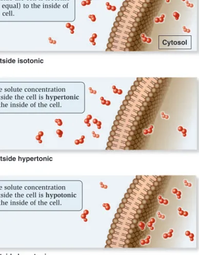
process, called osmosis, water diffuses across a membrane from the hypotonic compartment (a lower solute concentration) into the hypertonic compartment (a higher solute concentration). Animal cells, which are not surrounded by a rigid cell wall, must maintain a balance between the extracellular and intracellular solute concentrations; they are isotonic. Animal cells contain a variety of transport proteins that sense changes in cell volume and allow the necessary movements of solutes across the membrane to prevent osmotic changes and maintain normal cell shape. However, if animal cells are placed in a hypotonic medium, water will diffuse into them to equalize solute concentrations on both sides of the membrane. In extreme cases, a cell may take up so much water that it ruptures, a phenomenon called osmotic lysis (Figure 5.11a). Alternatively, if animal cells are placed in a hyper-tonic medium, water will exit the cells via osmosis and equalize solute concentrations on both sides of the membrane, causing them to shrink in a process called crenation.
How does osmosis affect cells with a rigid cell wall, such as bacteria, fungi, algae, and plant cells? If the extracellular fluid is hypotonic, a plant cell will take up a small amount of water, but the cell wall prevents osmotic lysis from occurring (Figure 5.11b). Alternatively, if the extracellular fluid surrounding a plant cell is hypertonic, water will exit the cell and the plasma membrane will pull away from the cell wall, a process called plasmolysis. If plas-molysis is severe, it could result in cell death.
Some freshwater microorganisms, such as amoebae and par-amecia, are found in extremely hypotonic environments where the external solute concentration is always much lower than the concentration of solutes in their cytosol. Because of the great ten-dency for water to move into the cell by osmosis, such organisms contain one or more contractile vacuoles to prevent osmotic lysis. A contractile vacuole takes up water from the cytosol and peri-odically discharges it by fusing the vacuole with the plasma mem-brane (Figure 5.12).
5.3
Reviewing the Concepts
• Biological membranes exhibit selectively permeability. Diffusion and facilitated diffusion, both examples of passive transport, occur when a solute moves from a region of high concentration to a region of lower concentration. Passive transport does not require an input of energy. Active transport is the movement of a sub-stance against a gradient and requires energy input (Figure 5.7). • The phospholipid bilayer is relatively impermeable to many
sub-stances (Figure 5.8).
• Living cells maintain an internal environment that is separated from their external environment. Transmembrane gradients, in which the concentration of a solute is higher on one side of a membrane than the other, are established across the plasma membrane and across organellar membranes (Figure 5.9). • In the process of osmosis, water diffuses through a membrane
from a solution that is hypotonic (lower concentration of dis-solved particles) into a solution that is hypertonic (higher con-centration of dissolved particles). Solutions with identical concentrations are isotonic. Some cells have contractile vacuoles to eliminate excess water (Figures 5.10, 5.11, 5.12).
Na electrochemical gradient is composed of both an electrical
gradient due to charge differences across the membrane along with a chemical gradient for Na.
One way to view the transport of solutes across membranes is to consider how the transport process affects the pre-existing gradients across membranes. Passive transport tends to decrease the magnitude of a pre-existing gradient. It is a process that is energetically favorable and does not require an input of energy (see Figure 5.7a, b). By comparison, active transport produces a chemical gradient and/or electrochemical gradient. The forma-tion of a gradient requires an input of energy.
Osmosis Is the Movement of Water Across Membranes
to Balance Solute Concentrations
Let’s now turn our attention to how gradients affect the movement of water across membranes. When the concentrations of dis-solved particles (solutes) on both sides of the plasma membrane are equal, the two solutions are said to be isotonic (Figure 5.10a). However, we have also seen that transmembrane gradients com-monly exist across membranes. When the concentration of sol-utes outside the cell is higher, it is said to be hypertonic relative to the inside of the cell (Figure 5.10b). Alternatively, the outside of the cell could be hypotonic—have a lower concentration of solutes relative to the inside (Figure 5.10c). Note that these two terms are always used relative to each other—if one region is hypertonic, the adjacent region must be hypotonic.
If solutes cannot readily move across the membrane, water will move and tend to balance the solute concentrations. In this
The solute concentration outside the cell is isotonic
(or equal) to the inside of the cell.
The solute concentration outside the cell is hypertonic
to the inside of the cell.
The solute concentration outside the cell is hypotonic
to the inside of the cell.
Solute
Cytosol (a) Outside isotonic
(b) Outside hypertonic
(c) Outside hypotonic
Cells are initially in an isotonic solution.
Cells undergo shrinkage (crenation) because water exits the cell.
Cells swell and may rupture (osmotic lysis) because water is taken into the cell.
Cell is initially in an isotonic solution. Place in hypertonic solution. Place in hypotonic solution. Place in hypertonic solution. Place in hypotonic solution. Cells maintain normal shape. Cells maintain normal shape. Plant cell Vacuole
Volume inside the plasma membrane shrinks, and the membrane pulls away from the cell wall (plasmolysis) due to the exit of water.
A small amount of water may enter the cell, but the cell wall prevents major expansion. Red blood cell
(a) Osmosis in animal cells (b) Osmosis in plant cells
H2O
H2O H2O
H2O
Figure 5.11
The phenomenon of osmosis. (a) In cells that lack a cell wall, such as animal cells, osmosis may promote cell swelling and possible rupture (osmotic lysis) or shrinkage (crenation). (b) In cells that have a rigid cell wall, such as plant cells, a hypotonic medium causes only a minor amount of expansion, whereas a hypertonic medium causes the plasma membrane to pull away from the cell wall.Concept Check:
Let’s suppose the inside of a cell has a solute concentration of 0.3 M, and the outside is 0.2 M. If the membrane is impermeable to solutes, which direction will water move?Filled contractile vacuole Vacuole after expelling water 60m 60m
Biology Principle
Figure 5.12
The contractile vacuole inParamecium caudatum. In the upper photo, a contractile vacuole is filled with water from radiating canals that collect fluid from the cytosol. The lower photo shows the cell after the contractile vacuole has fused with the plasma membrane (which would be above the plane of this page) and released the water from the cell.
Living Organisms Maintain Homeostasis
In this example, the paramecium maintains a relatively constant internal volume by using contractile vacuoles to remove excess water.
bro32271_ch05_087-110.indd 96
97
In this section, we will examine the two categories of transport proteins—channels and transporters—based on the manner in which they move solutes across the membrane.
Channels Provide an Open Passageway
for Solute Movement
The term channel refers to an open passageway for the facilitated diffusion of ions or molecules across a membrane. The type of channel shown in Figure 5.13 is a transmembrane protein. Solutes
move directly through this channel to get to the other side. When a channel is open, the transmembrane movement of solutes can be extremely rapid, up to 100 million ions or molecules per second!
Most channels are gated, which means they open to allow the diffusion of solutes and close to prohibit diffusion. The phe-nomenon of gating allows cells to regulate the movement of solutes. For example, gating may involve the direct binding of a molecule to the channel protein itself. These gated channels are controlled by the noncovalent binding of small molecules—called ligands—such as hormones or neurotransmitters. The ligands are often important in the transmission of signals between neurons and muscle cells or between two neurons.
5.3
Testing Your Knowledge
1. Carbon dioxide can move across a membrane without the aid of a transport protein. This movement is an example of
a. diffusion. d. active transport.
b. facilitated diffusion. e. both a and c.
c. passive transport.
2. Let’s suppose the inside of a cell is hypertonic relative to the out-side. If osmosis occurs, _______ will move ______ the cell to equalize solute concentrations.
a. solute molecules, into d. water, out of
b. solute molecules, out of e. both a and d
c. water, into
5.4
Transport Proteins
Learning Outcomes:
1. Outline the functional differences between channels and transporters.
2. Analyze the results of Agre and explain how they indicated that aquaporin is a water channel.
3. Compare and contrast uniporters, symporters, and antiporters.
4. Explain the concepts of primary active transport and secondary active transport.
5. Describe the structure and function of pumps.
Because the phospholipid bilayer is a physical barrier to the dif-fusion of most hydrophilic molecules and ions, cells can sepa-rate their internal contents from the external environment. However, this barrier also poses a potential problem because cells must take up nutrients from the environment and export waste products. How do cells overcome this dilemma? Over the course of millions of years, species have evolved a multitude of
transport proteins—transmembrane proteins that provide a passageway for the movement of ions and hydrophilic molecules across the phospholipid bilayer. Transport proteins play a cen-tral role in the selective permeability of biological membranes.
When a channel is open, a solute directly diffuses through the channel to reach the other side of the membrane.
Gate closed
Gate opened
Figure 5.13
Mechanism of transport by a channel protein.Concept Check:
What is the purpose of gating?Likewise, bladder and kidney cells, which play a key role in regu-lating water balance in the bodies of vertebrates, allow the rapid movement of water across their membranes. Based on these observations, researchers speculated that certain cell types might have channel proteins in their plasma membranes that enable the rapid movement of water.
One approach to characterizing a new protein is to first iden-tify a protein based on its relative abundance in a particular cell type and then attempt to determine the protein’s function. This
FEATURE INVESTIGATION
Agre Discovered That Osmosis Occurs More
Quickly in Cells with a Channel That Allows
the Facilitated Diffusion of Water
As discussed earlier in this chapter, osmosis is the flow of water to balance solute concentrations. Water can slowly cross biological membranes by diffusion through the phospholipid bilayer. How-ever, in the 1980s, researchers discovered that certain cell types allow water to move across the plasma membrane at a much faster rate than would be predicted by diffusion. For example, water moves very quickly across the membrane of red blood cells, which causes them to shrink and swell in response to changes in extracellular solute concentrations (refer back to Figure 5.11a).
named this protein CHIP28, which stands for channel-forming integral membrane protein with a molecular mass of 28,000 dal-tons (Da). During the course of their studies, they also identified and isolated the gene that encodes CHIP28.
In 1992, Agre and his colleagues conducted experiments to determine if CHIP28 functions in the transport of water across membranes (Figure 5.14). Because they already had isolated the rationale was applied to the discovery of proteins that allow the
rapid movement of water across membranes. Peter Agre and his colleagues first identified a protein that was abundant in red blood cells and kidney cells but not found in high amounts in many other cell types. Though they initially did not know the function of the protein, its physical structure was similar to other proteins that were already known to function as channels. They
Place oocytes into a hypotonic medium and observe under a light microscope. As a control, also place oocytes that have not been injected with CHIP28 mRNA into a hypotonic medium and observe by microscopy.
Oocyte rupturing Oocyte
Control CHIP28
Inject the CHIP28 mRNA into frog eggs (oocytes). Wait several hours to allow time for the mRNA to be translated into CHIP28 protein at the ER membrane and then moved via vesicles to the plasma membrane.
Add an enzyme (RNA polymerase) and nucleotides to a test tube that contains many copies of the CHIP28 gene. This results in the synthesis of many copies of CHIP28 mRNA.
HYPOTHESIS CHIP28 may function as a water channel.
KEY MATERIALS Prior to this work, a protein called CHIP28 was identified that is abundant in red blood cells and kidney cells. The gene that encodes this protein was cloned, which means that many copies of the gene were made in a test tube.
Experimental level Conceptual level
1 2 3 4 THE DATA Enzymes and nucleotides CHIP28 DNA RNA polymerase CHIP28 mRNA
Frog oocyte CHIP28 protein
CHIP28 protein Ribosome Control CHIP28 mRNA CHIP28 protein is inserted into the plasma membrane.
Nucleus Cytosol
Control CHIP28
3–5 minutes
Figure 5.14
The discovery of water channels (aquaporins) by Agre and colleagues.bro32271_ch05_087-110.indd 98
MEMBRANE STRUCTURE, TRANSPORT, AND CELL JUNCTIONS 99
results are consistent with the hypothesis that CHIP28 functions as a channel that allows the facilitated diffusion of water across the membrane. Many subsequent studies confirmed this obser-vation. Later, CHIP28 was renamed aquaporin to indicate its newly identified function of allowing water to diffuse through a channel in the membrane. More recently, the three-dimensional structure of aquaporin was determined (see chapter-opening fig-ure). In 2003, Agre was awarded the Nobel Prize in Chemistry for this work.
Experimental Questions
1. What observations about particular cell types in the human body led to the experimental strategy of Figure 5.14? 2. What characteristics of CHIP28 made Agre and associates
speculate that it may transport water? In your own words, briefly explain how they tested the hypothesis that CHIP28 has this function.
3. Explain how the results of the experiment of Figure 5.14 support the proposed hypothesis.
SOURCE Preston, G. M., Carroll, T. P., Guggino, W. B., and Agre, P. 1992. Appearance of water channels in Xenopus oocytes expressing red cell CHIP28 protein. Science 256:385–387.
CONCLUSION The CHIP28 protein, now called aquaporin, allows the rapid movement of water across the membrane. 5
6
gene that encodes CHIP28, they could make many copies of this gene in a test tube (in vitro) using gene cloning techniques (see Chapter 16). Starting with many copies of the gene in vitro, they added an enzyme to transcribe the gene into mRNA that encodes the CHIP28 protein. This mRNA was then injected into frog oocytes, chosen because frog oocytes are large, easy to inject, and lack pre-existing proteins in their plasma membranes that allow the rapid movement of water. Following injection, the mRNA was translated into CHIP28 proteins that were inserted into the plasma membrane of the oocytes. After allowing sufficient time for this to occur, the oocytes were placed in a hypotonic medium. As a control, oocytes that had not been injected with CHIP28 mRNA were also exposed to a hypotonic medium.
As you can see in the data, a striking difference was observed between oocytes that expressed CHIP28 versus the control. Within minutes, oocytes that contained the CHIP28 protein were seen to swell due to the rapid uptake of water. Three to five min-utes after being placed in a hypotonic medium, they actually lysed! By comparison, the control oocytes did not swell as rapidly, and they did not rupture even after 1 hour. Taken together, these
Transporters Bind Their Solutes and Undergo
Conformational Changes
Let’s now turn our attention to a second category of transport proteins known as transporters.* These transmembrane
pro-teins bind their solutes in a hydrophilic pocket and undergo a conformational change that switches the exposure of the pocket from one side of the membrane to the other side (Figure 5.15). For example, in 1995, Robert Brooker and colleagues proposed that a transporter called lactose permease, which is found in the bacte-rium E. coli, has a hydrophilic pocket that binds lactose. They fur-ther proposed that the two halves of the transporter protein come together at an interface that moves in such a way that the lactose-binding site alternates between an outwardly accessible pocket and an inwardly accessible pocket, as shown in Figure 5.15. This idea was later confirmed by studies that determined the structure of the lactose permease and related transporters.
Transporters provide the principal pathway for the uptake of organic molecules, such as sugars, amino acids, and nucleotides. In animals, they also allow cells to take up certain hormones and neurotransmitters. In addition, many transporters play a key role
Hydrophilic pocket
Solute
For transport to occur, a solute binds in a hydrophilic pocket exposed on one side of the membrane. The transporter then undergoes a conformational change that switches the exposure of the pocket to the other side of the membrane, where the solute is then released.
Conformational change
Biology Principle
Structure Determines Function
Two structural features—a hydrophilic pocket and the ability to switch back and forth between two conformations—allow transporters to move ions and molecules across the membrane.
Figure 5.15
Mechanism of transport by a transporter, also called a carrier.* Transporters are also called carriers. However, this term is misleading because transporters do not physically carry their solutes across the membrane.
in export. Waste products of cellular metabolism must be released from cells before they reach toxic levels. For example, a transporter removes lactic acid, a by-product of muscle cells during exercise. Other transporters, which are involved with ion transport, play an important role in regulating internal pH and controlling cell vol-ume. Transporters tend to be much slower than channels. Their rate of transport is typically 100 to 1,000 ions or molecules per second.
Transporters are named according to the number of solutes they bind and the direction in which they transport those solutes (Figure 5.16).
• Uniporters: bind a single ion or molecule and transport it across the membrane.
• Symporters: bind two or more ions or molecules and transport them in the same direction.
• Antiporters: bind two or more ions or molecules and
transport them in opposite directions.
Active Transport Is the Movement
of Solutes Against a Gradient
As mentioned, active transport is the movement of a solute across a membrane against its concentration gradient—that is, from a region of low concentration to higher concentration. Active trans-port is energetically unfavorable and requires an input of energy. Two general types are found:
• Primary active transport: involves the functioning of a
pump—a type of transporter that directly uses energy to transport a solute against a concentration gradient. Figure 5.17a
shows a pump that uses energy in the form of ATP to transport H against a gradient. Such a pump establishes an
H electrochemical gradient across a membrane. • Secondary active transport: involves the use of a
pre-existing gradient to drive the active transport of another
(a) Uniporter
(b) Symporter
Two solutes move in opposite directions.
(c) Antiporter
A single solute moves in one direction.
Two solutes move in the same direction.
Figure 5.16
Types of transporters based on the direction of transport.A pump actively exports H against a gradient.
Extracellular
environment An H
/sucrose symporter uses the H gradient to transport sucrose against a concentration gradient into the cell.
(a) Primary active transport (b) Secondary active transport
ATP ADP
H Sucrose
H
Cytosol
Pi
Figure 5.17
Types of active transport. (a) During primary active transport, a pump directly uses energy, in this case from ATP, to transport a solute against a concentration gradient. The pump shown here uses ATP to establish an H electrochemical gradient. (b) Secondary active transport viasymport involves the use of this gradient to drive the active transport of a solute, such as sucrose.
bro32271_ch05_087-110.indd 100
MEMBRANE STRUCTURE, TRANSPORT, AND CELL JUNCTIONS 101
solute. For example, an H/sucrose symporter uses an H
electrochemical gradient, established by a pump, to move sucrose against its concentration gradient (Figure 5.17b). In this regard, only sucrose is actively transported. Hydrogen ions move down their electrochemical gradient.
Symporters enable cells to actively import nutrients against a gra-dient. These proteins use the energy stored in the electrochemical gradient of H or Na to power the uphill movement of organic solutes such as sugars, amino acids, and other needed molecules. Therefore, with symporters in their plasma membrane, cells can scavenge nutrients from the extracellular environment and accumulate them to high levels within the cytoplasm. H/solute symporters are more common in bacteria, fungi, algae, and plant cells, because H pumps are found in their plasma membranes. In animal cells, a pump that exports Na maintains a Na gradient across the plasma membrane. Na/solute symporters are preva-lent in animal cells.
ATP-Driven Ion Pumps Generate Ion
Electrochemical Gradients
The phenomenon of active transport was discovered in the 1940s based on the study of the transport of sodium ions (Na) and
potassium ions (K). In animal cells, the concentration of Na
is lower inside the cell than outside, whereas the concentration of K is higher inside the cell than outside. After analyzing the
movement of these ions across the plasma membrane of muscle
cells, neurons, and red blood cells, researchers determined that the export of Na is coupled to the import of K. In the late 1950s, Danish biochemist Jens Skou proposed that a single transporter is responsible for this phenomenon. He was the first to describe
an ATP-driven ion pump, which was later named the Na/
K-ATPase. This pump can actively transport Na and K against their gradients by using the energy from ATP hydrolysis. The plasma membrane of a typical animal cell contains thousands of Na/K-ATPase pumps that maintain large concentration gradi-ents in which the concentration of Na is higher outside the cell and the concentration of K is higher inside the cell.
Let’s take a closer look at the Na/K-ATPase that Skou dis-covered. Every time one ATP is hydrolyzed, the Na/K-ATPase functions as an antiporter that pumps three Na into the extra-cellular environment and two K into the cytosol (Figure 5.18a). Because one cycle of pumping results in the net export of one positive charge, the Na/K-ATPase also produces an electri-cal gradient across the membrane. For this reason, it is electri-called an
electrogenicpump, because it generates an electrical gradient. By studying the interactions of Na, K, and ATP with the Na/K-ATPase, researchers have pieced together a molecu-lar road map of the steps that direct the pumping of ions across the membrane (Figure 5.18b). The Na/K-ATPase can alternate between two conformations, designated E1 and E2. In E1, the ion-binding sites are accessible from the cytosol—Na binds tightly to this conformation, whereas K has a low affinity. In E2, the ion-binding sites are accessible from the extracellular environment— Na has a low affinity, and K binds tightly.
3 Na Na/K-ATPase 2 K ATP ADP Pi Low [Na] Nerve cell
(a) Active transport by
the Naⴙ/ Kⴙ-ATPase (b) Mechanism of pumping 3 Nabind from cytosol. ATP is hydrolyzed. ADP is released and phosphate (P) is covalently attached to the pump, switching it to the E2 conformation. 3 Naare released outside of the cell. 2 Kbind from outside of the cell. Phosphate (Pi) is released, and the pump switches to the E1 conformation. 2 K are released into cytosol. The process repeats. E1 E2 E2 E1 ADP ATP P Pi 3 Na 3 Na 2 K 1 2 3 4 Extracellular environment Extracellular environment Cytosol Cytosol High [K] 2 K 3 Na 3 Na High [Na] Low [K] High [Na] Low [K]
Figure 5.18
Structure and function of the Na/K-ATPase. (a) Active transport by the Na/K-ATPase. Each time this protein hydrolyzes one ATPmolecule, it pumps out three Na and pumps in two K. (b) Pumping mechanism. This figure illustrates the protein conformational changes
between E1 and E2. As this occurs, ATP is hydrolyzed to ADP and phosphate. During the process, phosphate is covalently attached to the protein but is released after two K bind.
The pumping mechanism of the Na/ K-ATPase occurs as
follows (Figure 5.18b):
1. Three Nabind to the E1 conformation from the cytosol.
ATP is hydrolyzed to ADP and phosphate. Temporarily, the phosphate is covalently bound to the pump, an event called phosphorylation. The pump switches to the E2 conformation.
2. The three Na are released into the extracellular
environment, because they have a lower affinity for the E2 conformation.
3. Two K bind from the outside.
4. The binding of two K causes the release of phosphate. The
pump switches back to the E1 conformation. Because the E1 conformation has a low affinity for K, the two K are
released into the cytosol. The Na/K-ATPase is now ready
for another round of pumping.
The Na/K-ATPase is a critical ion pump in animal cells
because it maintains Na and K gradients across the plasma
membrane. Many other types of ion pumps are also found in the plasma membrane and in organellar membranes. Ion pumps play the primary role in the formation and maintenance of ion gradients that drive many important cellular processes (Table 5.2). ATP is commonly the source of energy to drive ion pumps, and cells typically use a substantial portion of their ATP to keep them working. For example, neurons use up to 70% of their ATP just to operate ion pumps! As discussed in Chapter 32, neurons need to maintain Na and K gradients so they can conduct impulses
known as action potentials.
5.4
Reviewing the Concepts
• Two classes of transport proteins are channels and transporters. • Channels form an open passageway for the direct diffusion of
solutes across the membrane. One example is aquaporin, which allows the movement of water. Most channels are gated, which allows cells to regulate the movement of solutes (Figures 5.13, 5.14).
• Transporters, which tend to be slower than channels, bind their solutes in a hydrophilic pocket and undergo a conformational change that switches the exposure of the pocket to the other side of the membrane. They can be uniporters, symporters, or antiporters (Figures 5.15, 5.16).
• Primary active transport involves pumps that directly use energy to generate a solute gradient. Secondary active transport uses a pre-existing gradient (Figure 5.17).
• The Na/K-ATPase is an electrogenic pump that uses energy in the form of ATP to transport ions across the membrane (Figure 5.18, Table 5.2).
5.4
Testing Your Knowledge
1. Which of the following is not a characteristic of channels?
a. They may be gated.
b. They form an open passageway for the movement of solutes.
c. They are usually faster than transporters.
d. They bind their solutes in a hydrophilic pocket, much like an enzyme.
e. They are transmembrane proteins.
2. Let’s suppose the Na/K-ATPase was in an environment in
which Na was inside the cell, ATP was inside the cell, but no K
was outside the cell. Under these conditions, the Na/K-ATPase
would be stuck in which conformation?
a. E1 with phosphate covalently bound
b. E2 with phosphate covalently bound
c. E1 without phosphate covalently bound
d. E2 without phosphate covalently bound
e. Could be a or c.
5.5
Intercellular Channels
Learning Outcome:
1. Outline the structure and function of gap junctions and plasmodesmata.
Thus far, we have learned that cells produce many types of transport proteins that allow the transport of solutes across the plasma membrane. Some transport solutes into the cell and others facilitate the export of solutes. The cells of multicellular organisms also may have intercellular channels that allow the direct movement of substances between adjacent cells. In this
Table 5.2
Important Functions of Ion
Electrochemical Gradients
Function Description
Transport of ions and molecules
Symporters and antiporters use H and Na
gradients to take up nutrients and export waste products (see Figure 5.17). Production of energy
intermediates
In the mitochondrion and chloroplast, H
gradients are used to synthesize ATP. Osmotic regulation Animal cells control their internal volume by
regulating ion gradients between the cytosol and extracellular fluid.
Neuronal signaling Na and K gradients are involved in
conducting action potentials, the signals transmitted by neurons.
Muscle contraction Ca2 gradients regulate the ability of muscle
fibers to contract.
Bacterial swimming H gradients drive the rotation of bacterial
flagella.
bro32271_ch05_087-110.indd 102
MEMBRANE STRUCTURE, TRANSPORT, AND CELL JUNCTIONS 103
section, we will examine gap junctions that often occur between adjacent animal cells and plasmodesmata that occur between plant cells.
Gap Junctions in Animal Cells Provide a Passageway
for Intercellular Transport
A type of junction found in animals is called a gap junction,
because a small gap occurs between the plasma membranes of cells connected by these junctions (Figure 5.19). Gap junctions are abundant in tissues and organs where the cells need to commu-nicate with each other. For example, cardiac muscle cells, which cause your heart to beat, are interconnected by many gap junc-tions. Because gap junctions allow the passage of ions, electrical changes in one cardiac muscle cell are easily transmitted to an adjacent cell that is connected via gap junctions. This is needed for the coordinated contraction of cardiac muscle cells.
In vertebrates, gap junctions are composed of many integral membrane proteins called connexins. Invertebrates have a struc-turally similar protein called innexin. Six connexin proteins in one vertebrate cell form a channel called a connexon. A connexon in one cell aligns with a connexon in an adjacent cell to form an intercellular channel (see inset to Figure 5.19). The term gap junc-tion refers to an organized associajunc-tion in which many connexons are close to each other in the plasma membrane and form many intercellular channels.
The connexons allow the passage of ions and small mol-ecules, including amino acids, sugars, and signaling molecules such as Ca2 and cAMP. In this way, gap junctions allow adjacent
cells to share metabolites and directly signal each other. At the same time, gap junction channels are too small to allow the pas-sage of RNA, proteins, or polysaccharides. Therefore, cells that communicate via gap junctions still maintain their own distinc-tive set of macromolecules.
Plasmodesmata Are Channels Connecting
the Cytoplasm of Adjacent Plant Cells
In 1879, Eduard Tangl, a Russian botanist, observed intercellular connections in the seeds of the strychnine tree and hypothesized that the cytoplasm of adjacent cells is connected by ducts in the cell walls. He was the first to propose that direct cell-to-cell com-munication integrates the functioning of plant cells. The ducts or intercellular channels that Tangl observed in plant cells are now known as plasmodesmata (singular, plasmodesma).
Plasmodesmata are functionally similar to gap junctions in animal cells because they allow the passage of ions and molecules between the cytosol of adjacent plant cells. However, the structure of plasmodesmata is quite different from that of gap junctions. As shown in Figure 5.20, plasmodesmata are channels in the cell walls of adjacent cells. At these sites, the plasma membrane of one cell is continuous with the plasma membrane of the other cell, which permits the diffusion of molecules from the cytosol of one cell to the cytosol of the other. In addition to a cytosolic connection, plasmodesmata also have a central tubule, called a desmotubule, connecting the smooth ER membranes of adjacent cells.
Plasmodesmata can change the size of their opening between closed, open, and dilated states. In the open state, they allow the passage of ions and small molecules, such as sugars and ATP. In this state, plasmodesmata play a similar role to gap junctions between animal cells. Plasmodesmata tend to close when a plant is wounded. The closure of plasmodesmata between adjacent cells helps to prevent the loss of water and nutrients from the wound site.
Researchers have recently discovered that unlike gap junc-tions between animal cells, plasmodesmata can dilate to allow the passage of macromolecules and even viruses between adja-cent plant cells. Though the mechanism of dilation is not well understood, the wider opening of plasmodesmata is important for the passage of proteins and mRNA during plant development.
Connexon Intercellular gap Small solute Gap junction Gap junction 30 nm
Figure 5.19
Gap junctions between adjacent cells that line the intestine. Gap junctions form intercellular channels that allow the passage of small solutes with masses less than 1,000 Da. A connexon consists of six proteins called connexins. Two connexons align to form an intercellular channel. The micrograph shows a gap junction, which is composed of many connexons, between intestinal cells.5.5
Reviewing the Concepts
• Gap junctions are intercellular channels composed of connex-ons, which permit the direct passage of ions and small mole-cules between adjacent animal cells (Figure 5.19).
• The plasma membranes and ER of adjacent plant cells are connected via plasmodesmata that allow the passage of ions and molecules between the cytosol of adjacent plant cells (Figure 5.20).
5.5
Testing Your Knowledge
1. Which of the following is not a characteristic of gap junctions?
a. They allow the passage of small molecules between adjacent cells.
b. They are composed of membrane proteins called connexins that form connexons.
c. They are found between adjacent plant cells.
d. They are found between adjacent animal cells.
e. All of the above are correct.
5.6
Exocytosis and Endocytosis
Learning Outcomes:
1. Describe the steps in exocytosis and endocytosis.
2. Compare and contrast three types of endocytosis.
We have seen that most small substances are transported via channels and transporters, which provide a passageway for the movement of ions and molecules directly across the membrane. Eukaryotic cells have two other mechanisms, exocytosis and endocytosis, to transport larger molecules such as proteins and polysaccharides, and even very large particles. Both mechanisms involve the packaging of the transported substance, sometimes called the cargo, into a membrane vesicle or vacuole. Table 5.3
describes some examples.
Exocytosis
During exocytosis, material inside the cell is pack-aged into vesicles and then excreted into the extracellular envi-ronment (Figure 5.21). These vesicles are usually derived from the Golgi apparatus. As the vesicles form, a specific cargo is loaded into their interior and a protein coat forms around the emerging vesicle. The assembly of coat proteins on the surface of the Golgi membrane causes the bud to form. Eventually, the bud separates from the membrane to form a vesicle. After the vesicle is released, the coat is shed. Finally, the vesicle fuses with the plasma mem-brane and releases the cargo into the extracellular environment.Endocytosis
During endocytosis, the plasma membrane invag-inates, or folds inward, to form a vesicle that brings substances into the cell. Three types of endocytosis are receptor-mediated endocytosis, pinocytosis, and phagocytosis.• In receptor-mediated endocytosis, a receptor in the plasma membrane is specific for a given cargo (Figure 5.22). Receptor-mediated endocytosis allows cells to take up substances (referred to as cargo) that may not be very concentrated in the extracellular environment, such as cholesterol. Cargo molecules binding to their specific receptors stimulate many receptors to aggregate, and then the coat proteins bind to the membrane. The protein coat causes the membrane to invaginate and form a vesicle. Once it is released into the cell, the vesicle sheds its coat. In most cases, the vesicle fuses with an internal membrane organelle, such as a lysosome, and the receptor releases its cargo. Depending on the cargo, the lysosome may release it directly into the cytosol or digest it into simpler building blocks before releasing it.
• Pinocytosis (from the Greek, meaning cell-drinking) involves the formation of membrane vesicles from the plasma membrane as a way for cells to internalize the extracellular fluid. This allows cells to sample the
Plasma Plasma membrane membrane Plasma membrane Plasma membrane Desmotubule passing through a plasmodesma Plasmodesmata Smooth endoplasmic reticulum Cell walls of
adjacent plant cells
Middle lamella Cytosol Cell 1 Cytosol Cell 2 0.6 m
Figure 5.20
Structure of plasmodesmata. Plasmodesmata are cell junctions connecting the cytosol of adjacent plant cells, allowing water, ions, and molecules to pass from cell to cell. At these sites, the plasma membrane of one cell is continuous with the plasma membrane of an adjacent cell. In addition, the smooth ER from one cell is connected to that of the adjacent cell via a desmotubule.BioConnections:
Look ahead to Figure 29.19. How do plasmodesmata play a role in the movement of nutrients through a plant root?bro32274_ch05_087-110.indd 104