RESEARCH
Development of a high resolution melting
analysis assay for rapid identification of JAK2
V617F missense mutation and its validation
Alireza Moradabadi
1, Alireza Farsinejad
2, Behzad Khansarinejad
3and Ahamd Fatemi
2*Abstract
Background: Myeloproliferative neoplasms (MPN) are heterogeneous diseases that classified by the presence of Philadelphia chromosome into Philadelphia chromosome negative (Ph-neg) and positive (Ph-pos) myeloprolifera-tive neoplasms. In ph-neg group A somatic point mutation (c.1849G>T) in the JAK2 gene, part of the JAK2-STAT signal-transduction pathway, causes substitution of phenylalanine for valine (V617F) in the JAK2 protein and has been identified. This mutation was seen in PV by 65% to 97% and ET (30–57%) and primary myelofibrosis (35–95%). Highly sensitive methods have been used to determine the presence of the JAK2V617F mutation instead of direct sequenc-ing. We aimed to assess JAK2 exon14 mutations by high-resolution melting (HRM) analysis, which allows variation screening in compare to other method for detecting mutation.
Methods: The mutation analysis included 45 individuals who were subjected for diagnosis of ph-neg MPN. Genomic DNA was isolated and different methods are performed.
Results: PCR RFLP, ARMS PCR and HRM method has a detection sensitivity comparable with conventional methods (Qiagen) to identify the mutations and sequencing.
Conclusions: For HRM analysis is cost-effective and beside that it is enzyme independence method also this method able to show amount of the mutant allele carried in samples and it’s helpful for treatments follow-up and determining MRD for them.
Keywords: JAK2 V617F, ARMS PCR, PCR RFLP, HRM, Diagnosis methods
© The Author(s) 2019. This article is distributed under the terms of the Creative Commons Attribution 4.0 International License (http://creat iveco mmons .org/licen ses/by/4.0/), which permits unrestricted use, distribution, and reproduction in any medium, provided you give appropriate credit to the original author(s) and the source, provide a link to the Creative Commons license, and indicate if changes were made. The Creative Commons Public Domain Dedication waiver (http://creat iveco mmons .org/ publi cdoma in/zero/1.0/) applies to the data made available in this article, unless otherwise stated.
Introduction
Myeloproliferative neoplasms (MPN) are heterogeneous diseases that are characterized by increased pan-cellular production in hematopoietic organs, mainly in bone mar-row. The majority of the cells that increase in numbers are non-lymphoid cells and platelets in peripheral blood [1–4]. MPNs are classified by the presence of Philadel-phia chromosome into PhiladelPhiladel-phia chromosome nega-tive (Ph-neg) and posinega-tive (Ph-pos) myeloproliferanega-tive neoplasms. A somatic point mutation (c.1849G>T) in the JAK2 gene, part of the JAK2-STAT signal-transduction
pathway, causes substitution of phenylalanine for valine (V617F) in the JAK2 protein and has been identified in Ph-neg MPNs especially in polycythemia vera (PV) [1–5].
This mutation is involved in the pathogenesis of PV. The mutation is also present in essential thrombocythemia (ET) and primary myelofibrosis but is not specific for this group of disease and some patients with this disease do not have this mutation. Estimation of the frequency of this mutation has been variable in different studies. Highest percentages were seen in PV by 65% to 97% [6], and the percentage of ET (30–57%) and primary myelofi-brosis (35–95%) are slightly lower than the PV cases. The variation of the reported percentages is mainly caused by the sensitivity of the detection method as the higher the sensitivity of the method, the higher the frequency of reported mutations in PV [2–5, 7–10]. The sensitivity of
Open Access
*Correspondence: [email protected]
2 Department of Hematology and Medical Laboratory Sciences, Faculty
the method should be carefully considered as with using too sensitive methods the rate of false positive results increases, and with using a low sensitivity method there would be increased false negative results [3, 7, 9, 11–13]. Recent molecular methods including ARMS PCR, PCR– RFLP and HRM are highly sensitive and have been used for detection of JAK2 exon 14 (V617F) mutation. Correct diagnosis of this mutation is very important as it is very rare in other similar disorders such as myelodysplasia, acute leukemia and other neoplasms without a history of MPNs. There are also some other mutations in exon 12 of the JAK2 gene that is involved in the pathogenesis of PV. Approximately 3% of PV cases have one of these muta-tions. In addition, new mutations such as C616Y, D620E, and C618R have been detected in patients with myelo-proliferative neoplasms (MPN) [14, 15].
The JAK2 V617F mutation is associated with consti-tutive activation of the tyrosine kinase in the absence of cytokines, resulting in cell proliferation and survival [1]. Therefore, the JAK2V617F mutation has an important role in the pathogenesis of MPN related disease and also the clinical manifestation of them [5, 16–21].
Highly sensitive methods have been used to deter-mine the presence of the JAK2V617F mutation instead of direct sequencing [3, 7, 9–13, 22]. These methods are ARMS PCR, PCR RFLP, and HRM Real-time PCR. The aim of this study was to develop an HRM method for the detection of JAK2 exon 14 V617F mutation and compare its results with some conventional molecular assays.
Materials and methods
Patients
A total number of 45 individuals who were subjected for diagnosis of MPN such as PV and ET, Erythrocytosis or primary non-myeloproliferative Erythrocytosis, and some cases of secondary thrombocytosis, were enrolled in this study. The study approved in Kamran university of medical science ethical committee and The Ethics Approval Code is IR.KMU.REC.1395.812. The peripheral blood samples were obtained from Emam-Reza labora-tory, Arak, Iran, between October 2015 and December 2016. The samples of healthy individuals with normal hemoglobin and platelet levels were subjected as the con-trol group. The JAK2 mutational analysis was performed on extracted DNA from whole peripheral blood.
DNA extraction
Genomic DNA was isolated by using the QIAamp DNA Mini Kit (QIAGEN Germany) based on the manufacturer protocol. The DNA concentration was determined by Nano Drop® at a wavelength of 260, and the absorption ratio of a pure sample DNA at a wavelength of 260 nm to 280 nm was calculated (260/280 ratio).
HRM analysis of JAK2 exon 14 mutations
Specific primers were designed to amplify the exon 14 mutated region of the JAK2 gene. The designed primers have the following sequence and were synthesized by Tib Molbiol (Berlin, Germany):
HRM. F: 5′-TTG AAG CAG CAA GTA TGA TG-3′, HRM. R: 5′CTT ACT CTC GTC TCC ACA G-3′
HRM Real-time PCR was performed using 10 μl of Type-It Master Mix (QIAGEN, Germany), 2 μl of DNA, 0.7 μl of each of forward and reverse primers (10 pmol) and 6.6 μl of nuclease-free water in a total volume of 20 μl. Thermal cycling conditions included an initial activation step at 95 °C for 5 min followed by 40 cycles including a denaturation step at 94 °C for 20 s and a com-bined annealing/elongation step at 60 °C for 30 s.
The reaction took place in the LIGHTCYCLER® 96 System (Roche Diagnostics, Mannheim, Germany). For HRM analysis, the PCR products were melted by warm-ing up the temperature from 40 to 95 °C at a ramp rate of 0.007 °C s−1 by 20 fluorescence acquisition every degree
of temperature. Dissociation of fluorescent dye from double-stranded DNA occurred with an increase in perature. The normalized graph and the normalized tem-perature-shifted difference graph (difference graph) from the gene scanning analysis were used to analyze the data. These data come from synthetic DNA samples with 75%, 50%, 25% and 0% allele burden for JAK2V617F mutation (obtained from Bird company, Italy). HRM data were analyzed using the LIGHTCYCLER® 96 software.
ARMS PCR analysis of JAK2 exon 14 mutations
Primers for the JAK2 gene were designed, one specific for the mutant sequence and the other for the normal sequence. Sequences were as follows:
ARMS. F1: 5′ TGG TTT TAT ATT ATG GAG TAT GTT 3′
ARMS. F2: 5′ TGG TTT TAT ATT ATG GAG TAT GTG 3′
ARMS. R: 5′ TGG GCA TTG TAA CCT TCT ACT 3′
PCR RFLP analysis of JAK2 exon 14 mutations
The designed primers to amplify the exon 14 mutated region of the JAK2 gene have the following sequences:
PCR RFLP. F: 5′-AGG ACT TTT CTG AGG ATA CA-3′
PCR RFLP. F: 5′-ATA GTT TAC ACT GAC ACC TA-3′
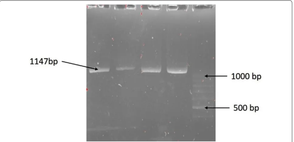
A segment of the JAK2 gene of about 1147 bp was amplified. PCR–RFLP was performed using 12.5 µl of master mix (amplicon), 1 μl of DNA, 1 μl of each of forward and reverse primers (10 pmol) and 9.5 μl of nuclease-free water in a total volume of 25 μl. Next, the samples were placed in 1000 °C. Touch thermal cycler Bio-Rad, programmed in the following cycle: initial denaturation at 95 °C for 5 min, 35 cycles of denatura-tion at 94 °C for 20 s, annealing at 52 °C for 30 s and primer extension step at 72 °C for 30 s. Ultimately, the final primer extension step was done at 72 °C for 5 min. A part of the PCR product in association with a 100 bp marker were run on agarose gel 2% containing 0.1 µg/ ml SYBR Safe color. After that, the remaining of ampli-fied sample was digested by BsaXI (NEB Cat No: R0609S) restriction enzyme, which cuts the specific site related to the JAK2V617F mutation. Recognition site of the enzyme is shown in Fig. 1. The change of this sequence occurs in the JAK2 V617F mutation with the guanine to thymine substitution at nucleotide 1849 in exon 14 of the JAK2 gene.
Comparison with standard methods
The test and control samples were analyzed using Ipso-gen JAK2 Mutant Kit (QiaIpso-gen, Germany), as the refer-ence method.
Results
ARMS‑PCR results
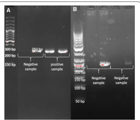
In the ARMS-PCR assay, the forward mutant-specific primer and reverse primer amplified a 250 bp product and showed a mutated allele in the positive sample. In another tube, the forward wild type-specific primer and reverse primer amplified a 250 bp product showing pres-ence of the wild type allele (Fig. 2). ARMS-PCR Results of JAK2 mutation detection was the same as the stand-ard method in 47 samples. The positive sample shows heterozygote allele burden in this assay. In this assay, the standard positive sample that shows 50% and 75% allele burden subjected and the result show heterozygosity.
PCR–RFLP results
PCR–RFLP requires generation of an amplified prod-uct. Forward and reverse primers were used to amplify an 1147 bp product (Fig. 3). Products were then used for cleavage with BsaXI enzyme. With the wild type allele, the 2 recognition sites in the amplified product make 130 bp, 140 bp and 807 bp cleaved products, and also two 30 bp products that are not detected in the gel. In the mutant allele one of the cleavage sites is omit-ted by the mutation, resulting in absence of the 140 bp product and a new 950 bp product. If a heterozygote sample is digested by BsaXI the 130 bp, 140 bp and 807 bp cleaved products are generated by the wild type allele and the mutant allele gives 130 bp and 950 bp cleaved products. At the end after electrophoresis the 130 bp, 140 bp, 807 bp and 950 bp product are seen in the polyacrylamide gel stained by Silver staining method (Fig. 4).
Real time PCR and HRM results
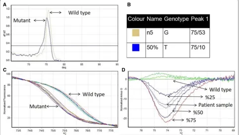
Primer pairs of HRM amplified an 80 bp product, whether the sample was positive or negative for the JAK2V617F mutation (c.1849G>T). In HRM analysis, positive and negative sample have different melting peaks (Fig. 5a, b). This shows c.1849G>T replacement
Fig. 1 Recognition site of BsaXI enzyme
Fig. 3 Amplified product before enzyme digestion shows 1147 bp in safe stain gel agarose
caused a decrease in melting peak. To further differen-tiate the mutant and wild type, a high-resolution differ-ence plot of the HRM assay was plotted by subtracting the melting curve of each species from the baseline (or reference) curve. The baseline curve shape is con-structed by negative sample and 0% synthetic DNA for JAK2 V617F. Based on the patterns of the difference plots, the assay was able to discriminate JAK2 V617F mutant allele from wild type and also could differenti-ate allele burden in the sample by the distance of differ-ent melt curves from the baseline (Fig. 5d).
Sensitivity and specificity of methods
The sensitivity and specificity of Ipsogen JAK2 Mutant Kit (Qiagen, Germany) confirmed by sequencing, HRM, PCR–RFLP and, ARMS PCR methods, calculate in 45 cases were 20 and 24 cases were negative and positive, respectively, the positive and negative cases confirmed by Ipsogen JAK2 Mutant Kit (Qiagen, Germany) con-firmed by sequencing as gold standard method. It means all positive sequenced samples were also positive in other methods, like negative cases. These data are summarized in Table 1.
Fig. 5 a, b Melting peak in wild type and mutant sample for the JAK2 V617F mutation shows different temperature (75/53 and 75/10, respectively).
c The normalized fluorescence plot which synchronization samples in maximum and minimum fluorescence emission. d Normalized and temperature-shifted difference plot of the various kinds of allele burden mutation in compare of wild type (G allele)
Table 1 Sensitivity and specificity of Ipsogen JAK2 Mutant Kit (Qiagen, Germany) confirmed by sequencing, HRM, PCR– RFLP and, ARMS PCR methods
Sample Mutant Wild type Sensitivity (%) Specificity (%) No. tests Positive No. tests Negative
Ipsogen JAK2 mutant Kit (Qiagen,
Germany) confirmed by sequencing 25 25 20 20 100 100
ARMS 25 25 20 20 100 100
PCR–RFLP 25 25 20 20 100 100
Discussion
Myeloproliferative neoplasms (MPN) are the hetero-geneous disease which classified by the existence of Philadelphia chromosome. In patients with negative Philadelphia chromosome(ph-neg), there is a somatic mutation (c.1849G>T) in JAK2 gene which constitutively activate JAK2-STAT signal-transduction pathway. Detec-tion of this point mutaDetec-tion is critical in diagnosis and treatment of the patients.
In the present study, Results of JAK2 V617F Mutation detection by PCR–RFLP, ARMS-PCR and HRM meth-ods were similar to gold standard methmeth-ods including Real Time PCR and sequencing. In comparison, each of these methods had some advantages and disadvantages.
The advantages and disadvantages of each tests are different due to the difference between the basis of each method, ARMS-PCR depend on primers and the set up performance, PCR–RFLP depend on the enzyme and the control sequence and the HRM depend on the instrument and the ability of the laboratory to set up the method. The sequencing is gold standard method in diagnosis of mutation Lin and et al. [23] find the Limit of detection (mutant concentration in %) for AS-PCR and HRM 2.5 and 6% of the whole DNA, respectively. In our study the HRM, PCR–RFLP and, ARMS-PCR per-formed and the HRM can find the allele burden as 25, 50,75%, less than, more them or between them. On the other hand, both PCR–RFLP and ARMS- PCR methods depend on operator and its set up. HRM analysis is an efficient and sensitive PCR–based approach for deter-mining the gene mutation with capability to differentiate heterozygous and homozygous mutations. Furthermore, this method provides the possibility of allele burden measurement in clinical evaluations.
Hong-Cui Cao et al. compared the different approaches to identify JAK2V617F mutations and introduced PCR– RFLP method as a definite and appropriate way to detect this mutation. Also they use the ARMS PCR and other molecular methods to detect mutations and selected sanger sequencing as reference method to identify the mutation. In our study, a modified PCR–RFLP could recognize the presence of mutations in patients as well as the sequencing method [24]. In another study by Tze-Kiong Er and colleagues in order to identify the JAK2 V617F mutation, PCR RFLP method was considered as a reference method for the approval of other methods. They have introduced PCR RFLP as an appropriate but operator-dependent method to identify this mutation [25]. In present study, PCR RFLP method has a detec-tion sensitivity comparable with convendetec-tional meth-ods (Qiagen) to identify the mutations and sequencing. Advantages of the present modified PCR–RFLP method include the presence of internal controls to verify the
performance of the enzyme cleavages as well as being cost-effective and affordable compared to the sequencing method and other methods such as HRM, which need to special equipment. However, this method is only capable of detecting just homozygous and hetero-zygote caus-ing states. Since the DNA samples were extracted from peripheral blood and this mutation is not present in lym-phoid cells, all the samples show the heterozygous state. In a study by Amy Jones et al. to evaluate the methods of identifying mutations in JAK2 V617F, techniques such as ARMS PCR were introduced as an appropriate and cost-effective method in order to examine the exact mutation [7]. Results obtained by the mentioned method in our study were the same as using PCR RFLP and sequenc-ing techniques. The present ARMS PCR method as well as the PCR–RFLP is cost-effective and beside that it is enzyme independence method. This method’s drawback could be that can only detect homozygous and hetero-zygote as well as PCR RFLP. In a study by Hong-Cui Cao et al. HRM method was identified as an easy, sensitive and reliable way to detect JAK2V617F mutation. Since this technique was performed using REAL TIME PCR and Reaction was conducted in a closed tube, contamina-tion error is minimized. Another benefit of this method of identification is about the amount of the mutant allele carried in samples taken from patients, means somehow allele burden, that makes doctors able to not only rec-ognize homozygous and heterozygous states but also to determine the percentage of mutations. This advantage allows the physician to examine the amount of carried mutant allele during and after treatment as well as in the detection of minimal residual disease (MRD). According to a study by Serge Carillo and his colleagues in which the mutation detection rate reaches up to 1% of mutant DNA in samples carrying the mutant allele, the NESSTED HRM method has been used to identify mutations and had a sensitivity of 100% and specificity of 96.7%.
In conclusion, given the particular sensitivity of HRM and many advantages in identifying JAK2V617F muta-tions, it can be a good way to identify mutations in patients with suspected myeloproliferative diseases using DNA extracted from the peripheral blood. In addition, due to the sensitivity of this method it can be useful in following the patients’ response to the treatment and determining MRD for them.
Authors’ contributions
AM performed the experiments, analyzed the data and wrote the manu-script. Alireza Farsinejad gave conceptual advice. BK gave conceptual advice, analyzed the data and wrote the manuscript. Ahamd Fatemi supervised the project and analyzed the data. All authors read and approved the final manuscript.
Author details
1 Student Research Committee, Faculty of Allied Medicine, Kerman University
•fast, convenient online submission
•
thorough peer review by experienced researchers in your field
• rapid publication on acceptance
• support for research data, including large and complex data types
•
gold Open Access which fosters wider collaboration and increased citations maximum visibility for your research: over 100M website views per year
•
At BMC, research is always in progress.
Learn more biomedcentral.com/submissions
Ready to submit your research? Choose BMC and benefit from: Laboratory Sciences, Faculty of Allied Medicine, Kerman University of
Medi-cal Sciences, Kerman, Iran. 3 Molecular and Medicine Research Center, Arak
University of Medical Sciences, Arak, Iran.
Acknowledgements
Hereby, the efforts of staff of stem cell research center and pathology group in Kerman University of Medical Sciences and Emam-Reza laboratory, Arak, Iran, who had a role in the fruition of this study, are appreciated with lots of grati-tude. Also we acknowledge Dr. Anna L. Godfrey in University of Cambridge due to guide us in writing article.
Competing interests
The authors declare that they have no competing interests.
Availability of data and materials
Please contact author for data requests.
Consent for publication
All authors consent for publication in experimental hematology and oncology journal.
Ethics approval and consent to participate
The study approved in Kamran university of medical science ethical commit-tee and The Ethic Approval Code is IR.KMU.REC.1395.812.
Funding
The present study was supported by the grant No. 95000484 from Kerman University of Medical Sciences.
Publisher’s Note
Springer Nature remains neutral with regard to jurisdictional claims in pub-lished maps and institutional affiliations.
Received: 6 February 2019 Accepted: 20 April 2019
References
1. James C, Ugo V, Le Couedic JP, et al. A unique clonal JAK2 mutation leading to constitutive signalling causes polycythaemia vera. Nature. 2005;434(7037):1144–8. https ://doi.org/10.1038/natur e0354 6. 2. Kralovics R, Passamonti F, Buser AS, et al. A gain-of-function mutation of
JAK2 in myeloproliferative disorders. N Engl J Med. 2005;352(17):1779–90.
https ://doi.org/10.1056/nejmo a0511 13.
3. Levine RL, Wadleigh M, Cools J, et al. Activating mutation in the tyrosine kinase JAK2 in polycythemia vera, essential thrombocythemia, and myeloid metaplasia with myelofibrosis. Cancer Cell. 2005;7(4):387–97. 4. Baxter EJ, Scott LM, Campbell PJ, et al. Acquired mutation of the
tyrosine kinase JAK2 in human myeloproliferative disorders. Lancet. 2005;365(9464):1054–61. https ://doi.org/10.1016/s0140 -6736(05)71142 -9. 5. Tefferi A, Thiele J, Vardiman JW. The 2008 World Health
Organiza-tion classificaOrganiza-tion system for myeloproliferative neoplasms. Cancer. 2009;115(17):3842–7.
6. Murugesan G, Aboudola S, Szpurka H, et al. Identification of the JAK2 V617F mutation in chronic myeloproliferative disorders using FRET probes and melting curve analysis. Am J Clin Pathol. 2006;125(4):625–33. 7. Jones AV, Kreil S, Zoi K, et al. Widespread occurrence of the JAK2
V617F mutation in chronic myeloproliferative disorders. Blood. 2005;106(6):2162–8. https ://doi.org/10.1182/blood -2005-03-1320. 8. Jelinek J, Oki Y, Gharibyan V, et al. JAK2 mutation 1849G>T is rare in acute
leukemias but can be found in CMML, Philadelphia chromosome–nega-tive CML, and megakaryocytic leukemia. Blood. 2005;106(10):3370–3. 9. Zhao R, Xing S, Li Z, et al. Identification of an acquired JAK2 mutation in
polycythemia vera. J Biol Chem. 2005;280(24):22788–92.
10. Wolanskyj AP, Lasho TL, Schwager SM, et al. JAK2V617F mutation in essential thrombocythemia: clinical associations and long-term prognos-tic relevance. Br J Haematol. 2005;131(2):208–13.
11. Poodt J, Fijnheer R, Walsh I, et al. A sensitive and reliable semi-quantitative real-time PCR assay to detect JAK2 V617F in blood. Hematol Oncol. 2006;24(4):227–33.
12. Rapado I, Albizua E, Ayala R, et al. Validity test study of JAK2 V617F and allele burden quantification in the diagnosis of myeloproliferative dis-eases. Ann Hematol. 2008;87(9):741–9.
13. Sidon P, El Housni H, Dessars B, et al. The JAK2V617F mutation is detect-able at very low level in peripheral blood of healthy donors. Leukemia. 2006;20(9):1622.
14. Grünebach F, Bross-Bach U, Kanz L, et al. Detection of a new JAK2 D620E mutation in addition to V617F in a patient with polycythemia vera. Leu-kemia. 2006;20(12):2210.
15. Schnittger S, Bacher U, Kern W, et al. Report on two novel nucleotide exchanges in the JAK2 pseudokinase domain: D620E and E627E. Leuke-mia. 2006;20(12):2195.
16. Fröhling S, Lipka DB, Kayser S, et al. Rare occurrence of the JAK2 V617F mutation in AML subtypes M5, M6, and M7. Blood. 2006;107(3):1242–3. 17. Pesu M, O’Shea J, Hennighausen L, et al. Identification of an acquired
mutation in Jak2 provides molecular insights into the pathogenesis of myeloproliferative disorders. Mol Interventions. 2005;5(4):211.
18. Fröhling S, Scholl C, Gilliland DG, et al. Genetics of myeloid malignancies: pathogenetic and clinical implications. J Clin Oncol. 2005;23(26):6285–95. 19. Antonioli E, Guglielmelli P, Pancrazzi A, et al. Clinical implications of
the JAK2 V617F mutation in essential thrombocythemia. Leukemia. 2005;19(10):1847.
20. Cazzola M, Skoda R. Gain of function, loss of control-a molecular basis for chronic myeloproliferative disorders. Haematologica. 2005;90(7):871–4. 21. Tefferi A, Gilliland DG. JAK2 in myeloproliferative disorders is not just
another kinase. Cell Cycle. 2005;4(8):4053–6.
22. Link-Lenczowska D, Pallisgaard N, Cordua S, et al. A comparison of qPCR and ddPCR used for quantification of the JAK2 V617F allele burden in Ph negative MPNs. Ann Hematol. 2018;97(12):2299–308.
23. Lin CY, Ho CM, Tamamyan G, et al. Validating the sensitivity of high-resolution melting analysis for JAK2 V617F mutation in the clinical setting. J Clin Lab Anal. 2016;30(6):838–44. https ://doi.org/10.1002/jcla.21945 . 24. Cao A, Galanello R. Beta-thalassemia. Genet Med. 2010;12(2):61. 25. Er T-K, Lin S-F, Chang J-G, et al. Detection of the JAK2 V617F missense