Journal of Chemical and Pharmaceutical Research, 2015, 7(8):300-306
Research Article
ISSN : 0975-7384
CODEN(USA) : JCPRC5
Expression and immunogenicity of erythropoietin gene cloned in eukaryotic
expression vector (pVAX1 vector)
Usha Tiwari
1*, Bipin Chand Misra
2, Nitin Sharma
1, Mahesh Kumar
1, Neha K. Mehra
1and Pragya Pandey
11Division of Animal Cell Culture Laboratory, Institute of Biotechnology, G.B. Pant University of Agriculture & Technology, Patwadangar (Nainital), UK, India
2Department of Biotechnology and Microbiology, SCMAT, Chaubepur, Kanpur
_____________________________________________________________________________________________
ABSTRACT
Erythropoietin gene is immunogenic. In the present study the recombinant plasmid pVAX1.epo.hu transfected HeLa cells expressed protein which was confirmed by immunoperoxidase test, SDS-PAGE and western blotting. The cells were found to express the protein with development of purple color precipitate in Immunoperoxidase test. Intense purple coloration of cells was observed in HeLa cells transfected with pVAX1.epo.hu. The vector alone and untransfected healthy cells failed to show any coloration confirmed no expression of proteins. By SDS-PAGE and western blotting, the expressed erythropoietin (34 kDa) was confirmed. The in vivo immunological response was studied in mice.
Keywords: HeLa cells, Erythropoietin gene, pVAX1 Vector, Replicase Vector, Gene Expression
_____________________________________________________________________________________________
INTRODUCTION
Erythropoietin (epo) is a glycoprotein hormone responsible for the regulation of red blood cell production. This hormone triggers the proliferation, differentiation and maturation of bone marrow erythroid precursors into functional erythrocytes when blood oxygen availability is decreased, such as during hypoxia. epo binds to and activates the receptor on erythroid progenitor cells. The treatment of anemic patients with epo significantly reduces their dependence on blood transfusions and minimizes potential side effects such as iron overload, infections and adverse reactions to leukocyte antigens. In addition to its role in hematopoeisis, epo is neuroprotective in the nervous system and can also protect other organs [1]. Prior to the 1980s, human epo for the treatment of kidney failure and other related blood disorders had to be extracted from donors. However, under normal conditions, the expression of epo is generally low, meaning that many donors are necessary to obtain sufficient material for treatment. Successful cloning of the epo gene and subsequent expression in Chinese hamster ovary (CHO) cells led to the production of several types of commercially available recombinant human epo (r.epo.hu) for human use. Keeping the above facts in view, the present work was been undertaken to express the epo.hu gene in replicase based eukaryotic pVAX1 vector.
EXPERIMENTAL SECTION
1.1. Recombinant plasmid (pVAX1.epo.hu)
consistent with the Food and Drug Administration (FDA) document, “Points to Consider on Plasmid DNA Vaccines for Preventive Infectious Disease Indications”, published December 22, 1996 (see FDA “Points to Consider” below). Features of the vector allow high-copy number replication in E. coli and high-level transient expression of the protein of interest in most mammalian cells.
1.2. Cell Line
HeLa cell line was obtained from National centre for Cell Science (NCCS), Pune. HeLa cell line was used in the study for in vitro expression analysis of recombinant plasmids (pVAX1.epo.hu) and was maintained in GMEM (Micro lab), supplemented with 10% new born calf serum (Gibco, NY),100 µg/ml penicillin and streptomycin.
1.3. Conjugates
Rabbit anti-mouse HRP conjugated antibody was obtained from Bangalore Genei, Bangalore, (India).
1.4. In vitro expression analysis 1.4.1. Transfection of HeLa cells
Cells were trypsinised using trypsin-versenate solution (TVS) and then 4 ml of GMEM containing 10% FCS and penicillin and streptomycin (50 µl/ml) was added, to make cell suspension of 1X10⁵ cells/ml. Harvested exponentially growing cells by trypsinization and prepared cell suspension in growth medium. Prepared the calcium phosphate-DNA coprecipitate as follows: combined 50 µl of 2.5M CaCl2 with 10 µl of plasmid DNA in a sterile microfuge tube, Added 40 µl DW, kept at room temperature. Immediately transferred the calcium phosphate-DNA suspension using 20 µl suspension for each wells of microtitre plate. Added 100 µl of cell culture suspension in each wells of 96 wells microtitter plate. Rocked the plate gently to mix the medium, which will become yellow-orange and turbid. Carried out this step as quickly as possible because the efficiency of transfection declines rapidly once the DNA precipitate is formed. Kept control wells without transfection. Incubated at 370C in a humidified incubator with an atmosphere of 5% CO2 for 72 hours. Examined for gene expression by IPT.
1.4.2. Raising primary antibody against pVAX1.epo.hu in mice
Primary polyclonal antibody against the human erythropoietin gene was raised in mouse by hyper immunization of six mice with pVAX1.epo.hu plasmid. 50 µg of rplasmid DNA was injected intramuscularly in lateral region of thigh muscles of each mouse and repeated every week for four weeks consecutively. One week after last injection bled through inner canthus of eyes with a capillary and serum was prepared.
1.4.3. Immunoperoxidase test (IPT)
After 72 hours, transfected HeLa cells were washed with 1XPBS twice and fixed with 80% chilled acetone at 40C for 10 min and air-dried. Put a few drops of mouse anti epo.hu hyperimmune serum and incubated at 370C for 1 hr and again washed with PBS, added a few drops of horseradish peroxidase (HRPO) conjugated rabbit anti-mouse antibody to wells and incubated at 370C for 1 hr in humid chamber. The cells were again washed with PBS thrice and incubated with 2-3 drops of Nadi reagent for 5 min. After the development of color, cells were washed with PBS, dried in air and observed under microscope and photographed, protocol was carried out as per [2].
1.4.4. SDS-PAGE
HeLa cells were transfected in 96 well plates with pVAX1.epo.hu rplasmid and pVAX1 vector. After 48 hours of transfection cells were processed following the method [3].
Gel preparation: The gels consist of a lower resolving gel and an upper stocking gel that concentrate the sample
before its entry into the resolving gel. The buffer system was the discontinuous system of laemmli. Stored all solution in brown bottle at 4°C and SDS solution at room temperature. The volumes below were for slab gels of 15 ml vol with a 6 ml stacking gel. Volumes are gives in ml.
immediately dried on Whatman No.1 filter paper and stained for direct visualizing of polypeptide bands. Placed gel in a staining container with lid. Added 50 ml staining solution. Covered and placed at room temperature for 15 min. Poured off solution, replaced with 50ml staining solution + 5ml stain. Covered and placed at room temperature for overnight. The gel was turned blue at that time. Poured off the staining solution, added 100ml destaining solution covered and returned to room temperature for 15min. Gentle mixing improved the destaining process. Poured off the destaining solution and replaced. Continued to repeat this step many times. Completed destaining to produce a clear gel with blue polypeptide bands depends on gel thickness and required 24 h.
Table 1: Component of resolving and stacking gel
Reagents Resolving gel (10%) Stacking gel (4%)
Lower gel buffer 3.75 ………
Upper gel buffer ………. 1.25
Acrylamide stock 5.0 0.80
Distilled water 6.05 3.87
10 % SDS 0.15 0.06
10%ammonium persulphate 0.05 0.02
TEMED 0.07 0.004
1.4.5. Western blot analysis
After completion of Sodium dedecyl sulfate-polyacrylamide gel electrophoresis, the epo.hu protein bands from unstained gel were blotted on to nitrocellulose membrane using SNAP i.d. system. This Millipore‟s patent pending SNAP i.d. Protein Detection System provides a fast and convenient method for the detection of immune reactive proteins on western blots and the Millipore‟s protocol was followed.
System Set-up
Placed the SNAP i.d. system on a level bench top and attached the vacuum tubing to the back of the system by pushing the coupling epo.hu insert on the end of the tubing into the quick disconnect fitting at the back of the system base. Connected the other end of the tubing to a vacuum source, used a one-liter vacuum flask as a trap and a Millex-FA50 filter to protect the vacuum source from contamination.
Rolled gently with blot roller to remove any air bubbles trapped between the blot holder and the blot. Wet the inner white face of the blot holder with Milli-Q water, until it turns gray. Removed excess liquid using the blot roller, was preventing movement of the blot during assembly. Placed the pre-wet blot membrane in the center of the blot holder and the protein side down Open the blot holder lid.
Blot Assembly
Before starting the blot assembly procedure, prepared the antibody, blocking, and wash buffers solutions. Placed the spacer on to top of the blot, making sure it completely covers all edges. Rolled blot again to ensure completed contact of blot spacer with blot membrane, Closed the blot holder lid Squeezed firmly at the base of the tab area to secure lid. Opened lid of system by squeezing latch between thumb and forefingers and lifting upwards Placed blot holder in system chamber with the well side up, aligning the blot holder tabs with notches of chamber. Repeated the assembly procedure for all blots being processed Closed and latch the system lid.
Immunodetection Protocol
solution had been completely emptied from the blot holder. With vacuum running continuously, washed the blot with 10 ml of wash buffer, three sequential washes were required for optimal performance Removed blot holder from the system, placed it on the bench with the well-side down, and opened the lid. With forceps, removed and discarded the spacer. Removed blot and incubated with the appropriate detection reagent such as Immobilon® Western HRP Substrate, visualize. Discarded the single-use blot holder.
1.6. Immune response studies
1.6.1. Assessment of hematopoietic activity in vivo
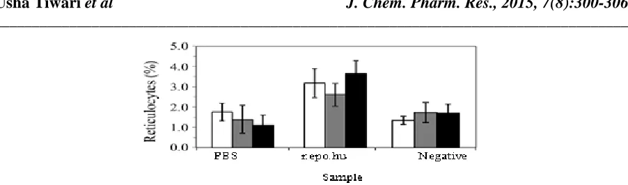
Female mice were used to test the hematopoietic activity of r.epo.hu. Each mouse was injected subcutaneously with 0.25 mL of sample (acetone-precipitated supernatant of r.epo.hu cultures) for three consecutive days. Three mice (6-8 weeks old) were used for each treatment and two groups of mice were used as controls (one of these two groups was treated with PBS and the other received no treatment). Blood samples were collected into 5% sodium EDTA on the fourth day after treatment. An equal volume of blood was mixed with new methylene blue and incubated at 37 °C for 1 h. Seven microlitres of this blood-dye mixture was then used to prepare smears on glass slides. Five slides were prepared for each mouse (total of 15 slides per treatment since there were three mice per group). Reticulocytes were counted with the aid of a microscope (at 100X magnification) in five randomly selected areas of each slide and their number expressed as a relative to the total number of red blood cells observed.
1.6.2. Statistical analysis
The results were expressed as the mean ± SEM, where appropriate. Statistical comparisons were done using Students t-test, with a value of p < 0.01 indicating significance.
RESULTS AND DISCUSSION
The expression of rplasmid (pVAX1.epo.hu) was checked by immuno assay in HeLa cell line and cells were found to express the epo.hu protein by development of the purple color precipitate. Intense purple coloration of cells was observed in which HeLa cells were transfected with pVAX1.epo.hu (Fig. 1). The vector alone and untransfected healthy cells (Fig. 2) failed to show any color indicating that the epo.hu gene was expressed in cells due to the presence of pVAX1.epo.hu plasmid. In this expression technique, 90% cells were found to express epo.hu protein.
Fig.2: Healthy Control HeLa cells showing no color reaction, 100X
The SDS-PAGE analysis of expressed epo.hu protein indicated a specific isolated thick band (Blue color) of 34 kDa (Fig. 3).The western blot analysis of expressed protein indicated a specific isolated band seen in nitrocellulose membrane of 34 kDa size (Fig. 4). Adaptive responses to hypoxia occur in many biological systems. A well-characterized example is the hypoxic induction of the synthesis of erythropoietin, a hormone which regulates erythropoiesis and hence blood oxygen content. The restricted expression of the erythropoietin gene in subsets of cells within kidney and liver has suggested that this specific oxygen-sensing mechanism is restricted to specialized cells in those organs. Using transient trasfection of reporter genes coupled with a transcriptional enhancer lying 3' to the erythropoietin gene, it was shown that an oxygen-sensing system similar, or identical, to that controlling erythropoietin expression is widespread in mammalian cells. The extensive distribution of this sensing mechanism contrasts with the restricted expression of erythropoietin, suggesting that it mediate other adaptive responses to hypoxia27. Hoc and Viet expressed epo.hu in E. coli cells, the expression of the epo.hu was analyzed by SDSPAGE and confirmed by western Blotting using anti epo.hu antibody. The Pichia pastoris expression system was used to produce recombinant human erythropoietin. The entire recombinant human erythropoietin (repo.hu) gene was constructed, cloned and expressed through the secretary pathway of the Pichia expression system. Recombinant erythropoietin was successfully expressed in Pichia pastoris 28. The estimated molecular mass of the expressed protein ranged from 32 kDa to 75 kDa, with the variation in size being attributed to the presence of repo.hu glycosylation analogs. A crude functional analysis of the soluble proteins showed that all of the forms were active in
vivo 29. Evidence from cell culture and animal experiments suggested a neuroprotective and neurotrophic function
Fig.3: SDS-PAGE of HeLa cells extract for epoexpression Lane M- Protein marker
Lane 1-transfected cells showing expressed protein band Lane 2-mock transfected cells extract showing no expression
34 kDa
20 kDa 30 kDa 60 kDa 1 2 3 4 M
Fig.4: Detection of epo protein using Western blotting Lane : M 1 kDa protein marker
Lane : 1 Mock transfected Lane : 2 Vector control Lane : 3 & 4 epo.hu protein
Fig. 5: Increase in the number of reticulocytes after injections of r.epo.hu.
CONCLUSION
The expression of rplasmid (pVAX1.epo.hu) was checked by immuno assay in HeLa cell lines and cells were found to express the epo.hu protein by development of a purple color precipitate. The expression of rplasmid was again checked by using SDS-PAGE and western blot analyses. The expressed epo.hu protein (34 kDa) was confirmed by SDS-PAGE and western blot analysis. In mice injected with r.epo.hu the number of reticulocytes increased by 3.16 ±0.64. These increases were significantly (p < 0.01) higher than those observed in mice injected with PBS and the non-treated negative control.
Acknowledgement
Authors thank Director IBT, IVRI and also thank Director, IBIT Izatnagar, Bareilly for providing facilities to carry out this work
REFERENCES
[1] Genc S, Koroglu TF and Genc K. Brain. Res., 2004, 1000, 19-31. [2] Nakane PK, Kawaoi A. J. Histochem. Cytochem., 1974, 22, 1084.
[3] Rodriguez R L and Tait R C, Recombinant DNA techniques. An introduction, Addison- Wesley PubI. Co.
London, 1983.
[4] Maxwell PH, Osmond MK, Pugh CW, Heryet A, Nicholls L G and Tan C. C. Kidney International, 1992, 44, 1149-1162.
[5] Hoc T C Y and Viet. Vista, 2006, 324, 10-15.
[6] Maleki A, Roohvand F, Tajerzadeh H, Khanahmad HB, Nobari M, Beiruti A and Najafabadi R, Avicenna
Journal of Medical Biotechnology, 2010, 2, 197-206.
[7] Dame C, Bartmann P, Wolber E, Fahnenstich H, Hofmann Dand Fandrey J, Brain Research Development Brain
Research, 2002, 29, 69-74.
[8] Sharma N, Kaushik P, Rai A, Maurya R, Pal P, Tiwari U. International J. Applied Bio. Pharma. Technol., 2010, 1, 339-348.