Journal of Chemical and Pharmaceutical Research, 2016, 8(3):405-423
Research Article
CODEN(USA) : JCPRC5
ISSN : 0975-7384
Synthesis and characterization of gold nanoparticles and their anticancer
activity using gamma radiation
Amro Hanora
1, M. M. Ghorab
2, Ahmed I. El-Batal
3and Farag M. Abo Mosalam
31Microbilogy and Immunology Dep., Faculty of Pharmacy, Suez Canal University, Egypt 2Pharmaceutical Dep., Faculty of Pharmacy, Suez Canal University, Egypt
3Drug Radiation Research Dep., Biotechnology Division, National Center for Radiation Research and Technology
(NCRRT), Atomic Energy Authority, Cairo, Egypt
_____________________________________________________________________________________________
ABSTRACT
Aqueous dispersed gold nanoparticles (AuNPs) were successfully synthesized by green chemical and biological synthesis processes using citrus pectin, sodium alginate, chitosan and aqueous extract of fermented fenugreek powder by gamma radiation. The prepared AuNPs are characterized by UV–vis spectroscopy, Transmission electronmicroscopy (TEM), Dynamic light scattering (DLS), X-ray diffraction (XRD) and Fourier transform infrared spectroscopy (FTIR) spectrum indicates the presence of different functional groups present in the biomolecule capping the nanoparticles. XRD analysis of the gold confirmed the formation of metallic gold nanoparticles. The results demonstrat that the green biological was superior than green chemical synthesis for the production of metal nanoparticles (NPs). This is resulted in production of smallest size particles and more effective as anticancer as follow:
AuNPs green chemical synthesis (Citrus Pectin 1%, metal concentration 1mM, pH 7 and radiation dose 5 kGy, size TEM (21nm), DLS (31nm), anticancer IC50 EAC=21.5µg/ml and CACO=24.4µg/ml)} and AuNPs green biological synthesis {(Aqueous extract fermented fenugreek powder, metal concentration 1mM, pH 7, 100OC, size TEM (12nm), DLS (23nm), anticancer IC50 EAC=4.8µg/ml and CACO= 5.45µg/ml)}. The nucleation and growth mechanism of gold nanoparticles is also discussed. The size of nanoparticles is influenced by certain parameters such as the choice of stabilizer, metal concentration, pH during synthesis and absorbed dose. Factorial design was used to study the effect of several parameters on AuNPs production especially effect of temperature and gamma radiation was investigated. The incorporated of AuNPs with reducing and stabilizing agents like citrus pectin, chitosan, sodium alginate and fermented fenugreek powder its increase the antioxidant activity.
Keywords: Natural polymers (chitosan, citrus pectin and sodium alginate), Radiation, Gold nanoparticles , Pleurotus ostreatus, fenugreek, anticancer and antioxidant activities.
_____________________________________________________________________________________________
INTRODUCTION
Fenugreek (Trigonella foenumgraecum), is a herb that is commonly found growing in the Mediterranean region of the world. While the seeds and leaves are primarily used as a culinary spice, it is also used to treat a variety of health problems in Egypt, Greece, Italy and South Asia. Fenugreek seeds have been found to contain protein, vitamin C, niacin, potassium, and diosgenin (which is a compound that has properties similar to estrogen). Other active constituents in fenugreek are alkaloids, lysine and L-tryptophan, as well as steroidal saponins (diosgenin, yamogenin, tigogenin, and neotigogenin) [1].
Naturally grown plants and various plant species can potentially serve as long asting and environmentally benign reservoirs relative to microorganisms. Gold NPs are probably the most attractive member of the noble metal nanoparticles family because of their potential applications in the field of chemical catalysis, nonlinear optics, surface enhanced Raman scattering, nanoelectronics, gene expression and disease diagnosis[2].
Solid state fermentation (SSF) of an edible plant matrix by filamentous fungi is a biotechnological strategy that may induce health beneficial naturally occurring antioxidant components including polyphenols during microbial fermentation [3].
Polymers such as chitosan, alginate…ect. can form various chemical bonds with metals components thus enhancing the stability of the nanoparticles; It has low toxicity and it is therefore safe for human applications as antimicrobial, anticancer. Gamma radiation has been proved to be a simple and efficient method for nanoparticles synthesis, requires an aqueous system, room temperature and ambient pressure [4]. The formation of NPs by radiation can be attributed to the radiolytic reduction which generally involves radiolysis of aqueous solutions that provides an efficient method to reduce metal ions.
In the radiolytic method, when aqueous solutions are exposed to gamma rays, they create solvated electrons, which reduce the metal ions and the metal atoms eventually coalesce to form aggregates [5].
Natural products have shown great promise in mitigating carcinogenesis and associated cellular aberrations, with substantial numbers of anticancer agents being derived from natural sources [6].
This study aims at the synthesis of gold Nanoparticles (AuNPs) using some natural polymers (Green chemical process) such as citrus pectin (CP), chitosan (Cs) and alginate (Alg) and fermented plant extract (green biological process) using aqueous fermented fenugreek (AEFF) by Pleurotus ostreatus.
Materials and Methods 1.Chemicals and materials
Citrus pectin powder (CPP) , Chloroauric acid, chitosan and sodium alginate were obtained from Sigma-Aldrich. All other chemicals and solvents used were of analytical grade.
2.Preparation of gold nanoparticle
2.1.Green chemical Synthesis of gold nanoparticles (AuNPs) using gamma radiation
a.By chitosan1%:
1gm chitosan was added to 80ml acetic acid (3%); completed to 100ml by deionized water. The mixture was stirred at room temperature and placed in dark for overnight to make it homogenous. Gold Chloride 1mM solution was mixed with chitosan 1% solution 1:1 v/v. The mixture was stirred at room temperature and (exposed to gamma ray at 0, 20, 40, 60, 80, and 100 kGy . This led to the immediate formation of AuNPs visualized as pink color solution. Then Au NPs were immediately characterized by UV-Visible spectroscopy.
b. By sodium alginate1% :
1gm of sodium alginate was added to 100ml of de ionized water. The mixture was stirred at room temperature and placed in dark for overnight to make it homogenous. Gold Chloride 1mM solution was mixed with sodium alginate1 % solution 1:1 v/ v. The mixture was stirred at room temperature and exposed to gamma ray at 0, 10, 15, 20, 25, 30, 35, 40, 45, and 50 kGy. This led to the immediate formation of Au NPs visualized as change of color solution. Then AuNPs were immediately characterized by UV-Visible spectroscopy.
c.By citrus pectin1%:
2.2.Green bilolgyical synthesis using gamma raradiation.
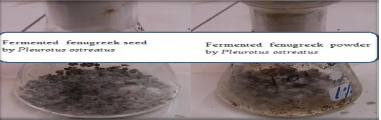
A. Fermented fenugreek (seed and powder): aqueous Extract of fermented fenuogreek preperd as follow;
Fungal strain; locally isolated fungal strains Pleurotus ostreatus was used in the study obtained from the culture collection in the Pharmaceutical Microbiology Laboratory Drug Radiation Research Department (NCRRT, Egypt). A strain was microscopically identified and kept on potato dextrose agar (PDA) at 4oC and periodically sub-cultured to maintain viability.
Fermentation medium (Solid state fermentation) ; Fermentation was carry out in 250ml Erlenmeyer flasks, where distilled water were added to fenugreek seed or powder (60% moisture content) and autoclaved at 121oC for 20min. Flasks inculcated by the fungus medium as a 2ml spore suspension (8 × 10⁶ spores/ml) and incubated at 30OC statically in complete darkness for 10days. After ten days, the whole contents of the flasks were soaked in de ionized water (1g fresh weight/10ml de.H2O) and put in a shaker at 200rpm (LAB-Line R Orbit Environ, U.S.A) for 2h and the centrifuged in cooling centrifuge (Hettich Universal 16 R, Germany) for 10min at 5oC. The supernatant was separate from sediment and used for experiment.
Gold Chloride 1mM solution was mixed with aqueous extract of fenugreek (fermented or unfermented seed and powder) 1:1 v/v . The mixture was stirred at room temperature and exposed to gamma ray at 0, 5, 10, 15, 20, and 25 kGy. This led to the immediate formation of AuNPs visualized as pink color solution. Then AuNPs were immediately characterized by UV-Visible
B. Experimental factorial design
Used to compare the effect of heat and radiation on the synthesis of gold nanoparticles by aqueous extract of fermented fenugreek powder (AEFFP); these factors were chosen as they were considered to have the most significant effect on the size of nanoparticles. The levels were selected based on knowledge acquired from initial experimental trials.
Effect of heat; the effects of the three variables in three level form namely: gold concentration (0.5 mM, 1mM and 2mM), pH of reaction mixture (4, 7, and 10) and temperature of reaction mixture (25 oC, 60 oC and 100 oC).
Effect of radiation; the effects of the two variables in three level form namely: gold concentration (0.5 mM, 1mM and 2 mM) and pH of reaction mixture (4, 7, and 10); all samples were exposed to 20kGy. The main effects of parameters on gold nanoparticles production were estimated by UV-visible response O.D.
3.Characterization of nanoparticles
The size, morphology and stability of the synthesized AuNPs were characterized using the following techniques.
UV-VIS spectral analysis:
For the preliminary determination of Au NPs, UV visible spectroscopy (T60 UV/Vis spectrometer); at a resolution of 1nm was performed to measure the Surface Plasmon Resonance (SPR) for the wave length ranging from 200-800 nm.
Transmission Electron Microscopy (TEM)
AuNPs suspension was loaded on carbon coated copper grids and solvent was allowed to evaporate by incubation at 37oC for 30min in an incubator. The size and morphology of the AuNPs were estimated by TEM (JEOL electron microscope JEM-100 CX) operating at 80 kV accelerating voltage.
Dynamic light scattering measurement (DLS)
Average particle size and size distribution were determined by the dynamic light scattering (DLS).technique (PSS-NICOMP 380-ZLS, USA); 250μl of suspension were transferred to a disposable low volume cuvette. After equilibration to a temperature of 25°C for 2 min, five measurements were performed using 12 runs of 10 s each.
Fourier transforms infrared spectroscopy (FTIR)
X-Ray diffraction (XRD)
XRD study of AuNPs was done to determine the structural characterization of the nanoparticles by using X-ray diffractometer (Shimadzu apparatus using nickel-filter and Cu-K a target, Shimadzu Scientific Instruments (SSI), Kyoto, Japan. Founded in 1875) operating with a Cu anode at 40Kv and 5omA in the range of 2θ value between 20° and 100° with a speed of 2°/min). The intensity of the diffracted X- rays was measured as a function of the diffracted angle 2θ.
4.Nitrate reductase assay.
Aqueous extract of fermented fenugreek powder (AEFFP) was considered for the assay of nitrate reductase according to the method of Harley [7]; 2mL of aqueous extract of fermented fenugreek powder was mixed with 2mL of the assay medium (30mM KNO3 and 5% iso-propanol in 0.1M phosphate buffer of pH 7.5) and incubated at 25 oC in the dark for 1hours and then 1mL of 50mM sulphanilamide and 1mL of 10mM NEED (N-(1-naphthyl)
ethylene diamine dihydrochloride) solutions were added. The intensity of the developed color was estimated in an UV–Vis spectrophotometer at 540nm.
5.Antioxidant assay.
Antioxidant activity of AuNPs synthesized by green chemical ( citrus pectin, sodium alginate and chitosan) and biological methods (fermented fenugreek powder and seed) was determined by 2,2' diphenyl-1- picryl hydrazyl (DPPH) radical scavenging; the method of Shimada [8] was used to determine the anti-oxidant activity. 4.0ml AuNPs was added to methanol solution of 10mM DPPH (1.0ml) on individual tubes. The mixture was shaken and left to stand at room temperature for 30minute. Absorbance of the resulting solution was than measured spectrophotometrically at 517nm. The inhibitory percentage of DPPH was calculated According to the following equation:
Scavenging effect (%) = [1- (absorbance sample/absorbance control ( ] ×100%
6.Anticancer (cytotoxic activity).
Ehrlich Ascites Carcinoma Cells (mouse tumor): Ehrlich Ascites Carcinoma Cells (EAC) was kindly supplied by the National Cancer Institute (NCI), Cairo University, Egypt, and was maintained by weekly intra-peritoneal transplantation of 2.5 x 10⁶ /ml cells in female Swiss albino mice.
A line of Ehrlich Ascites Carcinoma (EAC) cells was supplied from National Cancer Institute, Cancer Biology Department, and maintained by weekly I.P. transplantation of 2.5 x 10⁶ cells / mouse.
Cytotoxicity assay:
Assay of cytotoxic activity of AuNPs synthesized by citrus pectin 1%, sodium alginate 1% , chitosan 1% and aqueous extract of fermented fenugreek powder and determine the 50% inhibition of cell survival (IC50) for the best one; under investigation against Ehrlich Ascites Carcinoma cells
Procedure:
Ascites fluid was withdrawn under aseptic conditions (ultraviolet laminar flow system) from the peritoneal cavity of tumor bearing mice by needle aspiration after 7 days of EAC cells inoculation.
To adjust the number of EAC cells/ml, tumor cells obtained was diluted several times with normal saline. EAC viable cells were counted by trypan blue exclusion method where, 10μl trypan blue (0.05%) was mixed with 10μl of the cell suspensions. Within 5minutes, the mixture was spread onto haemocytometer, covered with a cover slip and then cells were examined under microscope. Dead cells are blue stained but viable cells are not [9].
Cells suspension was adjusted to contain 2.5 x 10⁶ viable cells/ml.
Trypan blue exclusion method [9]
1-In sterile test tubes the following were added:
Tested compound tubes Control tubes
EAC cells 100μl 100μl
RPMI medium 800μl 900μl
Tested compound 100μl
(of tested compound)
2-Cells were incubated for 1hour at 37 oC under a constant over lay of 5% CO2.
3-EAC viable cells were counted by trypan blue exclusion using haemocytometer as mentioned above. 4-Cell surviving fraction = (T/C)
Where, T represent the number of viable cells in a unit volume and C is the number of total (viable + dead) cells in the same unit volume.
IC50 estimation; The IC50 is the concentration of an inhibitor where the response (or binding) is reduced by half
IC50 assay was performed to determine the cytotoxic property of synthesized AuNPs against EAC and CACO cell
lines.
A line of Ehrlich Ascites Carcinoma (EAC) cells was supplied from National Cancer Institute, Cancer Biology Department, and maintained by weekly I.P. transplantation of 2.5 x 10⁶ cells / mouse.
A line of human colon adenocarcinoma (CACO) cells was obtained from the American Type Culture Collection (ATCC, Rockville, MD). The cells ware grown as monolayers in grow on RPMI-1640 medium supplemented with 10% inactivated fetal calf serum and 50ug/ml gentamycin. The cells were maintained at 37 oC in humidified atmosphere with 5% CO2 and were subculture two more three time a week.
The cells ware grown as monolayer’s in growth RPMI-1640 medium supplemented with 10% inactivated fetal calf serum and 50ug/ml gentamycin. The monolayers of 10000 cells adhered at the bottom of the walls in a 96-well microtiter plate incubated for 24h at 37oC at 37 oC in a humidified atmosphere with 5% CO2. The monolayers were then washed with sterile phosphate buffer saline (0.01 M Ph 7.2) and simultaneously the cell were treated with 100ul from different dilution of tested sample in fresh maintenance medium and incubated at 37oc. A control of untreated cells was made in the absence of tested sample. Positive control maintaining doxirubicin drug was also tested as reference drug for comparison. Six wells were used for each concentration of the test sample. Every 24h the observation under the inverted microscope was made. The Number of the surviving cells was determined by staining of the cells with crystal violet [10] [11]. followed by cell lysing using 33% glacial acetic acid and read the absorbance at 590 nm using ELISA reader (sunRise, TECAN, Inc,USA) after well mixing. The absorbance values from untreated cell were consided as 100% proliferation.
The number of viable cells was determined using ELISA reader as previously mentioned before and the percentage of viability was calculated as [1- (ODt/ODc] 100 where
ODt is the mean optical density of wells treated with the tested sample and ODt is the mean optical density of untreated sample. 50% inhibitory concentration (IC50) the concentration required to cause toxic effect in 50% of intact cell was estimated from graphic plots.
RESULTS AND DISCUSSION
1.
Characterization of gold nanoparticles 1.2. UV-Visible spectroscopy analysisGreen chemical Synthesis of gold nanoparticles (AuNPs) using gamma radiation
The appearance of change in color (Figure 1) and a strong absorption band at 550nm (Figure 2) is the indication of formation of gold nanoparticles. It is well known that gold nanoparticles exhibit pink to violet color in water; these colors arise due to phenomenon of surface plasmon excitations in the metal nanoparticles.
Optical properties of AuNPs were determined by the excitation of Plasmon resonances; λmax of AuNPs was found to be 550nm.The peak with very sharp and highest intensity which means there is more yield of gold nanoparticles and size distribution of the gold nanoparticles is narrow [12].
Table 1 shows the optical density (O.D) of AuNPs at λmax 550nm. With unirradiated reduction method, gold nanoparticles were generated with low O.D than irradiated reduction method. The advantage of gamma irradiation method for the synthesis of metallic nanoparticles lies in the fact that desired highly reducing radicals and free electron can be generated without formation of any byproduct.
The combined effect of both radiolytic reduction and presence of plant extract prevent of aggregates formation by "capping".
The free electron and primary radicals and scandary radical molecules produced in water upon gamma irradiation are e-aq, OH•, H•, H2 and H2O2. The OH and H radicals are capable to abstract hydrogen from the polymer
producing an polymer secondary radical;
We have employed polymers as a reducing agent to Au3+ ion and a stabilizer for AuNPs. We suggest the following provisional mechanism for gamma ray, which is consistent with similar studies on the irradiation reduction of metal nanoparticles in other solutions [13][14][15] [16].
(Eqs. (1) (6) wher RCOH = polymer structure
Figure2 and table 1 illustrated that, generation of AuNPs depend on types of polymer and dose of radiation; where citrus pectin AuNPs show high optical density (1.94) at 5kGy, chitosan AuNPs show high optical density (1.3) at 40kGy and sodium alginate AuNPs show high optical density (1.1) at 45kGy; the O.D of AuNPs synthesized by pectin is higher than chitosan and alginate; this may be atributted to formation of degraded unit of citrus pectin that providing the best growth of Au NPs with high stability. That prevent aggregation and agglomeration of synthesized gold nanoparticles.
Fig.2 UV- visible; gold nanoparticles synthesized by citrus pectin at (5kGy), chitosan at (40kGy) and alginate at (45kGy)
Table 1 optical density (O.D) of AuNPs synthesized by citrus pectin, chitosan and alginate at different doses of gamma radiation at λ max 550nm
Radiation doses
(kGy) Au NPs chitosan
Radiation doses (kGy)
Au NPs citrus pectin
Radiation doses
(kGy) Au NPs alginate
0 0.152 0 0.8 0 0.77
10 0.352 1 1.7 10 0.64
20 0.948 5 1.94 15 0.88
40 1.03 10 1.60 20 0.77
60 0.8643 15 1.01 25 0.874
80 0.422 20 1.03 30 0.99
100 0.367 25 0.9 35 0.412
40 0.662
45 1.11
50 0.87
Biological Synthesis of gold nanoparticles (AuNPs) using gamma radiation.
The fermentation of fenugreek by Pleurotus stratus (Figure 3) and gamma irradiation play an important role in the biological synthesis of gold nanoparticles. After mixing of gold chloride 1mM solution with aqueous extract of fenugreek (fermented or unfermented seed and powder) then exposed to gamma ray at 0, 5, 10, 15, 20, and 25kGy. Aqueous extract of fermented fenugreek powder was generated the gold nanoparticles at 20kGy with high O.D and sharp absorption band at range from 500 to 600nm than other (fermented seed, unfermented powder and unfermented seed) (Figure 5) this may be attributed to production of byproducts due to fermentation (amino acid, protein, polyphenols and other fenugreek powder contents) that providing the best growth of AuNPs with high stability. The content of Aqueous extract of fermentedfenugreekk powder prevent aggregation and agglomeration of synthesized AuNPs.
The appearance of change in colour (Figure 4) indicate formation of gold nanoparticles; where gold nanoparticles exhibit pink to violet color in aqueous solution. Gamma radiation synergism synthesis of gold nanoparticles lies in the fact that desired highly reducing radicals can be generated; the reduction of Au3+ ions takes place by free
electron and radicals Eqs. (1) to (6).
[image:7.612.117.498.567.687.2]Fig.4 image of gold nanoparticles synthesized by aqueous extract of fermented fenugreek using Pleurotus ostreatus at 20kGy
Fig.5 UV- visible; gold nanoparticles synthesized by aquoeus extract of fermented fenugreek and unfermented (seed and powder ) using
Pleurotus ostreatus at 20 kGy
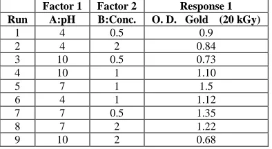
Experimental factorial design .
After carrying out 28 experiments in case of heat and 9 experiments in case of gamma irradiation effect (20kGy), reflecting different combinations of the variables (Table 2 and 3), the results revealed that run number 7 (pH 7, conc. 1 mM and 100oC) give the highest AuNPs O. D 2 (Figure 6) than others in case of heat effect and run number 5 (pH
7, conc. 1mM, 25oC and 20kGy) give the highest AuNPs optical density (O. D) 1.5 (Figure 7) than others in case
gamma radiation effect.
Figure 6 and 7 illustrated that, Peak area and O. D spectrum obtained from the reaction carried out at pH 7 is considerably higher than those obtained at other pHs (4 and 10), indicating its higher nanoparticle productivity; this is attributed to AuNPs tend to precipitation in alkaline pH values [17]. On the other hand acidic pH conditions causes a low negative value of the zeta potential so the gold tend to aggregate and precipitate [18].
The results (Figure 6 and 7) clearly indicated that 1mM concentration of Au3+ ions was most appropriate for the maximum synthesis of AuNPs. The reason for the decrease optical density with increasing AuCl3 concentration may also be because AuCl3 forms a coat on growing particles [19][15][20] and low concentration of gold chloride lead to amount of gold ion converted to metallic nanoparicles is low. The 100 oC was most appropriate for the maximum
synthesis of AuNPs (Figure 6 and 7); this is indicated to Au+ ions converted to Auo by increase temperature.
Figure 6, 7 and 8 illustrateds that temperature most effective than gamma irradiation for synthesis of gold nanoparticles, lies in the fact that high heating temperatures can significantly accelerate the nucleation and crystal growth of AuNPs. Therefore, the variation in the AuNPs obtained by different temperature using the same stabilizing and reducing agent, is believed to be due to their different perceptions of the temperature. This also may due to the thermal properties of gold, which make it easy to turn into a Nano by mode the heat.
Pleurotus stratus (2nm) its higher than optical density of AuNPs synthesized by citrus pectin (1.9); this is attributed to presence of byproducts by fermentation process (amigo acid, protein, enzymes, fibers, ….) that act as reducing agents and stabilizer. Prevent aggregation and agglomeration of synthesized metallic nanoparticles.
Table 2. Experimental factorial design experiments response in case of temperature effect
Factor 1 Factor 2 Factor 3 Response 1 Run pH Conc. mM Temp. oC O.D nm
1 10 0.5 60 0.9
2 7 2 100 1.08
3 10 2 100 0.8
4 4 1 60 0.5
5 10 2 60 0.7
6 10 1 100 1.5
7 7 1 100 2
8 7 0.5 25 0.5
9 7 1 60 1.85
10 4 2 25 0.5
11 7 2 60 0.6
12 10 1 25 0.4
13 10 1 60 0.67
14 4 0.5 60 0.74
15 4 2 60 0.65
16 7 0.5 60 0.76
17 4 1 100 1.7
18 10 0.5 100 0.8
19 10 2 25 0.43
20 10 0.5 25 0.48
21 4 2 100 0.68
22 4 1 25 0.52
23 4 0.5 25 0.49
24 7 2 25 0.73
25 7 1 25 1.7
26 7 0.5 100 1.34
27 4 0.5 100 0.86
Table 3. Experimental factorial design experiments response in case of gamma radiation effect
Factor 1 Factor 2 Response 1 Run A:pH B:Conc. O. D. Gold (20 kGy)
1 4 0.5 0.9
2 4 2 0.84
3 10 0.5 0.73
4 10 1 1.10
5 7 1 1.5
6 4 1 1.12
7 7 0.5 1.35
8 7 2 1.22
[image:9.612.209.406.449.557.2]Fig.6 UV- visible; gold nanoparticles synthesized by aqueous extract of fermented fenugreek powder using Pleurotus ostreatus (highest O.D experimental factorial design) in case of heat effect
Fig.7 UV- visible; gold nanoparticles synthesized by aqueous extract of fermented fenugreek powder using Pleurotus ostreatus (highest O.D experimental factorial design) in case of gamma radiation effect 20kGy
Fig.9 UV- visible; compare between gold nanoparticles synthesized by aqueous extract of fermented fenugreek powder using Pleurotus
ostreatus and AuNPs synthesized by citrus pectin
1.2. Transmission Electron Microscopy (TEM).
TEM images of AuNPs synthesized by citrus pectin at 5kGy and aqueous extract of fermented fenugreek powder using Pleurotus ostreatus at 100 oC Figure 10 and 11 respectively. The mean size of AuNPs citrus pectin 21nm and
AuNPs aqueous extract of fermented fenugreek powder 12nm. Figure 4a and b illustrated that size of AuNPs synthesized by biological method is smallest than that synthesized by chemical method, this is attributed to presence of byproducts during fermentation (amino acid, protein, enzymes, fibers, ….) that act as reducing agents and stabilizer.
[image:11.612.117.495.439.638.2]Fig.11 TEM Micrograph of AuNPs synthesized by aqueous extract of fermented fenugreek powder using Pleurotus ostreatus at 1000C
1.3. Dynamic light scattering (DLS).
The particle size of the synthesized AuNPs determined using DLS technique. DLS size range of AuNPs citrus pectin and AuNPs aqueous extract of fermented fenugreek powder were found to be 31.6nm and 23nm, (Figure 12 and 13), respectively. DLS size ranges of AuNPs was found to be greater than TEM size. This might be due to the fact that DLS measures hydrodynamic diameter of nanoparticles, where the amphiphilic nanoparticles were surrounded by water molecules [21]; may be attributed to be the cause of large size of capped formulation.
[image:12.612.118.503.452.595.2]Fig. 13 DLS graph of AuNPs synthesized by aqueous extract of fermented fenugreek powder using Pleurotus ostreatus at 1000C
1.4. Fourier Transform Infrared Spectroscopy (FTIR).
Figure 14 and 15 show the FTIR spectra of AuNPs citrus pectin and AusNPs aqueous extract of fermented fenugreek, respectively.
Fig. 14 shows the FT-IR spectra of citrus pectin and Au NPs prepared from citrus pectin, aiming to investigate the possible interaction between the citrus pectin molecules and Au NPs in the solution.
The FT-IR spectrum (Fig. 14) shows strong and wide stretching peak at approximately 3470 cm−1 can is ascribed to the OH stretching vibration. The absorption band at 1636 cm−1 can be attributed to the asymmetrical COO− stretching vibration and that at 1426 cm−1 to the symmetrical COO− stretching vibration [22].
The free COOH group, indicating the reduction of Au3+ to AuNPs [23] [24] in case of citrus pectin AuNPs is
slightly increase intensity with slightly shifts of the FT-IR spectrum peaks, meaning that the combination between Au nanoparticles and citrus pectin molecules is through affinity or electrostatic interaction. Therefore, the functional groups present in the citrus pectin molecules play a dual role of both reducing agent and stabilizing agent in synthesizing as well as protecting the as formed AuNPs. The AuNPs are subsequently capped and stabilized by citrus pectin because of its oxygen rich. Meanwhile, the hydroxyl groups in the polymeric chains and networks will also promote the stabilization of the AuNPs.
FTIR measurements were carried out to identify the possible biomolecules present in aqueous extract of fermented fenugreek powder which are responsible for the reduction and capping of Au NPs. The spectrum (Fig. 15) shows bands at 1715, 1641, 1350, 1234, 1077, 989 and 837 cm-1. The IR band due to C=O stretch is observed at 1715
cm-1[25]. The very strong absorption band at 1641 cm-1 is identified as the amide I and arises due to the carbonyl
stretching vibrations in the amide linkages of the proteins [26]. The band located at 1350 cm-1 is due to the C–N stretching or the O–H bending vibrations [27]. The absorption band at 1234 cm-1 is assigned as amide III band of
proteins present in the aqueous extract of fermented fenugreek powder [27]. The C–OH vibration of proteins is observed at 1077cm-1. The absorption band at 989 cm-1 is due to the C–O–C vibrations of proteins / polysaccharides
present in the aqueous extract of fermented fenugreek powder. It is well known that proteins can bind to Au NPs through free carboxylate group [28].
The presence of bands at 1641, 1234 and 1077 cm-1 indicates that Au NPs are possibly bound to proteins through
Fig. 14 FTIR spectra of AuNPs synthesized by citrus pectin
Fig. 15 FTIR spectra of AuNPs synthesized by by aqueous extract of fermented fenugreek powder using
Pleurotus ostreatus
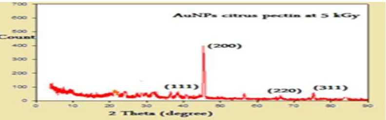
1.5
. X-Ray Diffraction (XRD)The crystal structure of AuNPs was further characterized by XRD analysis. The XRD patterns for the synthesized AuNPs by citrus pectin at 5kGy and aqueous extract of fermented fenugreek powder using Pleurotus ostreatus at 100oC are reproduced in the Figure16 and 17 respectively. The X-ray diffraction (XRD) spectrum (Figure 16 and 17) of the gold nanoparticles showed four main characteristic Bragg diffraction peaks positioned at 2θ values of 38.2298˚, 44.4052˚, 64.7315˚ and77.703˚ degrees for gold nanoparticles. All these reflections corresponded to (111), (200), (220) and (311) facets of the face centeredcubic (fcc) structure. These diffraction peaks were consistent with the standard database files of gold (JCPDS cardno. 04-0784), suggesting that the synthesized nanoparticles were of pure crystalline nature.
[image:14.612.110.506.563.687.2]Fig.17 XRD pattern the AuNPs synthesized by aqueous extract of fermented fenugreek powder using Pleurotus ostreatus at 100 0C
2.Nitrate Reductase activity.
Hence, the role of reductase in the fungal aqueous extract was investigated by nitrate reductase assay. aqueous extract of fermented fenugreek powder produced nanoparticles were stabilized by the proteins and reducing agents secreted by the fungus. Our study indicated that Pleurotus ostreatus didn’t secrete extracellular NADH-dependent nitrate reductase enzyme. That indicate other reducing factors may be responsible for the nanoparticles synthesis such as proteins, amino acid and fenugreek powder contents.
3.DPPH Free Radical Scavenging Activity% .
Table 4 and 5 shows the difference in antioxidant activities after and before AuNPs formation by green chemical and biological methods respectively. The study indicates an increase of antioxidant activities after AuNPs synthesis; This may be due to reduction of Au3+ ions into Auo metals that has scavenging activity.
The antioxidant of stabilizing and reducing agent are increased after gold nanoparticles synthesis; this confirm the gold nanoparticles has antioxidant activity and act as synergistic. AuNPs had good radical scavenging capabilities[30].
In this study the antioxidant activities decreased with radiation dose of increased; this attributed to exhaustion of antioxidant by generated free radical during exposed of aqueous solution to gamma rays Equ. (1) (2).
[image:15.612.72.549.460.582.2]The result confirm incorporation of AuNPs with natural polymers and aqueous extract of fenugreek improve its the scavenging activity.
Table 4 Scavenging activity of natural polymer (pectin, alginate and chitosan) and gold nanoparticles using DPPH for assay
Radiation dose
(kGy) Pectin
Au NPs Pectin
Radiation dose
(kGy) chitosan
Au NPs chitosan
Radiation dose
(kGy) Alginate
Au NPs Alginate
0 60.32 65.18 0 55.02 70.23 0 50.12 55.45
1 55.43 60.03 10 40.23 60.43 10 60.54 65.43
5 50.61 60.13 20 35.34 55.31 15 55.25 58.65
10 45.48 55.61 40 33.54 40.07 20 45.41 50.32
15 45.89 45.23 60 25.32 35.51 25 40.31 48.12
20 30.13 40.81 80 20.21 25.72 30 30.74 35.59
25 25.52 35.16 100 18.80 20.12 35 30.49 37.05
40 25.31 30.37
45 20.32 25.16
[image:15.612.104.508.635.704.2]50 20.13 23.79
Table 5 Scavenging activity of unfermented, fermented fenugreek (seed and powder) and AuNPs aqueous extract of fermented fenugreek powder using DPPH for assay
Radiation Dose (kGy) Seed Fermented seed Powder Fermented powder Au NPs Fermented powder
0 35.10 40.35 45.12 50.67 55.12
5 31.43 35.16 41.45 46.89 51.54
10 25.87 31.67 35.21 40.52 44.76
15 21.79 25.81 28.34 36.45 40.65
20 17.12 20.43 25.56 27.90 32.78
4.Anticancer (cytotoxic activity).
The preliminary tumor cytotoxic activities of AuNPs synthesized by citrus pectin, sodium alginate, chitosan and aqueous extract of fermented fenugreek powder were evaluated against EAC cells at 1concentrations using trypan blue 0.5% assay.
Table (6) summarize the cytotoxicity data of citrus pectin, sodium alginate, chitosan and aqueous extract of fermented fenugreek powder alone and after gold nanoparticles synthesis against EAC cells after I hour of incubation. The data were expressed as surviving percent. The results showed that EAC cells proliferation was significantly inhibited by AuNPs synthesized by citrus pectin and fermented fenugreek powder at 5kGy and 100oc respectivelly than others.
IC50 assay was performed to determine the cytotoxic property of AuNPs synthesized by citrus pectin and fermented
fenugreek powder at 5kGy and 100oC respectivelly against EAC and CACO cell lines.
Treatment of cancer cells with AuNPs at increasing concentrations (1.56 -50µg/ml; Table 7 and 8) showed cytotoxicity against EAC with 50% inhibition of cell survival (IC50) at concentration of 4.8 µg/ml (Figure 18)and
21.4µg/ml (Figure 19) for citrus pectin and fermented fenugreek powder at 5kGy and 100oC respectivelly .
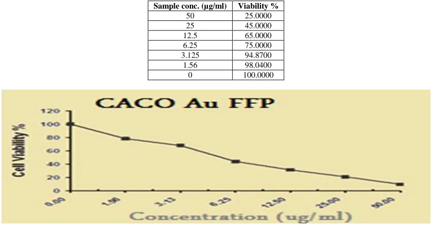
The in vitro cytotoxicity of the AuNPs was evaluated against CACO cell line at different concentrations (1.56 - 50µg/ml; Table 9 and 10); the result showed that CACO cells proliferation was significantly inhibited by AuNPs with the IC50 value 5.46 µg/ml Figure 20) and 24.4 μg/ml (Figure 21) for citrus pectin and fermented fenugreek powder at 5kGy and 100 oCrespectivelly.
In vitro cytotoxicity tests of the AuNPs citrus pectin and AuNPs fermented fenugreek it was observed that fermented fenugreek AuNPs more effective than AuNPs citrus pectin; this strongly indicated that incorporation of AuNPs modified the chemical nature of the AuNPs and cause change the interaction with other molecules such as proteins and others . The capping may prohibit the direct reaction of the cell surface and reduced the cytotoxic effect; AuNPs fermented fenugreek is more free than AuNPs citrus pectin so become very reactive, and cytotoxic to cancer cell.
Comparison of the reducing and stabilizing agents revealed that citrus pectin and fermented fenugreek produced more pronounced response and sensitivity to the Cytotoxicity. The cell viability results indicate that gold nanoparticles are toxic to the EAC and CACO cells. The incorporation of surface functionalities via citrus pectin and fermented fenugreek renders these nanoparticles highly biocompatible. The cell survival at different concentrations of gold nanoparticles stabilized with different capping agents showed variation with the increase in the concentration of the nanoparticles.
The gold nanoparticles of 21 nm and 12 nm are coated with citrus pectin (AuNPs CP) and fermented fenugreek powder (AuNPs FFP) respectivelly, taken with a series of increasing concentrations (3.25, 6.25, 12.5, 25, and 50 μg/ml). The AuNPs FFP show high cytotoxicity effect compared to AuNPs CP. This is mainly due to the fact that fermented fenugreek powder has cytotoxic effect more than citrus pectin so ac as synergism. More over the particle sizes are difference in AuNPs FFP (12 nm) than AuNps CP (21 nm).
Hence in this study the size dependent cytotoxicity and stabilizing agents are responsible for Cytotoxicity. These results are consistent with previous investigations [31] which demonstrated that the stabilizing agents has role for Cytotoxicity; Fenugreek extract has a very selective cytotoxicity against cancer cell lines such as T-cell lymphoma (TCP), B-cell lymphomas, Thyroid Papillary carcinoma (FRO) and breast cancer (MCF7). On the other hand, there was no significant cell cytotoxicity amongst normal cells, including human lymphocytes and meningioma, when treated with fenugreek [32]. This clearly indicates that fenugreek has selective cytotoxic effects against cancer cells.
Pectin seems to exert antitumor activity on different cell lines and these probably through different effects. These mechanisms dependon the structure of pectin or on the modified form of pectin that is likely to yield to various active fragments [33].
R. D
(kGy) CP Au NPs CP R. D
(kGy) Cs Au NPs C R.D.
(kGy) Alg Au NPs Alg kGy AEFFP Au NPs AEFFP
0 90.01 66 0 90.31 72 0 100 100 0 10.01 5
1 55.23 15 10 85.12 60 10 100 100 1 10.13 21
5 50.49 3 20 80.70 60 15 100 100 5 12.62 30
10 65.14 45 40 95.09 50 20 100 100 10 15.34 45
15 68.03 60 60 95.76 55 25 100 100 15 17.76 50
20 70.43 75 80 100.0 90 30 100 100 20 20.03 55
25 80.32 85 100 100.0 95 35 100 100 25 21.32 63
40 100 100
45 100 100
50 100 100
R.D = radiation dose, CP = Citrus pectin, Cs = Chitosan, Alg =Alginate, AEFFP = Aqueous extract fermented fenugreek powder
Fig.18 Cytotoxicity of AuNPs fermented fenugreek powder (AuNPs FFP) against Ehrlich Ascites Carcinoma (EAC) cells;IC 50 (4.8 µg/ml)
Table7. Surviving percent in Ehrlich Ascites Carcinoma (EAC) cells as affected by different concentrations of AuNPs fermented fenugreek powder (AuNPs FFP) after 1 hour incubation
Sample conc. (µg/ml) Viability %
50 8.0000
25 18.0000
12.5 30.0000
6.25 41.0000
3.125 65.0000
1.56 74.0000
0 100.0000
Table 8. Surviving percent in Ehrlich Ascites Carcinoma (EAC) cells as affected by different concentrations of AuNPs citrus pectin (AuNPs CP) after 1 hour incubation
Sample conc. (µg/ml) Viability %
50 25.0000
25 45.0000
12.5 65.0000
6.25 75.0000
3.125 94.8700
1.56 98.0400
0 100.0000
[image:18.612.104.514.374.593.2]Fig.20 Cytotoxicity of AuNPs fermented fenugreek powder(AuNPs FFP) against (CACO) cells;IC50 (5.46 µg/ml)
Table9. Surviving percent in (CACO) cells as affected by different concentrations of AuNPs fermented fenugreek powder (AuNPs FFP) after 1 hour incubation
Sample conc. (µg/ml) Viability %
50 9.71
25 20.95
12.5 31.28
6.25 43.97
3.125 67.85
1.56 78.19
0 100.00
Table 10. Surviving percent in (CACO) cells as affected by different concentrations of AuNPs citrus pectin (AuNPs CP) after 1 hour incubation
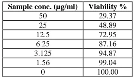
Sample conc. (µg/ml) Viability %
50 29.37
25 48.89
12.5 72.95
6.25 87.16
3.125 94.87
1.56 99.04
0 100.00
CONCLUSION
Aqueous extract of fermented fenugreek by Pleurotus ostreatus reduce gold ions to AuNPs best than natural polymers and gamma irradiation improve its synthesis, AuNPs can be incorporated with natural polymers such as citrus pectin, alginate, chitosan and fermented fenugreek powder. AuNPs inhibit the cancer cell with potent effect on EAC than CACO. Synthesized AuNPs has potential effect as antioxidant.
Acknowledgements
The authors would like to thank the Nanotechnology Research Unit (P.I. Prof. Dr. Ahmed El-Batal), Drug Radiation Research Department, National Centre for Radiation Research and Technology (NCRRT), Egypt, for financing and supporting this study under the project ”Nutraceuticals and Functional Foods Production by using Nano/Biotechnological and Irradiation Processes".
REFERENCES
[1] S A Aromal; D Philip, Spectrochimica Acta Part A: Molecular and Biomolecular Spectroscopy, 2012, 97 (2012) 1–5.
[2] M C Daniel; D Astruc, Chem. Rev., 2014, 104: 293-346. [3] P P McCue; K Shetty, Proc. Biochem., 2005, 40:1791-1797.
[4] J Li; B Kang; S Chang; Y Dai, Radiation Physics and Chemistry,2012, 76, 290–1194. [5] J L Marignier; J Belloni; M O Delcourt; J P Chevalier, Nature,1985, 317:344-345.
[6] S Nobili; D Lippi; E Witort; M Donnini; L Bausi; E Mini; S Capaccioli, Pharmacological Research, 2009, 59: 365–378.
[7] P Mukherjee; S Senapati; D Mandal; A M Ahmad; I Khan; R Kumar; M Sastry, Chem Bio Chem.pp.
2002,461-463.
[8] K Shimada; K Fujikawa; K Yahara, T Nakamura, J. Agric. Food Chem., 1992, 40, 945–948) [9] D A Ribeiro; M E Marques; D M Salvadori , Braz Dent J.,2006, 17(3):228-232
[10] T Mosann, J.Immunol.Methods, 1983, 65:55-63.
[11] Gangadevi and Muthumary, A frican Journal of biotechnology, 2007, 6:182-1386. [12] L Taihua; P G Hyun; H C Seong, Materials Chemistry and Physics, 2007,105 325–330. [13] A I EL-Batal; M R Nora; S Y Aymen; A A Magdy, Biotechnology Reports, 2015, 5: 31–39.
[14] A I EL-Batal; M H Bakry; A F Ayman; B Ahmad; S E Gharieb,. British Journal of Pharmaceutical Research,
2014, 4(11): 1341-1363.
[15a] A I El-Batal; A F El-Baz; F M Abo Mosalam; A A Tayel, J. Chem. Pharm. Res., 2013, 5(8):1-15.
[16] M Z Kassaeea; A Akhavana; N Sheikhb; R Beteshobabrud, Radiation Physics and Chemistry, 2008, 77, 9, 1074–1078 .
[17] K Eha; M Sathishkumar; JK Mao; IS wak; SY Yan, Process biochemistry, 2010, 162, 989-943.
[18] D S Aji; S Basavaraja; R Deshpande; MB Mahesh; B K Prabhakar; A Venkataraman, Colloids and surface B Biointerfaces.2009, 86, 88-92.
[19] F E Ashraf; AIB EL-Batal; MA Farag; AT Ahmed; MS Yousria; TY Shang,. Journal of Basic Microbiology,
[20] S Gurunathan; K Kalishwaralal; R Vaidyanathan; V Deepak; K Ram; S Pandian;J Muniyandi; N Hariharan; SE Hyun, Colloids and Surfaces B. Biointerfaces,2009, 74 328–335.
[21] A Akhgari;A H Garekani; F Sadeghi; M Azimaien, International Journal of Pharmaceutics, 2015, 305(1-2): p. 22-30.
[22] C Delattre; L Rios; C Laroche; NHL Le; D Lecerf; L Picton, International Journal of Biological Macro-molecules, 2009, 45, 458–462.
[23] Y Shi; X Sun, Nanjing: Jiangsu Science Technology Press. (1988).
[24] C Sun; R Qu; H Chen; C Ji; C Wang; Y Sun, CarbohydrateResearch, 2008, 343, 2595–2599. [25] D Philip, Physica E., 2010, 42 .1417–1424.
[26] K B Narayanan; N Sakthivel , Materials Research Bulletin.,2011, 46 -1708–1713. [27] A Bankar; B Joshi; A R Kumar, S Zinjarde, Colloids Surf. B, 2010, 80 45–50. [28] S L Smitha ; D Philip; K G Gopchandran, Spectrochim. Acta A., 2009, 74 .735–739. [29] S B Bukhari; M I Bhanger; S Memon, J. Anal. Environ. hem., 2008, 9 78– 83.
30] J Djajadistra; S Urnamasari; P A Pujiyanto, international Journal of Pharmacy and Pharmaceutical Sciences,
2014, 6- 0975-1491.
[31] S Vijayakumar; S Ganesan, Journal of Nanomaterials, 2012, 734398, 9.
[32] A Alsemari; F Alkhodairy; A Aldakan; M Al-Mohanna; E Bahoush; Z Shinwari; A Alaiya, Complementary and Alternative Medicine . 2014, 1472-6882-14-114.