BIROn - Birkbeck Institutional Research Online
Ferrè, E.R. and Longo, Matthew R. and Fiori, F. and Haggard, P.
(2013) Vestibular modulation of spatial perception.
Frontiers in Human
Neuroscience 7 , 00660. ISSN 1662-5161.
Downloaded from:
Usage Guidelines:
Please refer to usage guidelines at or alternatively
IN HUMAN NEUROSCIENCE
Vestibular modulation of spatial perception
Elisa Raffaella Ferre, Matthew Longo, Federico Fiori and Patrick Haggard
Journal Name: Frontiers in Human Neuroscience
ISSN: 1662-5161
Article type: Original Research Article
Received on: 06 Jun 2013
Accepted on: 23 Sep 2013
Provisional PDF published on: 23 Sep 2013
Frontiers website link: www.frontiersin.org
Citation: Ferre ER, Longo M, Fiori F and Haggard P(2013) Vestibular modulation of spatial perception. Front. Hum. Neurosci. 7:660. doi:10.3389/fnhum.2013.00660
Article URL: http://www.frontiersin.org/Journal/Abstract.aspx?s=537& name=human%20neuroscience&ART_DOI=10.3389
/fnhum.2013.00660
(If clicking on the link doesn't work, try copying and pasting it into your browser.)
Copyright statement: © 2013 Ferre, Longo, Fiori and Haggard. This is an open-access article distributed under the terms of the Creative Commons Attribution License (CC BY). The use, distribution or reproduction in other forums is permitted, provided the original author(s) or licensor are credited and that the original publication in this journal is cited, in accordance with accepted academic practice. No use, distribution or reproduction is permitted which does not comply with these terms.
Vestibular modulation of spatial perception
Elisa R. Ferrè1*, Matthew R. Longo2, Federico Fiori2,3 and Patrick Haggard1
1 Institute of Cognitive Neuroscience, University College London, London, UK
2 Department of Psychological Sciences, Birkbeck, University of London, London, UK
3 Department of Brain and Behavioural Sciences, University of Pavia, Pavia, Italy
Correspondence:
Elisa Raffaella Ferrè
Institute of Cognitive Neuroscience
17 Queen Square
London WC1N 3AR, UK
Telephone: +44 (0) 20 7679 1149
Email: [email protected]
Abbreviations:
USN: unilateral spatial neglect; GVS: galvanic vestibular stimulation; CVS: caloric vestibular
stimulation.
Keywords:
Abstract
Vestibular inputs make a key contribution to the sense of one’s own spatial location. While
the effects of vestibular stimulation on visuo-spatial processing in neurological patients have been
extensively described, the normal contribution of vestibular inputs to spatial perception remains
unclear. To address this issue, we used a line bisection task to investigate the effects of galvanic
vestibular stimulation (GVS) on spatial perception, and on the transition between near and far
space. Brief left-anodal and right-cathodal GVS or right-anodal and left-cathodal GVS were
delivered. A sham stimulation condition was also included. Participants bisected lines of different
lengths at six distances from the body using a laser pointer. Consistent with previous results, our
data showed an overall shift in bisection bias from left to right as viewing distance increased. This
pattern suggests leftward bias in near space, and rightward bias in far space. GVS induced strong
polarity dependent effects in spatial perception, broadly consistent with those previously reported in
patients: left-anodal and right-cathodal GVS induced a leftward bisection bias, while right-anodal
and left-cathodal GVS reversed this effect, and produced bisection bias toward the right side of the
space. Interestingly, the effects of GVS were comparable in near and far space. We speculate that
vestibular-induced biases in space perception may optimize gathering of information from different
1. Introduction
The sense of one’s own position, orientation and motion in three-dimensional space derives
from the integration of a variety of signals, including muscles, joints, vision, touch, and vestibular
inputs (Lackner and DiZio, 2005). The vestibular system contains two distinct structures: the
semicircular canals, which detect changes in angular acceleration, and the otolith organs, which
detect changes in linear acceleration and gravity. Both semicircular canals and otolith organs
constantly provide information to the brain regarding our body’s position and movement. Thus, the
vestibular signals are crucial to spatial perception (Villard et al., 2005; Clement et al., 2009;
Clement et al., 2012).
Several studies focussed on the vestibular contribution to spatial perception in neurological
patients. Patients with unilateral spatial neglect (USN) fail to detect objects or to perform
movements in the space contralateral to the cerebral lesion. The classic lesion site is the parietal
lobe of the right hemisphere (Bisiach and Vallar, 2000; Vallar, 1998; for review see Kerkhoff,
2001). Line bisection is one of the most common tests for assessing USN (Albert, 1973). Patients
are instructed to visually examine a horizontal line, generally presented on a sheet of paper aligned
with the patient's trunk midline, and to indicate its centre using a pencil. USN patients locate the
bisection point shifted toward the ipsilesional side of the space, so that right hemisphere damaged
patients produce a characteristic rightward error in bisection (Daini et al., 2002; Doricchi and
Angelelli, 1999; Heilman and Valenstein, 1979; Milner et al., 1993; Schenkenberg et al., 1980).
Vestibular stimulation was one of the first sensory stimulations used in order to modulate left
USN (Silberfenning, 1941). Rubens (1985) applied cold caloric vestibular stimulation (CVS) to the
auditory canal of the left ear in right brain-damaged patients. This transiently improved signs
related to USN. More recently, Rorsman and colleagues (1999) reported a reduction of USN in a
visuo-motor task (line cancellation task) during left-anodal and right-cathodal galvanic vestibular
vestibular reflex and the spontaneous recovery. Similarly, left-anodal and right-cathodal GVS
ameliorates visuo-constructive deficits in the Rey figure (Wilkinson et al., 2010) and the rightward
bias in the bisection task (Utz et al., 2011). The recovery of USN (Cappa et al., 1987; Bisiach et al.,
1991; Rode and Perenin, 1994), and the demonstration of contralateral cortical activation after
vestibular stimulation (Fasold et al., 2002) suggested an interaction between vestibular stimulation
and spatial perception: amelioration of USN may depend on the activation of cortical areas
receiving vestibular projections in the right hemisphere.
In contrast to the clear effects in patients, the contribution of vestibular inputs to space
perception in normal cognition remains unclear. On the one hand, vestibular stimulation might act
on non-specific mechanisms, such as general attention or arousal. On the other hand, vestibular
inputs might directly affect spatial processing. Several previous studies investigated low-level
visuo-vestibular mechanisms for orienting the gaze (Angelaki and Cullen, 2008), or perceiving the
subjective visual vertical (Bohmer and Mast, 1999). Rorden and colleagues (2001) found no shifts
of visuo-spatial attention following CVS in a Posner-like task (Posner, 1980). In contrast, natural
vestibular activation induced by passive whole-body rotation influenced the allocation of spatial
attention toward the side of rotation (Figliozzi et al., 2005). However, this form of vestibular
stimulation will inevitably also activate other afferents, including those from cutaneous and
proprioceptive receptors. Thus, differences between the types of vestibular stimulation used and the
consequent activations of vestibular and other afferents might explain the contrasting findings
(Lopez et al., 2012). No previous study has demonstrated a laterality-specific shift of spatial
representation in healthy participants using purely vestibular stimulation.
In the present study, therefore, we examined whether vestibular stimulation alters the
perception of position along the left-right spatial dimension. Further, we investigated whether
vestibular stimulation also influences the transition between near and far space, i.e. depth or
participants bisected lines located at several distances using a laser pointer. In standard
paper-and-pencil line bisection tasks, healthy participants generally mis-bisect horizontal lines slightly to the
left, a phenomenon known as ‘pseudoneglect’ (Bowers and Heilman, 1980; Bradshaw et al., 1987;
Chokron and Imbert, 1993; Manning et al., 1990; McCourt and Jewell, 1999; Jewell and McCourt,
2000). A number of studies have demonstrated that the leftward bias in near space shifts gradually
with increased viewing distance to become a rightward bias in far space (e.g., McCourt and
Garlinghouse, 2000; Gamberini et al., 2008; Longo and Lourenco, 2006, 2007; Lourenco and
Longo, 2009; Varnava et al., 2002). This rightward transition occurs between distances within
arm’s reach, outside of arm’s reach, as well as distances crossing this boundary, suggesting that
there is no discrete border of near space (Longo and Lourenco, 2006). Nevertheless, the rate at
which the transition occurs is correlated both with arm length (Longo and Lourenco, 2007) and with
self-reported claustrophobic fear (Lourenco et al., 2011), suggesting that the ‘size’ of near space can
be quantified in terms of how rapidly bisection biases change with viewing distance.
We delivered binaural GVS to non-invasively activate the vestibular organs (i.e., both otoliths
and semicircular canal afferents, Stephan et al., 2005). An anode and cathode were placed on the
left and right mastoid, or vice versa. Perilymphatic anodal currents hyperpolarize the trigger site
and lead to inhibition, whereas cathodal currents depolarize it resulting in excitation (Goldberg et
al., 1984). This induces a polarity-dependent ‘virtual rotation vector’ (Day and Fitzpatrick, 2005)
which can influence orientation perception and posture. More surprisingly, GVS also causes
polarity-dependent modulation of sensory and cognitive functions (see Utz et al., 2010 for a
review). These behavioural effects are consistent with neuroimaging evidence revealing
asymmetrical cortical vestibular projections in the non-dominant hemisphere (Dieterich et al.,
2003). Here we hypothesised that left-right spatial perception would be affected by GVS: we
predicted that left-anodal and right-cathodal GVS would induce a leftward bias in the line bisection
and left-cathodal, would induce a rightward bias by activating the left hemisphere. An additional
point of interest would be any interaction between vestibular stimulation and viewing distance –
such as a difference between the effects of GVS on bisection in near compared to far space.
2. Material and Methods
2.1. Participants
Fourteen naïve right-handed paid participants volunteered in the study (9 male, ages mean ±
SD: 26.7 ± 4.19 years). Participants with a history of visual, vestibular or auditory disorders were
excluded. Informed consent was obtained prior to participation in the experiment. The
experimental protocol was approved by University College London research ethics committee.
2.2. Galvanic Vestibular Stimulation
Bipolar GVS was used to deliver a boxcar pulse of 1 mA with 8 s of duration, via a
commercial stimulator (Good Vibrations Engineering Ltd., Nobleton, Ontario, Canada). Carbon
rubber electrodes (area 10 cm2) were placed binaurally over the mastoid processes and fixed in
place with adhesive tape. The areas of application were first cleaned with cotton wool soaked in
surgical spirit, and electrode gel was applied to reduce the impedance. The left-anodal and
right-cathodal configuration is named ‘L-GVS’ following previous convention (Ferrè et al., 2013). The
inverse polarity, namely left-cathodal and right-anodal configuration, is named ‘R-GVS’ (Figure
1b). This GVS configuration induces sensations of head movement, illusory perception of motion
and it evokes postural movements in the direction of the anodal ear (Day et al., 1997). A skin
tingling sensation is reported to be stronger on the cathodal side. Importantly, no long-lasting
effects have been described delivering low intensity (1mA) and short duration (8s) bipolar GVS. A
sham stimulation, ‘PSEUDO-GVS’, based on that used by Lopez et al. (2010), was applied
with left anodal and right cathodal configuration (Figure 1b). This causes a similar tingling skin
sensation to real GVS but without stimulating the vestibular organs. It functions as a control for
non-specific alerting effects. In our experiment, such non-vestibular effects could include skin
sensations generated by the GVS electrodes, and also the knowledge that an unusual stimulation is
occurring.
2.3. Stimuli and Procedure
Verbal and written instructions about the task were given to participants at the beginning of
the session. Participants performed a line bisection task during L-GVS, R-GVS or PSEUDO-GVS.
Electrodes for GVS and PSEUDO-GVS were placed at the beginning of the session and remained
in place for the entire duration of the experiment (Figure 1b). The electrodes and the polarity of
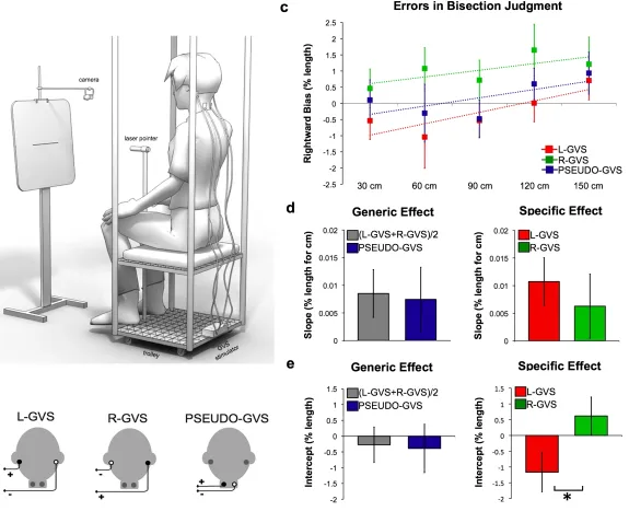
stimulation were selected under randomised computer control.
To reduce the postural consequences of vestibular input, the experiment was conducted in a
comfortable sitting position. This also reduced the tendency to tilt towards the anodal side during
GVS. Participants were seated on a movable custom-built trolley whose seat was 71 cm above the
floor. They were required to bisect lines presented at different distances in space (Figure 1a). The
participant’s head was in a neutral posture, i.e. neither tilted nor flexed, for all the duration of the
task. A laser pointer was attached to the head of a tripod fixed to the trolley 117 cm above the floor
(Figure 1a). Participants used their right hand to adjust the left-right position of the tripod head, so
as to bisect the line with the laser beam. Participants were instructed to move the beam downwards
to the floor on the left or the right side at random, after each bisection.
A panel holding the stimuli was placed in front of the participant (Figure 1a). A camera
(Logitech Webcam Pro 9000) was suspended on a tripod directly above the centre of the panel and
aligned with the lines. The camera was controlled by a computer custom-build program that
beam, and saved them for off-line coding. Stimuli consisted of lines of 10, 20 and 30 cm (1 mm in
height) centered on 29.7 cm x 42 cm sheets of paper attached horizontally to the panel. Each line
was presented 115.5 cm above the floor. The distance from the participant to the line could be 30
cm, 60 cm, 90 cm, 120 cm or 150 cm. These distances were controlled by an experimenter pushing
the trolley on which the participant was seated to the appropriate location, marked on the floor with
tape. As line length was held constant, across distances, angular size varied decreased with
increasing distance.
A total of ten blocks were administered, two for each distance from the participant to the line.
The order of blocks was randomized across participants. Each block comprised 9 trials in random
order, defined by factorial combination of line length (10 cm, 20 cm and 30 cm) and stimulation
(L-GVS, R-GVS and PSEUDO-GVS). On each trial, L-(L-GVS, R-GVS or PSEUDO-GVS was
delivered after 1 s from the beginning of the trial. Then, after 1 s, a tone signalled participants to
open their eyes, to point to the centre of the line with the laser beam, and maintain the pointing
location until a further tone occurred 6 s later. The images were captured during the interval of time
between the two tones. Participants were instructed to move the beam to the centre of the line, and
then hold it there without making further adjustments. This instruction was designed to prevent
participants from exploring the space.
*** Insert Figure 1 about here ***
2.4. Data Analysis
The pixel coordinates of each pointing laser beam, and each left and right extreme of the line
were measured on each image using ImageJ software (http://rsbweb.nih.gov/ij/). Errors in bisection
percentage of line length, and were calculated for each participant for each distance in each
experimental condition (L-GVS, R-GVS, PSEUDO-GVS).
We fitted a linear regression to model bisection error as a function of distance for each
condition and for each participant. Line bisection bias has been considered as the combination of
two factors (Longo and Lourenco, 2006). First, the slope of the relation between bisection error and
distance is a measure of the ‘size’ of near space, reflecting the rate at which the bisection bias shifts
rightward with increasing distance. Second, the intercept of the fitted lines represents the
leftward/rightward bias at hypothetical distance zero, and thus the general lateral shift of spatial
representation. Slope and intercept are logically independent (Longo and Lourenco, 2006):
experimental manipulations can induce a reduction/increase of slope without a corresponding
change in intercept or vice versa. Accordingly, estimates of slope and y-intercepts were used for
subsequent analyses.
Slope and intercept values were compared across different stimulation conditions using
planned contrasts. We hypothesised that vestibular stimulation might influence the slope and
intercept in two distinct ways (Ferrè et al., 2013a; Ferrè et al., 2013b). First, any activation of the
vestibular system might influence bisection independent of polarity and hemispheric effects,
perhaps because of generic effects such as general arousal. To test this generic hypothesis, we
compared the average of the L-GVS and R-GVS conditions to the PSEUDO-GVS condition, for
each dependent variable (slope and intercept). Second, we hypothesised that the effects of
vestibular stimulation could be specific to the hemisphere activated, and would therefore differ
between L-GVS and R-GVS conditions. Our planned contrasts thus reflect hypothesis about
plausible ways that vestibular stimulation might influence spatial perception. Distinguishing
generic and specific effects of an intervention is an established method in biosciences, and has been
3. Results
Analysis of regression slopes showed a systematic shift in the bisection bias toward the right
with increasing distance. This bias was found across all conditions (t(13) = 2.069, p=0.05) and it is
consistent with previous results (Gamberini et al., 2008; Longo and Lourenco, 2006, Longo and
Lourenco, 2007; Lourenco and Longo, 2009; Varnava et al., 2002; see data in Figure 1c).
To identify whether generic vestibular input influences the perception of space, we compared
(L-GVS+R-GVS)/2 to PSEUDO-GVS condition. This planned comparison revealed no significant
difference in slope values reflecting the transition between near and far space (t(13) = 0.264,
p=0.796). Similarly, intercept values representing the leftward/rightward bias were not different
between conditions (t(13) = 0.301, p=0.768) (Figure 1d).
To investigate the specific vestibular effect, we directly compared L-GVS to R-GVS
conditions. This contrast was designed to reveal how vestibular projections in each hemisphere
might influence the cognitive processes involved in space perception. No significant difference
were found in slope values (t(13) = 0.686, p=0.505). In contrast, intercept values revealed a
significant difference between L-GVS and R-GVS (t(13) = -3.613, p=0.003) (Figure 1e). L-GVS
induced a bias toward the left side of the space, while R-GVS toward the right. We additionally
compared each individual stimulation condition to PSEUDO-GVS. Neither GVS condition was
significantly different from the PSEUDO-GVS (p>0.05), suggesting that the effect lay in the
balance between the two hemispheric stimulations.
4. Discussion
Information from the vestibular peripheral organs in the inner ear is integrated with several
other classes of signals about the body. Low-level interactions between vestibular and visuo-spatial
information are essential in providing the organism with space representation (Villard et al., 2005;
environmental space has proved difficult to study. Here, we demonstrated that vestibular input in
general did not influence spatial processing: neither horizontal left/right spatial representation, nor
the transition between near and far space. In contrast, polarity-specific vestibular input had
differential effects on spatial perception, broadly consistent with those previously reported in USN
patients: left-anodal and right-cathodal GVS, which is considered to activate the vestibular
projections in the right hemisphere, induced a leftward bisection bias, while right-anodal and
left-cathodal GVS produced a bias towards the right side of space. These left-right biases caused by
GVS were comparable in near and far space.
GVS polarity-dependent differences in postural, sensorimotor and cognitive functions have
previously been demonstrated both in healthy volunteers and in brain damaged patients (Utz et al.,
2010). L-GVS decreases the firing rate of the vestibular nerve on the left side and increases it on
the right side (Day and Fitzpatrick 2005; Goldberg et al., 1984), while R-GVS has the opposite
effect. Neuroimaging studies have revealed asymmetrical cortical vestibular projections,
suggesting that the core region of the vestibular network is primarily located in the non-dominant
right hemisphere in right-handed subjects (Dieterich et al., 2003; Bense et al., 2001; Suzuki et al.,
2001; Janzen et al., 2008). Additionally, Fink et al. (2003) used fMRI to study the effects of bipolar
GVS. They found that left-anodal and right-cathodal L-GVS produced unilateral activation of the
right hemisphere vestibular projections, while the opposite polarity, i.e. left-cathodal and
right-anodal GVS, activated both left and right hemispheres (Fink et al., 2003).
Two alternative mechanisms could explain our results. First, R-GVS and L-GVS might
diffusely activate a large-scale hemispheric network for spatial attention. The activation of each
cerebral hemisphere would lead to a contralateral attentional bias (Corbetta et al., 1995;
Kinsbourne, 1987; Làdavas et al., 1989) as observed in our study. Alternatively, vestibular input
might project to specific cortical areas within each hemisphere involved in spatial processing.
studies identified a network of activations induced by vestibular stimulation, involving the posterior
and anterior insula, the temporoparietal junction, and the primary and secondary somatosensory
cortices (Fasold et al., 2002; Bottini et al., 1994; Emri et al., 2003); the primary motor cortex and
premotor cortex (Bense et al., 2001; Fasold et al., 2002); and the superior temporal gyrus and the
inferior parietal lobule (Bense et al., 2001; Bottini et al., 1994; Fasold et al., 2002). We speculate
that the leftward bias on bisection tasks caused by L-GVS would result from an over-excitation
cased by vestibular stimulation of the right posterior parietal cortex. Conversely, the rightward bias
induced by R-GVS could reflect the activation of the homologous areas in the left hemisphere.
However, it remains unclear if our results reflect activations which produce a diffuse imbalance
between hemispheres, or whether specific activations within each hemisphere are responsible.
Recent clinical studies in patients with peripheral vestibular disorders support the hypothesis
of spatial representation changes based on vestibular induced changes. Saj and colleagues (2013)
described severe horizontal deviations in the representation of body orientation after unilateral
vestibular loss. Interestingly, they found that only patients affected by left peripheral vestibular loss
showed changes in perception of body orientation in space, suggesting not only a role of the
vestibular system in the processing of space, but also a right hemispheric dominance.
Could the bias in bisection be a non-specific effect of GVS? For example, GVS influences
balance and postural control, producing compensatory postural sway in the direction of the anode
(Day and Fitzpatrick, 2005). However, an indirect effect on bisection by postural responses seems
unlikely. The effects induced by GVS on postural responses have been mainly demonstrated in
standing participants, while participants in our study were seated. Importantly, GVS was delivered
not at the beginning of bisection response, but several seconds before. Therefore, any postural
adjustments should have stabilised at the time of the bisection task. GVS also induced a tingling
skin sensation that could have acted as a cue for the participant. If so, GVS might change general
strongest. This would not explain the shift induced toward the anodal side in both GVS conditions
observed in our data. Thus, our results do not imply a general non-specific bias in bisecting, but
rather a high-level spatial modulation.
In conclusion, our results showed that vestibular inputs shift spatial attention towards one side
of the horizontal space, as result of the activation of the vestibular projections in the contralateral
hemisphere. Every movement of the head implies a new relation between the organism and the
surrounding space to acquire salient information from the environment. We suggest the vestibular
Conflict of Interest
The Authors declare that the research was conducted in the absence of any commercial or
Acknowledgements
ERF and PH were supported by EU Project VERE WP1. PH was additionally supported by a
Professorial Fellowship from ESRC. FF was partially supported by QUALIA (Associazione per la
References
Albert, M. L. (1973). A simple test of visual neglect. Neurology. 23, 658-664.
Angelaki, D. E., and Cullen, K. E. (2008). Vestibular system: the many facets of a multimodal
sense. Ann. Rev. Neurosci. 31, 125-150.
Bense, S., Stephan, T., Yousry, T.A., Brandt, T., and Dieterich, M. (2001). Multisensory cortical
signal increases and decreases during vestibular galvanic stimulation (fMRI). J. Neurophysiol.
85, 886-899.
Bisiach, E., and Vallar, G. (2000). Unilateral neglect in humans. In Boller, F., Grafman, J.,
Rizzolatti, G. Handbook of neuropsychology. Amsterdam, Netherlands: Elsevier Science
Publishers B.V. 459-502.
Bisiach, E., Rusconi, M. L., and Vallar, G. (1991). Remission of somatoparaphrenic delusion
through vestibular stimulation. Neuropsychologia 29, 1029-1031.
Bjoertomt, O., Cowey, A., and Walsh, V. (2002). Spatial neglect in near and far space investigated
by repetitive transcranial magnetic stimulation. Brain 125, 2012-2022.
Bohmer, A. and Mast, F. (1999). Assessing otolith function by the subjective visual vertical. Ann.
New York Acad. Sci. 871, 221-231.
Bottini, G., Sterzi, R., Paulesu, E., Vallar, G., Cappa, S. F., Erminio, F., Passingham, R. E., Frith, C.
D., and Frackowiak, R. S. (1994). Identification of the central vestibular projections in man:
A positron emission tomography activation study. Exp. Brain Res. 99, 164-169.
Bowers, D., and Heilman, K. M. (1980). Pseudoneglect: Effects of hemispace on a tactile line
bisection task. Neuropsychologia, 18(4), 491-498.
Bradshaw, J. L., Nettleton, N. C., Wilson, L. E., and Bradshaw, C. S. (1987). Line bisection by
left-handed preschoolers: a phenomenon of symmetrical neglect. Brain and Cogn. 6, 377-385.
Cappa, S., Sterzi, R., Vallar, G., and Bisiach, E. (1987). Remission of hemineglect and anosognosia
Chokron, S., and Imbert, M. (1993). Influence of reading habits on line bisection. Cogn. Brain Res.
1, 219-222.
Clement, G., Fraysse, M. J., and Deguine, O. (2009). Mental representation of space in vestibular
patients with otolithic or rotatory vertigo. Neuroreport 20, 457-461.
Clement, G., Skinner, A., Richard, G., and Lathan, C. (2012). Geometric illusions in astronauts
during long-duration spaceflight. Neuroreport 23, 894-899.
Corbetta, M., Shulman, G. L., Miezin, F. M., and Petersen, S. E. (1995). Superior parietal cortex
activation during spatial attention shifts and visual feature conjunction. Science 270, 802-802.
Daini, R., Angelelli, P., Antonucci, G., Cappa, S. F., and Vallar, G. (2002). Exploring the syndrome
of spatial unilateral neglect through an illusion of length. Exp. Brain Res. 144, 224-237.
Day, B. L., and Fitzpatrick, R. C. (2005). Virtual head rotation reveals a process of route
reconstruction from human vestibular signals. J. Physiol. 567, 591-597.
Day, B.L., Séverac Cauquil, A., Bartolomei, L., Pastor, M.A., and Lyon, I.N. (1997). Human
body-segment tilts induced by galvanic stimulation: a vestibularly driven balance protection
mechanism. J Physiol. 500 (Pt 3):661-72.
Dieterich, M., Bense, S., Lutz, S., Drzezga, A., Stephan, T., Bartenstein, P., and Brandt, T. (2003).
Dominance for vestibular cortical function in the non-dominant hemisphere. Cereb. Cortex
13, 994-1007.
Doricchi, F., and Angelelli, P. (1999). Misrepresentation of horizontal space in left unilateral
neglect: role of hemianopia. Neurology 52, 1845-1845.
Emri, M., Kisely, M., Lengyel, Z., Balkay, L., Marian, T., Miko, L., Berenyi, E., Sziklai, I., Tron,
L., and Toth, A. (2003). Cortical projection of peripheral vestibular signaling. J.
Fasold, O., von Brevern, M., Kuhberg, M., Ploner, C. J., Villringer, A., Lempert, T., and Wenzel, R.
(2002). Human vestibular cortex as identified with caloric stimulation in functional magnetic
resonance imaging. Neuroimage 17, 1384-1393.
Ferrè, E. R., Vagnoni, E., and Haggard, P. (2013a). Galvanic vestibular stimulation influences
randomness of number generation. Exp. Brain Res 224, 233-241.
Ferrè, E. R., Vagnoni, E., and Haggard, P. (2013b). Vestibular contributions to bodily awareness.
Neuropsychologia. 51(8):1445-1452.
Figliozzi, F., Guariglia, P., Silvetti, M., Siegler, I., and Doricchi, F. (2005) Effects of vestibular
rotatory accelerations on covert attentional orienting in vision and touch. J. Cogn. Neurosci.
17, 1638-1651.
Fink, G. R., Marshall, J. C., Weiss, P. H., Stephan, T., Grefkes, C., Shah, N. J., Zilles, K., and
Dieterich, M. (2003). Performing allocentric visuospatial judgments with induced distortion
of the egocentric reference frame: an fMRI study with clinical implications. Neuroimage 20,
1505-1517.
Gamberini, L., Seraglia, B., and Priftis, K. (2008). Processing of peripersonal and extrapersonal
space using tools: evidence from visual line bisection in real and virtual environments.
Neuropsychologia 46, 1298-1304.
Goldberg, J. M., Smith. C. E., and Fernandez, C. (1984). Relation between discharge regularity and
responses to externally applied galvanic currents in vestibular nerve afferents of the squirrel
monkey. J. Neurophysiol. 51, 1236-1256.
Heilman, K. M., and Valenstein, E. (1979). Mechanisms underlying hemispatial neglect. Ann.
Neurol. 5, 166-170.
Janzen, J., Schlindwein, P., Bense, S., Bauermann, T., Vucurevic, G., Stoeter, P., and Dieterich, M.
(2008). Neural correlates of hemispheric dominance and ipsilaterality within the vestibular
Jewell, G. and McCourt, M.E. (2000). Pseudoneglect: A review and meta-analysis of performance
factors in line bisection tasks. Neuropsychologia, 38, 93-110.
Kerkhoff, G. (2001). Spatial hemineglect in humans. Prog. Neurobiol. 63, 1-28.
Kinsbourne, M. (1987). Mechanisms of unilateral neglect. In: Neurophysiological and
Neuropsychological Aspects of Spatial Neglect. Jeannerod M,. Amsterdam: Elsevier Science
Publishers. 69-86.
Lackner, J. R., and DiZio, P. (2005). Vestibular, proprioceptive, and haptic contributions to spatial
orientation. Annu. Rev. Psychol., 56, 115-147.
Làdavas, E., Del Pesce, M., and Provinciali, L. (1989). Unilateral attention deficits and hemispheric
asymmetries in the control of visual attention. Neuropsychologia 27, 353-366.
Longo, M. R., and Lourenco, S. F. (2006). On the nature of near space: effects of tool use and the
transition to far space. Neuropsychologia 44, 977-981.
Longo, M. R., and Lourenco, S. F. (2007). Space perception and body morphology: extent of near
space scales with arm length. Exp. Brain Res. 177, 285-290.
Lopez, C., Blanke, O. and Mast, F.W. (2012) The human vestibular cortex revealed by
coordinate-based activation likelihood estimation meta-analysis. Neuroscience. 212:159-79.
Lopez, C., Lenggenhager, B. and Blanke, O. (2010) How vestibular stimulation interacts with
illusory hand ownership. Conscious Cogn.19(1):33-47.
Lourenco, S. F., and Longo, M. R. (2009). The plasticity of near space: evidence for contraction.
Cognition 112, 451-456.
Lourenco, S. F., Longo, M. R., and Pathman, T. (2011). Near space and its relation to
claustrophobic fear. Cognition 119, 448-453.
McCourt, M.E. and Garlinghouse, M. (2000). Asymmetries of visuospatial attention are modulated
by viewing distance and visual field elevation: Pseudoneglect in peripersonal and
McCourt, M.E. and Jewell, G. (1999). Visuospatial attention in line bisection: Stimulus modulation
of pseudoneglect. Neuropsychologia, 37, 843-855.
Manning, L., Halligan, P. W., and Marshall, J. C. (1990). Individual variation in line bisection: a
study of normal subjects with application to the interpretation of visual neglect.
Neuropsychologia 28, 647-655.
Milner, A. D., Harvey, M., Roberts, R. C., and Forster, S. V. (1993). Line bisection errors in visual
neglect: misguided action or size distortion? Neuropsychologia 31, 39-49.
Posner, M. I., (1980). Orienting of attention. Q J Exp Psychol 32, 3–25.
Rode, G., and Perenin, M. T. (1994). Temporary remission of representational hemineglect through
vestibular stimulation. Neuroreport 5, 869-872.
Rorden, C., Karnath, H. O., and Driver, J. (2001). Do neck-proprioceptive and caloric-vestibular
stimulation influence covert visual attention in normals, as they influence visual neglect?
Neuropsychologia 39, 364-375.
Rorsman, I., Magnusson, M., and Johansson, B. B. (1999). Reduction of visuo-spatial neglect with
vestibular galvanic stimulation. Scand. J. Rehab. Med. 31, 117-124.
Rubens, A. B. (1985). Caloric stimulation and unilateral visual neglect. Neurology 35, 1019-1024.
Saj, A., Honoré, J., Bernard-Demanze, L., Devèze, A., Magnan, J., and Borel, L. (2013). Where is
straight ahead to a patient with unilateral vestibular loss? Cortex 49, 1219-1228.
Schenkenberg, T., Bradford, D. C., and Ajax, E. T. (1980). Line bisection and unilateral visual
neglect in patients with neurologic impairment. Neurology 30, 509-517.
Schmidt, L., Utz, K.S., Depper, L., Adams, M., Schaadt, A.K., Reinhart, S., and Kerkhoff, G.
(2013) Now You Feel both: Galvanic Vestibular Stimulation Induces Lasting Improvements
in the Rehabilitation of Chronic Tactile Extinction. Front Hum Neurosci. 7:90.
Silberfenning, J. (1941). Contribution to the problem of eye movements. Disturbances of ocular
Stephan, T., Deutschländer, A., Nolte, A., Schneider, E., Wiesmann, M., Brandt, T., and Dieterich,
M. (2005). Functional MRI of galvanic vestibular stimulation with alternating currents at
different frequencies. Neuroimage 26, 721-732.
Suzuki, M., Kitano, H., Ito, R., Kitanishi, T., Yazawa, Y., Ogawa, T., Shiino, A., and Kitajima, K.,
(2001). Cortical and subcortical vestibular response to caloric stimulation detected by
functional magnetic resonance imaging. Cogn. Brain Res. 12, 441-449.
Utz, K. S., Dimova, V., Oppenländer, K., and Kerkhoff, G. (2010). Electrified minds: transcranial
direct current stimulation (tDCS) and galvanic vestibular stimulation (GVS) as methods of
non-invasive brain stimulation in neuropsychology—a review of current data and future
implications. Neuropsychologia 48, 2789-2810.
Utz, K. S., Keller, I., Kardinal, M., and Kerkhoff, G. (2011). Galvanic vestibular stimulation
reduces the pathological rightward line bisection error in neglect—a sham
stimulation-controlled study. Neuropsychologia 49, 1219-1225.
Vallar, G. (1998). Spatial hemineglect in humans. Trends Cogn. Sci. 2, 87-97.
Varnava, A., McCarthy, M., and Beaumont, J. G. (2002). Line bisection in normal adults: direction
of attentional bias for near and far space. Neuropsychologia 40, 1372-1378.
Villard, E., Garcia-Moreno, F. T., Peter, N., and Clement, G. (2005). Geometric visual illusions in
microgravity during parabolic flight. Neuroreport 16, 1395-1398.
Wilkinson, D., Zubko, O., DeGutis, J., Milberg, W., and Potter, J. (2010). Improvement of a figure
copying deficit during subsensory galvanic vestibular stimulation. J. Neuropsychol. 4,
Legend for figures
Figure 1. Effects of Galvanic Vestibular Stimulation (GVS) on spatial perception.
(a) Experimental set-up. (b) GVS polarities and electrodes configurations. (c) Errors in bisection
judgment. Raw data in each condition in function of the distances and fitted linear regression
(dashed lines). (d) Slope data as a function of GVS condition. (e) Intercept data as a function of