Open Access
Technical notes
Dual stack black blood carotid artery CMR at 3T: Application to
wall thickness visualization
Axel Bornstedt*, Mathias Burgmaier, Vinzenz Hombach, Nikolaus Marx and
Volker Rasche
Address: Department of Internal Medicine II - Cardiology, University Hospital Ulm, Albert-Einstein-Allee 23, Ulm, 89081, Germany
Email: Axel Bornstedt* - [email protected]; Mathias Burgmaier - [email protected]; Vinzenz Hombach - [email protected]; Nikolaus Marx - [email protected]; Volker Rasche - [email protected]
* Corresponding author
Abstract
Background: The increasing understanding of atherosclerosis as an important risk factor for the development of acute ischemic events like ischemic stroke has stimulated increasing interest in non-invasive assessment of the structure, composition and burden of plaque depositions in the carotid artery wall. Vessel wall imaging by means of cardiovascular magnetic resonance (CMR) is conventionally done by 2D dual inversion recovery (DIR) techniques, which often fail in covering large volumes of interest as required in plaque burden assessment. Although the technique has been extended to 2D multislice imaging, its straight extension to 3D protocols is still limited by the prolonged acquisition times and incomplete blood suppression. A novel approach for rapid overview imaging of large sections of the carotid artery wall at isotropic spatial resolutions is presented, which omits excitation of the epiglottis. By the interleaved acquisition of two 3D stacks with the proposed motion sensitized segmented steady-state black-blood gradient echo technique (MSDS) the coverage of the carotid artery trees on both sides in reasonable scan times is enabled.
Results: 10 patients were investigated with the proposed technique and compared to conventional transversal DIR turbo spin and gradient echo approaches centered at the height of the carotid bifurcation. In all MSDS experiments sufficient black-blood contrast could be obtained over the entire covered volumes. The contrast to noise ratio between vessel and suppressed blood was improved by 73% applying the motion sensitizing technique. In all patients the suspicious areas of vessel wall thickening could be clearly identified and validated by the conventional local imaging approach. The average assessable vessel wall segment length was evaluated to be 18 cm. While in 50% of the cases motion artifacts could be appreciated in the conventional images, none were detected for the MSDS technique.
Conclusion: The proposed technique enables the time efficient coverage of large areas of the carotid arteries without compromising wall-lumen CNR to get an overview about detrimental alterations of the vessel wall. Thickening of the vessel wall can be identified and the suspicious segments can be targeted for subsequent high-resolution CMR. The exclusion of the epiglottis may further facilitate reduction of swallowing induced motion artifacts.
Published: 10 November 2009
Journal of Cardiovascular Magnetic Resonance 2009, 11:45 doi:10.1186/1532-429X-11-45
Received: 8 April 2009 Accepted: 10 November 2009
This article is available from: http://www.jcmr-online.com/content/11/1/45
© 2009 Bornstedt et al; licensee BioMed Central Ltd.
Background
The well-established understanding of atherosclerosis as an important risk factor for the development of acute ischemic events has stimulated increasing interest in non-invasive assessment of the structure, composition and burden of plaque depositions in the carotid artery wall. Over the last decade, cardiovascular magnetic resonance (CMR) has proven to contribute substantially to the non-invasive assessment and characterization of atheroscle-rotic lesions, especially in the aorta and the carotid arteries [1-3].
For vessel wall imaging, black-blood contrast is required for ensuring sufficient contrast between the vessel wall and blood. Most commonly, suppression of the blood sig-nal is achieved by applying a double-inversion recovery (DIR) black-blood preparation prepulse [4]. Most pub-lished approaches combine the DIR preparation with two-dimensional (2D) imaging by means of multi spin-echo (MSE) techniques [1-3]. Without compromising spatial resolution due to severe T2 apodization, this technique allows for the acquisition of a single slice in the minute time range, while providing sufficient signal to noise ratio (SNR) and lumen-wall contrast (CNR). Different image contrasts including T2 - (T2W), proton density (PDW) and, limited by the required inversion recovery time for blood signal nulling and heart rate, T1 - weighting (T1W) can be realized by slight modifications of the sequence timing. While the DIR technique has been extended to enable multislice acquisitions [5], its application for three-dimensional (3D) coverage of large sections of the arteries at sufficient isotropic spatial resolution has been limited. The resulting long acquisition times often cause image-distorting motion artifacts [6] due to swallowing, arterial pulsation and breathing. Incomplete blood sup-pression [7,8] caused by insufficient exchange of the blood in the entire volume may mimic incorrect wall thickening. Time-efficient 3D imaging of the carotid and aortic arteries has been achieved by applying segmented gradient echo techniques [9-11]. Swallowing motion arti-facts have been addressed by utilization of navigators monitoring the position of the epiglottis [10,12] and arte-rial pulsation artifacts can be reduced by synchronizing the data acquisition with the heart beat. Improvement of blood suppression has been obtained by phase-sensitive reconstruction techniques [13] and diffusion-prepared or motion sensitized preparation techniques [10,9,14], all relying rather on the motion of the blood than on com-plete blood exchange between preparation and readout.
A major limitation of arterial wall imaging by CMR still results from the long acquisition times, which do not allow covering large sections of the arteries at sufficient spatial resolution for plaque characterization. Fast three-dimensional imaging methods, providing an overview on
the arterial tree with sufficient spatial resolution and con-trast, appear attractive for targeting the lesion of interest and planning of the subsequent high-resolution CMR plaque characterization protocol.
We recently developed a 3D localized volume-selective sequence for arterial wall imaging [15]. By combining a 2D pencil beam excitation with motion sensitized black-blood prepared imaging, large segments of the carotid artery tree could be covered in reasonable image acquisi-tion times. Although the moacquisi-tion sensitizing prepulse causes a preweighting of the spins similar to a T2 prepara-tion pulse of about 15 ms length, an excellent wall lumen contrast could be maintained and the reduced spatial res-olution was still sufficient for assessment of the vessel wall thickness. Motion artifacts due to swallowing could not be observed, most-likely due to the local character of the excitation, which ensures that the region of the epiglottis does not contribute to the final MR image. Limitations of this technique rise from the rather long duration of the 2D excitation pulses, which is required to avoid substantial side-loops in the 2D excitation profile and the restriction to acquire a single carotid artery tree, only.
In this contribution, a new dual-stack approach is sug-gested, in which two parasagittally aligned 3D volumes (stacks) are acquired temporally interleaved. Black-blood contrast is achieved by motion-sensitized preparation, which is applied prior to the acquisition of each tempo-rally interleaved stack. As in the local excitation approach, the proposed technique omits excitation of the epiglottis and is supposed to show similar advantages regarding swallowing artifacts. The efficiency of the motion-sensitiz-ing magnetization preparation dual stack gradient echo (MSDS) sequence is demonstrated in patients for the rapid overview imaging of long segments of the carotid artery wall for both sides simultaneously. The applicabil-ity of the technique for identification of thickened vessel wall segments was investigated in direct comparison to the conventional high-spatial resolution protocols.
Methods
The method was evaluated in 10 patients with a proven vascular disease defined as arteriosclerosis confirmed by the presence of coronary artery disease, peripheral artery disease or carotid plaque (10 males, mean age 58 +/- 7 y, mean weight 83 +/- 6 kg) without any contraindications for CMR. Written informed consent was obtained from all subjects before CMR and the protocol was approved by the ethics committee of the University.
dedicated two segment four-element carotid coil (Philips Research Europe, Germany). Either segment comprises two independent coil elements with spatial extent of 65 × 50 mm2 each. The two segments were located on either side of the neck and fixated with a Vac-Lok neck cushion (Medtec, USA), which also immobilized the head of the patient to a great extent.
Localizer
For localization, a multislice gradient-echo technique acquiring three orthogonal slabs with transversal, sagittal, and coronal slice orientation was applied. The resulting scout images were used for planning of the subsequent scans.
Coil-Sensitivity Reference Scan
A coil-sensitivity reference scan was acquired, which was needed for the homogenization of the image signal and the vendor specific parallel imaging reconstruction, respectively.
Angiograms
For targeting the position of the carotid arteries and bifur-cations, fast thick slab phase contrast angiograms (see Table 1 for sequence details) were obtained in coronal and sagittal orientation.
Volume Imaging of the Carotid Arteries
A 3D motion sensitized, fat suppressed, segmented, spoiled gradient echo dual stack sequence (MSDS) was developed. As reported previously [9,10,14,15],
suppres-sion of blood signal was accomplished by a motion sensi-tizing spin preparation. The preparation comprised a 90°x-180°y-90°-x-driven equilibrium technique with inte-grated balanced magnetic field gradients for dephasing the spins of moving blood. Suppression of residual trans-versal magnetization was achieved by subsequently applied spoiler gradients. The motion sensitizing or as previously called diffusion weighting strength (B-value) of the preparation pulse was chosen according to [10]. Overall preparation duration resulted as 21.5 ms for a cumulated B-value of 17.1 s mm-2 in all three directions
applying a 15 mT m-1 gradient amplitude for 4.5 ms
[image:3.612.57.554.478.729.2]before and after the 180° refocusing pulse (slew rate 200 mT/(m ms), total gradient area 137 ms mT/m, first gradi-ent momgradi-ent 1029 mT ms2/m). The left and right carotid artery tree was covered by two independently planned sag-ittal volumes of dimension 150 × 30 × 200 mm at iso-tropic spatial resolution between 0.83 mm3 and 1 mm3. Planning was done based on the phase contrast angi-ograms as shown in figure 1. During planning, it was ensured that the pharynx was not contained in the cov-ered volumes of interest (VOI). To allow sufficient recov-ery of longitudinal magnetization, subsequent segments of the same stack were either acquired at a fixed user-defined spacing TRS of 700 - 1000 ms or a single RR inter-val. The acquisition of the second stack was interleaved and started TRS/2 after starting the acquisition of the first stack. The preparation was applied once prior to the acquisition of each segment (Figure 1 bottom) to ensure sufficient blood suppression in both stacks. Sequence details are provided in Table 1.
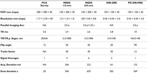
Table 1: Scan parameters for the acquisitions performed.
PCA Survey
MSDS 1.0 mm
MSDS 0.8 mm
DIR-GRE PD DIR-MSE
FOV mm (mps) 300 × 300 × 50 150 × 200 × 30 150 × 200 × 30 150 × 150 × 20 150 × 150 × 20
Resolution mm (mps) 1.17 × 2.34 × 50 1.0 × 1.0 × 1.0 0.8 × 0.8 × 0.8 0.45 × 0.45 × 2.0 0.45 × 0.45 × 2.0
Parallel Imaging Acc. NA 2.0 p 2.0 p/1.25 s NA 2.0 p
TE ms 5.6 2.4 2.6 2.8 10
TR/TR p. Segm. ms 20/NA 5.2/1000 5.5/1000 5.4/1HB NA/3 HB
Flip angle 15 20 20 20 90
Turbo factor NA 40 40 30 12
Signal Averages 1 4 6 2 1
Acq. Duration ms NA 206 222 161 125
For comparison, a high-resolution proton-density weighted 3D double-inversion recovery multi spin echo sequence (DIR-MSE) and a 3D double-inversion recovery segmented gradient echo sequence (DIR-GRE) in axial view orientation were performed centered around the bifurcation of the respective carotid artery. For suppres-sion of swallowing motion artifacts, a pencil beam navi-gator gating technique, initially developed for cardiac respiratory motion suppression [16], was applied in which the position of the epiglottis was monitored. Sequence details are provided in Table 1.
Data Analysis
For image analysis, the data was transferred to a dedicated CMR image processing workstation (ViewForum, R2.6, Philips Medical Systems). The multi planar reformat tool was applied to define a path along the centerline of the common, external and internal carotid arteries (ACC, ACE and ACI). The endpoints of the paths were identified either at the location where the vessel exceeds the imaging volume or by a drop of the contrast CW between vessel wall and suppressed blood signal below 3
( ). The length of the vessels was
meas-ured individually for the ACC (starting from the carina
downwards) and the ACE/ACI (starting from the central ostia upwards).
Comparison of the suggested MSDS technique and the conventional DIR-MSE technique was done after axial reformatting of the MSDS data.
The effectiveness of the blood suppression of the motion sensitizing prepulse in comparison to the standard DIR technique was evaluated using the DIR-GRE and the MSDS data sets, as both sequences are based on nearly identical gradient echo acquisitions. Contrast to noise ratios, normalized to the voxelsize, were determined as follows: CNRnorm = (Swall - Slumen)/(stddevlumen * voxelsize). The ROIs were drawn in the vicinity of the carotid bifurca-tion at corresponding locabifurca-tions to ensure that the coil sen-sitivity and intrinsic SNR properties were identical.
The performance of the new technique for the reduction of swallowing motion induced artifacts was qualitatively assessed by direct comparison of images acquired with the dual-stack approach with images acquired by the DIR-MSE and DIR-GRE acquisition techniques.
Ultrasound Acquisition
Morphology of the ACC, ACI and ACE were displayed real-time with a high-resolution 9 MHz broadband linear array transducer (iU22 Ultrasound System, Philips
Elec-CW Signalhigh
Signallow
= −1
Example of the location of the two stacks, planned on the thick-slab PCA images and acquisition order in MSDS
Figure 1
tronics, Bothell, WA, USA). Representative pictures were taken and compared to CMR.
Results
The imaging experiments could be completed successfully in all subjects. In all experiments sufficient suppression of the blood signal could be obtained over the imaged VOI, while preserving the signal of the vessel wall. The CNRnorm
was 52 +/- 7 for the MSDS measurements, while it was 30 +/- 14 for the DIR-GRE; indicating the superior blood sig-nal suppression and robustness of the motion sensitizing technique. Figure 2 shows two examples of reformatted images acquired at spatial resolutions of 0.83 and 1.0
mm3, demonstrating the obtainable image quality and
coverage of the suggested technique. The arrows indicate atherosclerotic, partly calcified lesions, which were exem-plarily confirmed by ultrasound. The inset demonstrates a vessel length measurement on a curved MPR indicating (crosses) a path of 110 mm length. Figure 3 shows a direct comparison of the original MSDS data to the ultrasound image at the according position, showing a severely calci-fied lesion in the left carotid artery bifurcation.
Visual inspection of the endovascular vessel wall revealed sufficient delineation over the entire accessible course of the carotid artery, while the contrast between the epivas-cular wall and the surrounding tissue was less pro-nounced, especially in the more distal sections of the ACE
and ACI. No distinct motion artifacts (e. g. ghosting) introduced by swallowing could be observed.
The average length of the vessel wall, which could clearly be depicted, was 38.8 8.1 mm for the ACC, 77.5 +/-14.6 mm for the ACI and 62.8 +/- 9.4 mm for the ACE. The overall assessable length (ACC + ACI + ACE) resulted to 179.1 +/- 18.1 mm for each of the 24 carotid vessel trees analyzed. The stopping criteria for the path length of the ACC was the drop of contrast and SNR respectively in 100% of the cases since the placement of the coil was restricted by the musculature of the neck. ACE/ACI paths could be followed to the border of the VOI in 83% of the cases.
In all patients vessel wall thickening or plaque could be seen in the DIR-MSE technique and could also be clearly identified from the MSDS images, indicating the applica-bility of the MSDS technique to fast identification of the location of atherosclerotic lesions. In one case, the full extent of the wall thickening was missed in the DIR-MSE images due to insufficient coverage of the distal section of the ACI. A direct comparison of DIR-MSE and MSDS images is provided in figure 4. Please note the superior blood suppression in the MSDS technique even so a sub-stantially larger section of the carotid arteries was covered. Quantitative assessment of the vessel wall thickness in MSDS was limited by the achievable spatial resolution.
Examples of dual stack (L: left side, R: right side) carotid imaging (reformats) acquired in two patients (left patient 0.83 mm3,
right patient 1.0 mm3 resolution) Figure 2
Examples of dual stack (L: left side, R: right side) carotid imaging (reformats) acquired in two patients (left patient 0.83 mm3, right patient 1.0 mm3 resolution). The images clearly reveal vessel wall thickening (arrow heads) as
While 1.0 mm3 to 0.83mm3, as reported here, appeared generally feasible, the acquisition of 0.73 mm3 resolution datasets, tested in volunteers, yielded only satisfying results in subjects with vessels located in close proximity to the skin surface and thus the coil.
None of the MSDS images showed swallowing related image artifacts, where for the acquisition of the conven-tional double-inversion recovery multi-spin echo (DIR-MSE) and respective gradient-echo (DIR-GRE) tech-niques, navigators had to be used for avoiding severe motion artifacts caused by swallowing. Despite the navi-gator technique, in 50% of the DIR-MSE and DIR-GRE images motion artifacts were still observable in 50% of the cases.
Discussion
In this work, a 3D motion-sensitized segmented dual-stack gradient echo (MSDS) technique was developed for fast black blood CMR of large sections of the carotid arter-ies. The motion-sensitized preparation revealed the potential for providing excellent blood signal suppression and contrast to noise ratios over large segments of the carotid arteries without obvious loss of signal intensity of stationary tissue and enabled dual stack acquisition with-out prolongation of the image acquisition time. Sufficient wall lumen contrast was achieved in the ACC, the ACE and the ACI enabling a clear delineation of the vessel wall over a mean length 179 +/- 18 mm. The interleaved acqui-sition of the right and left carotid artery tree by the sug-gested dual stack approach enabled rapid acquisition of
Original CMR slice data cropped (a) and zoomed to lesion, indicated by arrows (b)
Figure 3
Original CMR slice data cropped (a) and zoomed to lesion, indicated by arrows (b). The ultrasound image (9 MHz) at the according position, showing the severely calcified lesion in the left carotid artery bifurcation, is depicted in (c). Images correspond to the patient depicted on the right in Fig. 2.
Comparison of PD TSE DIR BB (top center) with motion sensitized dual stack (MSDS) GRE
Figure 4
an overview visualization of the carotid artery tree at iso-tropic spatial resolutions between 0.83 mm3 and 1 mm3 in less than 10 minutes. Compared to the alternative approach of acquiring a single coronal slab, the dual-stack approach is supposed to be less sensitive to motion arti-facts caused by swallowing [6], since the region of the epi-glottis is not included in the excited VOI and hence does not contribute to the final image. Cancellation of signal from blood by motion-sensitized preparation clearly holds the potential to generate black blood over large seg-ments of the carotid arteries, which cannot be achieved by the competing DIR technique, since the blood is not totally exchanged over the large volumes addressed in this contribution. Even though, blood in the carotid arteries is mainly flowing in FH direction, best blood suppression was achieved by applying motion sensitizing gradients in all three spatial directions simultaneously.
The most promising application of the proposed MSDS technique is supposed to rise in the field of rapid global assessment of vessel properties and wall thickness in the carotid arteries for rapid identification of those locations along the vessel wall showing local thickening. Here, especially the isotropic spatial resolution, which enables almost lossless reformatting of the data along the center-line is supposed to be beneficial for identification of ves-sel wall thickening. Due to some limitations in spatial resolution, the proposed technique may not yet be suited to replace the well-established acquisition protocols for plaque characterization published earlier [1,3] and closer inspection of suspicious locations on the vessel wall with multi-contrast high resolution imaging is required for more detailed plaque analysis.
The application of MSDS for high-resolution imaging may become feasible with the advent of dedicated coils provid-ing improved SNR in combination with better parallel imaging capabilities. Due to the coverage of large seg-ments of the carotid arteries by the MSDS approach, patient motion between subsequent scans will not cause a targeted lesion to leave the VOI, as may be the case in con-ventional axially aligned DIR-MSE imaging approaches due to the limited coverage of the vessel in feet-head (FH) direction. This property of the MSDS technique may have further advantages with respect to longitudinal monitor-ing of plaque progression, since the larger covered VOI is supposed to ensure that the location of the lesion is cov-ered completely even in case the VOI is not being planned perfectly the same. Furthermore, registrations based on a large VOI are supposed to be more precise than registra-tions performed on a small VOI. The suggested motion-sensitized dual-stack approach is not limited to gradient echo techniques and can be combined with other approaches for generating variable image contrast
Conclusion
In conclusion, the 3D MSDS technique appears to be a very promising technique for rapid artifact-free overview imaging of the vessel wall of the carotid arteries with excellent black-blood contrast. Its application to high-res-olution multi-contrast imaging is, in principle, possible but remains to be proven.
Competing interests
The University Hospital Ulm and Philips Medical Systems have a collaborative research and development agreement for cardiovascular magnetic resonance imaging. No other financial conflicts of interest are identified.
Authors' contributions
AB, VH, NM and VR have made substantial contributions to conception and design of the study and have been involved in drafting the manuscript or revising it. AB and VR have developed the methods and carried out the data analysis. MB was responsible for the ultrasound studies and revised the manuscript. All authors read and approved the final manuscript.
Acknowledgements
Supported by the Else Kröner - Fresenius Stiftung (P28/2004)
Unrestricted grant from Takeda Pharmaceutical Company Limited
References
1. Yuan C, Mitsumori LM, Beach KW, Maravilla KR: Carotid athero-sclerotic plaque: noninvasive MR characterization and iden-tification of vulnerable lesions. Radiology 2001, 221:285-99. 2. Saam T, Hatsukami TS, Takaya N, Chu B, Underhill H, Kerwin WS,
Cai J, Ferguson MS, Yuan C: The vulnerable, or high-risk, athero-sclerotic plaque: noninvasive MR imaging for characteriza-tion and assessment. Radiology 2007, 244:64-77.
3. Watanabe Y, Nagayama M, Suga T, Yoshida K, Yamagata S, Okumura A, Amoh Y, Nakashita S, Van Cauteren S, Dodo Y: Characteriza-tion of atherosclerotic plaque of carotid arteries with his-topathological correlation: vascular wall MR imaging vs. color Doppler ultrasonography (US). J Magn Reson Imaging 2008, 28:478-85.
4. Edelman RR, Chien D, Kim D: Fast selective black blood MR imaging. Radiology 1991, 181:655-60.
5. Yarnykh VL, Yuan C: Multislice double inversion-recovery black-blood imaging with simultaneous slice reinversion.
Journal of Magnetic Resonance Imaging 2003, 17:478-483.
6. Boussel L, Herigault G, de la Vega A, Nonent M, Douek PC, Serfaty JM: Swallowing, arterial pulsation, and breathing induce motion artifacts in carotid artery MRI. J Magn Reson Imaging 2006, 23:413-5.
7. Luk-Pat GT, Gold GE, Olcott EW, Hu BS, Nishimura DG: High-res-olution three-dimensional in vivo imaging of atherosclerotic plaque. Magn Reson Med 1999, 42:762-71.
8. Crowe LA, Gatehouse P, Yang GZ, Mohiaddin RH, Varghese A, Char-rier C, Keegen J, Firmin DN: Volume-selective 3D turbo spin echo imaging for vascular wall imaging and distensibility measurement. J Magn Reson Imaging 2003, 17:572-80.
9. Koktzoglou I, Li D: Diffusion-prepared segmented steady-state free precession: Application to 3D black-blood cardiovascu-lar magnetic resonance of the thoracic aorta and carotid artery walls. J Cardiovasc Magn Reson 2007, 9:33-42.
10. Koktzoglou I, Li D: Submillimeter isotropic resolution carotid wall MRI with swallowing compensation: imaging results and semiautomated wall morphometry. J Magn Reson Imaging 2007,
Publish with BioMed Central and every scientist can read your work free of charge
"BioMed Central will be the most significant development for disseminating the results of biomedical researc h in our lifetime."
Sir Paul Nurse, Cancer Research UK
Your research papers will be:
available free of charge to the entire biomedical community
peer reviewed and published immediately upon acceptance
cited in PubMed and archived on PubMed Central
yours — you keep the copyright
Submit your manuscript here: BioMedcentral 11. Lin H, Flask CA, Dale BM, Duerk JL: Rapid dark-blood carotid
vessel-wall imaging with random bipolar gradients in a radial SSFP acquisition. J Magn Reson Imaging 2007, 25:1299-304. 12. Crowe LA, Keegan J, Gatehouse PD, Mohiaddin RH, Varghese A,
Symmonds K, Cannell TM, Yang GZ, Firmin DN: 3D volume-selec-tive turbo spin echo for carotid artery wall imaging with nav-igator detection of swallowing. J Magn Reson Imaging 2005,
22:583-8.
13. Crowe LA, Varghese A, Mohiaddin RH, Yang GZ, Firmin DN: Elimi-nation of residual blood flow-related signal in 3D volume-selective TSE arterial wall imaging using velocity-sensitive phase reconstruction. J Magn Reson Imaging 2006, 23:416-21. 14. Wang J, Yarnykh VL, Hatsukami T, Chu B, Balu N, Yuan C: Improved
suppression of plaque-mimicking artifacts in black-blood carotid atherosclerosis imaging using a multislice motion-sensitized driven-equilibrium (MSDE) turbo spin-echo (TSE) sequence. Magn Reson Med 2007, 58:973-81.
15. Bornstedt A, Bernhardt P, Hombach V, Kamenz J, Spiess J, Subgang A, Rasche V: Local excitation black blood imaging at 3T: applica-tion to the carotid artery wall. Magn Reson Med 2008,
59:1207-11.