© 2017, IRJET | Impact Factor value: 5.181 | ISO 9001:2008 Certified Journal
| Page 1693
Tumor segmentation using Improved watershed transform for the
application to mammogram image compression
Amrutha Varshini N
1, K S Babu
21
MTech Student, Biomedical Signal Processing and Instrumentation, Dept. of IT, SJCE, Mysuru,
Karnataka, India
2
Assistant Professor, Dept. of IT, SJCE, Mysuru, Karnataka, India
---***---Abstract -In this work, an automatic image segmentation method is used for the tumor segmentation from mammogram images by means of improved watershed transform using prior information. The segmented regions are then applied to perform a loss and lossless compression for the storage efficiency according to the importance of region data. These are mainly performed in two procedures, including region segmentation and region compression. In the first procedure, an Improved watershed transform based on intrinsic prior information is then adopted to extract tumor boundary. Finally, the tumor regions are detected , and are segmented in mammogram. In the second procedure, Discrete Cosine Transform(DCT) is applied on the regions with different compression rates according to the importance of region data so as to simultaneously reserve important tumor features and reduce the size of mammograms for storage efficiency. Experimental results show that the proposed method gives promising results in the compression applications.
Key Words: Image segmentation, Mammogram, Tumor,
Improved watershed transform, Discrete Cosine Transform, Compression.
1.INTRODUCTION
Breast cancer is the most common one for women in world-wide. The women disease incidence rate that the breast cancer leaps to first poses the quite big threat to the domestic women. The Digital X-ray mammography is an effective method to achieve the goal of early diagnoses. Accordingly, the accurate segmentation of tumor in mammogram images in very important.
Image segmentation plays an important role and is an essential process in medical images. General segmentation is the process of partitioning the image into disjointed regions so as to the characteristics of each region are homogeneous. A large variety of image segmentation methods have been presented. Amongthese methods, the watershed that is a region-based approach is a traditional but popular method from mathematical morphology. The region-based approaches group similar pixels into a region based on some pixel information. The advantages of watershed approach are that it is fast, it can be parallelized, and it produces a complete division of image even if its contrast is low. Many
techniques related to the watersheds have been presented in recent decades. The main drawback of watershed algorithm is thatit produces over-segmented results. That is, when the watershed obtains catchment basins from the gradient of image, the results of watershed contain too many small regions. Moreover, it is sensitive to noise. Local variations of the image can significantly change the results. In addition, it is poor detection in significant areas with low contrast boundaries. If the signal to noise ratio is not high enough at the contour, the watershed transform will be unable to detect it accurately. Accordingly, the improved version of watershed algorithm may be overcome the intrinsic problems. In addition, various kinds of pre-processing have been developed to solve the problems of over-segmentation, such as the median filter and anisotropic diffusion filter. In this study, an automatic image segmentation method based on improved watershed transform using prior information is proposed for the tumor segmentation from mammogram images.
Moreover, for medical applications, we should be very cautious to retain sufficient image information in supporting different diagnosis purposes so we will go for image compression.
Image compression is very important for efficient transmission and storage of images. Demand for communication of multimedia data through the telecommunications network and accessing the multimedia data through Internet is growing explosively .With the use of digital cameras, requirements for storage, manipulation, and transfer of digital images, has grown explosively . These image files can be very large and can occupy a lot of memory. A gray scale image that is 256 x 256 pixels has 65, 536 elements to store, and a a typical 640 x 480 colour image has nearly a million. Downloading of these files from internet can be very time consuming task. Image data comprise of a significant portion of the multimedia data and they occupy the major portion of the communication bandwidth for multimedia communication. Therefore development of efficient techniques for image compression has become quite necessary.
© 2017, IRJET | Impact Factor value: 5.181 | ISO 9001:2008 Certified Journal
| Page 1694
categories: lossless and lossy image compression. JPEGprocess is a widely used form of lossy image compression that centres around the Discrete Cosine Transform. The DCT works by separating images into parts of differing frequencies. During a step called quantization, where part of compression actually occurs, the less important frequencies are discarded, hence the use of the term “lossy”. Then only the most important frequencies that remain are used to retrieve the image in the decompression process. As a result, reconstructed images contain some distortion, but these levels can be adjusted during the compression stage.
2.METHODOLOGY
To achieve the objective, the appearance model has built. this appearance model comprises of four major steps:
Image preprocessing: Preprocessing images commonly involves removing low-frequency background noise, normalizing the intensity of the individual particles images, removing reflections, and masking portions of images.
Feature extraction: It is a type of dimensionally reduction that efficiently represents interest parts of an image as a compact feature vector.
Segmentation: Image segmentation is the process of partitioning a digital image into multiple segments(set of pixels, also known as super-pixels).
Compression: It is a technique that reduces the size of an image file without affecting or degrading its quality to a greater extent. Compression applied to digital images, to reduce their cost for storage or transmission.
Fig- 1: System overview
2.1 Pre-Processing
Preprocessing mainly involves those operations that are normally necessarily prior to the main goal analysis and extraction of the desired information and normally geometric corrections of the original actual image. These improvements includes correcting the data for irregularities and unwanted atmospheric noise, removal of non-brain
element image and converting the data so they correctly reflected in the original image. As soon as mammogram image has been acquired, a preprocessing is performed to remove noise and clean up the image background.
2.2 Segmentation
Segmentation is an image processing operation which aims to partition an image into homogenous regions composed of pixels with the same characteristics according to predefined criteria. In breast mammogram analysis, image segmentation is commonly used for measuring and visualizing the breasts anatomical structures, for analyzing the micro calcifications and detecting the cancerous cells in mammogram.
Image segmentation aims at partitioning an image into meaningful parts having similar features and properties. Watershed executes a main role in image segmentation field. the results of segmentation are used to border detection and object recognition. In this context, the standard Euclidean distance helps in finding the neighbouring pixels. A weighted distance measure utilizing pixel coordinates ,pixel intensity, and image texture is also used.
2.2.1 Improved watershed transform
An improved watershed transform based on intrinsic prior information is adopted to extract tumor boundary from the breast. We first introduce important definitions of watershed transform about the lower slope and lower neighbours. Let f be a gray image. The lower slope of f at a pixel p, LS(p) is defined,
LS(p)= max ((fp)- f(q) / d(p,q ))
Where N(p) is set of neighbours of p, and d(p,q) is the Euclidean distance between p and q. To define a steepest slope relation between pixel is necessary for lower slope, which will be used to calculate the watershed transform.
LN(p)= N(p) (fp-fp0)
The set of lower neighbours is the subset of neighbouring pixels for which the directed gradient to the pixel p equals the lower slope.
In practical applications, the watershed transform is not calculated directly on the image, but rather on the absolute value of its gradient, which has high values at the contours. Gradient estimation at the centre of the pixels reduces the original resolution of images.
LS(p)= max f(p)- f(q)/ feðp; qÞ
where feðp; qÞ is to quantify the probability of having an
© 2017, IRJET | Impact Factor value: 5.181 | ISO 9001:2008 Certified Journal
| Page 1695
The improved watershed transform produces apossibility for different applications, depending on the amount of knowledge available on the objects. A function is used to measure the difference in class probability between two neighbouring pixels for generic image segmentation. Normal distributions are assumed for the objects in the image, for which mean and variance are calculated using a set of seed pixels for each class. The seeds are selected using automatic techniques.
2.3 Feature Extraction
1. Mean
The mean, m of the pixel values in the defined window, estimates the value in the image in which central clustering occurs. The mean can be calculated using the formula:
Where p(i,j) is the pixel value at point (i,j) of an image of size MxN.
2. Standard Deviation
The standard deviation ,is the estimate of mean square deviation of gray pixel value p(i,j) from its mean value m. Standard deviation describes the dispersion within the local region. It is determined by the formula,
3. Variance
Variance is the square root of standard deviation. The formula for finding Variance is:
Where SD is the Standard Deviation.
2.4 Lossy compression
In the technique of Lossy compression, it decreases the bits by recognizing the not required information and by eliminating it. The system of decreasing the size of the file of data is commonly termed as the data-compression, though its formal name is the source-coding that is coding get done at source of data before it gets stored or sent. In these methods few loss of the information is acceptable. Dropping non-essential information from the source of data can save the storage area. The Lossy data-compression methods are aware by the researches on how the people anticipate data in the question. As an example, the human eye is very sensitive to slight variations in the luminance as compare that there are so many variations in the colour. The Lossy image compression technique is used in the digital cameras, to raise
the storage ability with the minimal decline of the quality of picture. Similarly in the DVDs which uses the lossy MPEG-2 Video codec technique for the compression of the video.
2.4.1 Discrete Cosine Transform
Original image is divided into blocks of 8 x 8.
Pixel values of a black and white image range from 0-255 but DCT is designed to work on pixel values ranging from -128 to 127 .Therefore each block is modified to work in the range and is used to calculate DCT matrix.
DCT is applied to each block by multiplying the modified block with DCT matrix on the left and transpose of DCT matrix on its right.
Each block is then compressed through quantization.
Quantized matrix is then entropy encoded.
Compressed image is reconstructed through reverse process.
Inverse DCT is used for decompression.
3.RESULTS
Fig-2: Input image
© 2017, IRJET | Impact Factor value: 5.181 | ISO 9001:2008 Certified Journal
| Page 1696
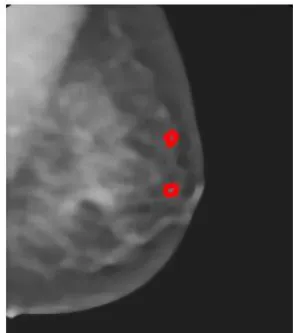
[image:4.595.76.250.330.516.2]Fig-4: Cancer spot segmented
Fig-5: Compressed image
Fig-6: Reconstructed original image
3.1 Performance evaluation
Recall or Sensitivity( Tpr)
Recall or Sensitivity is the proportion of Real Positive cases that are correctly Predicted Positive. This measures the Coverage of the Real Positive cases by the +P (Predicted Positive) rule. Its desirable feature is that it reflects how many of the relevant cases the +P rule picks up. In a Medical context Recall is moreover regarded as primary, as the aim is to identify all Real Positive cases.
Precision( Tpa)
Precision or Confidence denotes the proportion of Predicted Positive cases that are correctly Real Positives. It can however analogously be called True Positive Accuracy(TPA) , being a measure of accuracy of Predicted Positives in contrast with the rate of discovery of Real Positives (TPR).
F- measure:
[image:4.595.308.559.508.655.2]A measure that combines precision and recall is the harmonic mean of precision and recall, the traditional F-measure or balanced F-score.
[image:4.595.88.235.566.733.2]© 2017, IRJET | Impact Factor value: 5.181 | ISO 9001:2008 Certified Journal
| Page 1697
Chart -1: Preformance plot
4. CONCLUSIONS
This work presents an effective method for detection and segmentation of tumor in mammogram images. Here a region based segmentation and compression method is used. The mammogram images are detected and segmented by means of improved watershed transform using prior information. The segmented regions are then applied to loss or lossless compression for the storage efficiency according to the importance of region data.
The experimental results shows that it is an effective method for tumor segmentation. After segmentation, mammogram images are further compressed. The Discrete Cosine Transform( DCT) is used with different compression rate to reserve important details and reduce the size of the mammogram image for storage or transmission. This experiment results also shows that the presented method can reconstruct very well in the applications of mammogram image compression.
REFERENCES
[1] Boren Li and Mao Pan, “An Improved Segmentation of High Spatial Resolution Remote Sensing Image using Marker-based Watershed Algorithm,‟‟ pp. 98-104, IEEE,2012.
[2] Xiaoyan Zhang, Lichao Chen, Lihu Pan and Lizhi Xiong, “Study on the Image Segmentation Based On ICA and Watershed Algorithm,” Fifth International Conference on Intelligent Computation Technology and Automation, pp. 978-912, IEEE 2012.
[3] Zhonglin Xia, Danfeng Hu and Xueyan Hu, “Watershed Algorithm Based On Segmentation,”, Xian institute of posts and telecommunications, pp.103-107, 2011.
[4] Li Cheng, Li Yan and Fan Yan Shangchun, “CCD infrared image segmentation using watershed algorithm,” Third
International Conference on Measuring Technology and Mechatronics Automation, vol.1, pp.680-683, IEEE,2011.
[5] Quan Longzhe, “Automatic Segmentation method of Touching Corn Kernels in Digital Image Based on Improved Watershed Algorithm,” pp.34-37, IEEE, 2011.
[6] GuiMei Zhang and Ming-Ming Zhou, “Labelling watershed algorithm based on Morphological Reconstruction in Colour Space,” pp.51-55, IEEE, 2011.
[7] Jun Tang, “A Colour Image Segmentation algorithm Based on Region Growing,” vol.6, pp.634-637, IEEE, 2010.
[8] Gupta, Maneesha, and Amit Kumar Garg. "Analysis Of Image Compression Algorithm Using DCT." Georgian Electronic Scientific Journal: Computer Science and Telecommunications 3 (2012).